Wrist and Hand: Difference between revisions
Kim Jackson (talk | contribs) No edit summary |
Kim Jackson (talk | contribs) No edit summary |
||
| Line 4: | Line 4: | ||
'''Top Contributors''' - {{Special:Contributors/{{FULLPAGENAME}}}} | '''Top Contributors''' - {{Special:Contributors/{{FULLPAGENAME}}}} | ||
</div> | </div> | ||
<div class="noeditbox">This page is currently undergoing work, but please come back later to check out new information!!</div> | |||
== Introduction == | == Introduction == | ||
Revision as of 16:00, 25 July 2018
Original Editors - Rachael Lowe
Top Contributors - Kim Jackson, Rachael Lowe, Lucinda hampton, Laura Ritchie, George Prudden, Joao Costa, Wendy Snyders, Admin, Cheryl Rentchler, Mande Jooste, Tony Lowe, Evan Thomas, Nikhil Benhur Abburi and Abdallah Ahmed Mohamed
Introduction[edit | edit source]
The upper limb has sacrificed locomotor function and stability for mobility, dexterity and precision. The hand sits at the end of the upper limb and is a combination of complex joints whose function is to manipulate, grip and grasp- this is made possible by the opposing movement of the thumb.[1]
Bony Anatomy[edit | edit source]
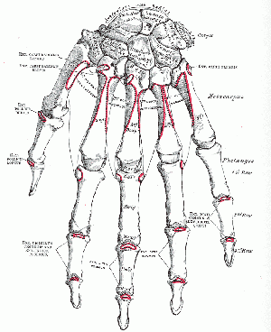
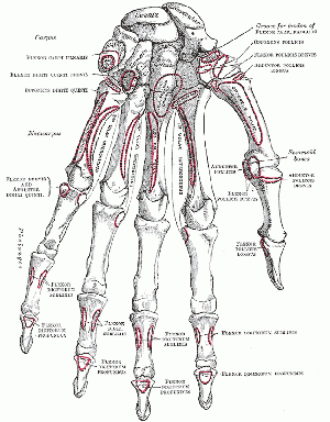
The hand and wrist have a total of 27 bones arranged to roll, spin and slide[2]; allowing the hand to explore and control the environment and objects. The hand is divided into three regions[3]:
- Proximal region of the hand is the carpus (wrist)
- The middle region the metacarpus (palm)
- The distal region the phalanges (fingers).
The Carpus[edit | edit source]
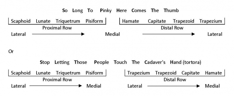
The carpus consists of eight bones, sitting in two rows, with four bones in each row (figure 1). The carpus controls length-tension relationships in the multiarticular hand muscles and to allow fine adjustment of grip.[4]
Three of the bones in the proximal row articulate with the radius forming the radiocarpal joint and distally with the distal carpal forming the midcarpal joint. The four carpal bones in the distal row articulate with the bases of the five metacarpal bones forming the carpometacarpal joints[5]. The joints formed between the carpal bones are known as the intercarpal joints and most are of the plane synovial type[1], as the bones interlock with each other the rows are sometimes referred to as two single synovial joints[1].
The arrangement of the bones and ligaments allows very little movement between bones[1], but they do slide contributing to the finer movements of the wrist[6]. The exception to this is the capitate which has a larger range of movement[1]. The small bones are named after the shape they resemble.
Proximal Row[5][edit | edit source]
- Scaphoid – (boat like)– anterior surface palpable tubercle. Articulates proximally with radius, medially with lunate and distally with the head of the capitate. It is a common site of fracture- 70% of all carpal fractures[5], often injured by a fall onto an outstretched limb
- Lunate – (moon shaped) – Its palmar surface is smooth and convex and is larger than its dorsal surface. Proximally it articulates with the radius and articular disc, medially with the triquetrum, laterally with the scaphoid and distally with the head of the capitate
- Triquetrum – (three cornered) – Nestles in the space between the lunate and hamate. When the hand is adducted it enters the radiocarpal joint.
- Pisiform – (pea shaped) = a small round bone found in the tendon of flexor carpi ulnaris. It articulates with the palmar surface of the triquetrum. The anterior surface projects distally and laterally forming the medial part of the carpal tunnel.
Distal Row[5][edit | edit source]
- Trapezium – four sided figures with no two sides parallel – the most irregular, with a palpable tubercle and groove anterior medially. It articulates proximally with the scaphoid and medially with the trapezoid. Its articular surface is saddle-shaped and contributes to the mobility of the carpometacarpal joint of the thumb
- Trapezoid – four sided figure with two parallel sides – Articulates distally with the second metacarpal, laterally with the trapezium, proximally with the scaphoid and medially with the capitate
- Capitate - head shaped – The largest of all the carpal bones sitting centrally and articulating with the lunate and scaphoid, medially with the hamate and laterally with the trapezoid. The distal surface articulates mainly with the base of the third metacarpal but also by narrow surfaces with the bases of the second and fourth metacarpals.
- Hamate – hooked – This is wedge shaped with a curved palpable hook projecting from the palmar surface near the base of the fifth metacarpal.
The following pneumonic makes it easy to remember the position of each bone:
The Carpal Tunnel – formed by the anterior concave space formed by the pisiform and hamate – on the ulnar side and the scaphoid and trapezium – on the radial side, with a roof like covering of the flexor retinaculum (strong fibrous bands of connective tissue). The long flexor tendons of the digits and thumb and the median nerve pass through the carpal tunnel
The Metacarpus[edit | edit source]
The metacarpus, the palm of the hand, which is made up of five bones – the metacarpals. The bones are numbered laterally, from the thumb, 1 – 5. Each bone is long with a proximal quadrilateral base, a shaft (body) and a distal rounded head. The base of the first metacarpal is saddle-shaped and articulates with he trapezium. The base of the second metacarpal articulates with the trapezium, trapezoid and capitate. The base of the third metacarpal articulates with the capitate. The bases of the fourth and fifth metacarpal articulate with the hamate. The bases of the second to fifth metacarpals also articulate with each other.
The heads of the metacarpals, commonly known as knuckles, are smooth and rounded and extend onto the palmar surface – these become visible when the fist is clenched[3]. The head of the first metacarpal is wider than the others, having two sesamoid bones, usually found in the short tendons crossing the joint, which articulate with the palmar part of the joint surface. The heads fit into a concavity on the base of the proximal phalanx at the metacarapophlangeal joints.
The Phalanges[edit | edit source]
The phalanges, the fingers, consist of 14 long bones. Apart from the thumb (the pollex) each phalanx has three bones, the distal, middle and proximal phalanx – the thumb has only two distal and proximal. As with the metacarpals the phalanges are numbered 1-5 starting at the thumb. The proximal phalanx is large and is concave for articulation with the head of the metacarpal. The shaft is curved along its length being convex dorsally. It is convex from side to side on its dorsal surface and flat on the palmar surface. The distal end, the head, is smaller and convex to articulate with the next bone in sequence. In order from the thumb digits are also known as the index finger, middle finger, ring finger and little finger.
Clinical Examination[edit | edit source]
- Wrist & Hand Examination
- Special Tests
- Outcome Measures
Conditions[edit | edit source]
- Carpel Tunnel Syndrome
- De Quervains
- Wrist Sprain
- Carpel Instability
- Colles Fracture
- Smiths Fracture
- Scaphoid Fracture
- Wrist & Hand Osteoarthritis
- Rheumatoid Arthritis
- Complex Regional Pain Syndrome
- Triangular Fibrocartilage Complex Injuries
- Gamekeeper’s Thumb
- Blackberry Thumb
- Lunate Instability
- Hamate Fracture
- Ape Hand
- Benediction Hand
- Claw Hand
- Dupuytren’s Contracture
- Metacarpal Fractures
- Extensor Mechanism Injuries
- Flexor Tendon Injuries
- Ulnocarpal Impaction Syndrome
- Lunotriquetral Ligament Tears
Procedures[edit | edit source]
References[edit | edit source]
- ↑ 1.0 1.1 1.2 1.3 1.4 Palastanga N, Soames R. Anatomy and Human Movement: Structure and Function. 6th Ed. London: Churchill Livingstone, 2012.
- ↑ Maitland, G.D. Maitland's Peripheral Manipulations. 3rd Edition Edinburg: Elsevier Butterworth-Heinemann, 1999.
- ↑ 3.0 3.1 Physical Examination of the Spine and Extremities. Hoppenfield, S. New York: Appleton-Century-Crofts, 1976.
- ↑ Levangie PK, Norkin CC. Joint Structure and Function: A Comprehensive Analysis. 5th Ed. Philadelphia: F A Davis Company, 2011
- ↑ 5.0 5.1 5.2 5.3 Principles of Anatomy & Physiology. Tortora GJ, Derrickson B. 13th Ed. NJ: John Wiley & Sons, Inc, 2012.
- ↑ Cael C. Functional Anatomy: Musculoskeletal Anatomy, Kinesiology, an Palpation for Manual Therapists. Lippincott Williams & Wilkins, 2009.


