Subtalar Joint: Difference between revisions
No edit summary |
No edit summary |
||
| Line 18: | Line 18: | ||
# The posterior subtalar joint: Allows the to glide backward (posterior articulation) | # The posterior subtalar joint: Allows the to glide backward (posterior articulation) | ||
The talus and calcaneous bones are held in place by strong but flexible connective ligaments. The main ligament that attaches these bones is called the interosseous talocalcaneal ligament. Four other weaker ligaments provide the joint with added stability<ref>Very well health Subtalar joint Available: https://www.verywellhealth.com/what-is-the-subtalar-joint-1337686<nowiki/>(accessed 5.6.2022)</ref>. The interosseous talocalcaneal ligament, acts to bind the talus and calcaneus together. It lies within the sinus tarsi (a small cavity between the talus and calcaneus), and is particularly strong; providing the majority of the ligamentous stability to the joint<ref name=":0" /><ref>Kenhub Subtalar joint Available: https://www.kenhub.com/en/library/anatomy/subtalar-joint<nowiki/>(accessed 5.6.2022)</ref>. | The talus and calcaneous bones are held in place by strong but flexible connective ligaments. The main ligament that attaches these bones is called the interosseous talocalcaneal ligament. Four other weaker ligaments provide the joint with added stability<ref name=":1">Very well health Subtalar joint Available: https://www.verywellhealth.com/what-is-the-subtalar-joint-1337686<nowiki/>(accessed 5.6.2022)</ref>. The interosseous talocalcaneal ligament, acts to bind the talus and calcaneus together. It lies within the sinus tarsi (a small cavity between the talus and calcaneus), and is particularly strong; providing the majority of the ligamentous stability to the joint<ref name=":0" /><ref>Kenhub Subtalar joint Available: https://www.kenhub.com/en/library/anatomy/subtalar-joint<nowiki/>(accessed 5.6.2022)</ref>. | ||
In between the calcaneus and talus is the synovial membrane. This tissue secretes fluid to lubricate the joint space, protecting the cartilage and bones from damage. | In between the calcaneus and talus is the synovial membrane. This tissue secretes fluid to lubricate the joint space, protecting the cartilage and bones from damage. | ||
| Line 32: | Line 32: | ||
* Pronation requires a combination of dorsiflexion, abduction, and eversion. | * Pronation requires a combination of dorsiflexion, abduction, and eversion. | ||
* Supination requires a combination of plantar flexion, adduction and inversion | * Supination requires a combination of plantar flexion, adduction and inversion<ref name=":1" /> | ||
== Physiotherapy Relevance == | |||
The subtalar joint is vital to mobility. It is prone to wear and tear, trauma, and joint-specific disorders. Any damage done to the subtalar joint and any connective tissues that support it can trigger pain, lead to foot deformity (often permanent), and affect gait and mobility. The damage may be broadly described as capsular or non-capsular. | |||
== References == | == References == | ||
Revision as of 03:21, 5 June 2022
Original Editor - Lucinda hampton
Top Contributors - Lucinda hampton, Vidya Acharya, Abbey Wright and Kim Jackson
Introduction[edit | edit source]
The subtalar (ST) joint is an articulation between two of the tarsal bones in the foot, the talus and calcaneus. The joint is classed structurally as a synovial joint, and functionally as a plane synovial joint[1].
The ST joint allows the foot side-to-side (inversion/eversion), pivot to change directions, and stay balanced as we move across uneven terrain. Without this joint, a person would constantly roll their ankles when running, jumping, or walking.
Structure[edit | edit source]
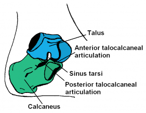
The ST joint is multi-articular joint, with three articulated facets that provide a surface for the joint to glide:
- The anterior subtalar joint : Allows joint to glide forward (anterior articulation)
- The medial subtalar joint: Allows joint to glide side-to-side (eversion/inversion)
- The posterior subtalar joint: Allows the to glide backward (posterior articulation)
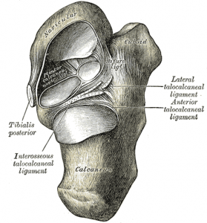
The talus and calcaneous bones are held in place by strong but flexible connective ligaments. The main ligament that attaches these bones is called the interosseous talocalcaneal ligament. Four other weaker ligaments provide the joint with added stability[2]. The interosseous talocalcaneal ligament, acts to bind the talus and calcaneus together. It lies within the sinus tarsi (a small cavity between the talus and calcaneus), and is particularly strong; providing the majority of the ligamentous stability to the joint[1][3].
In between the calcaneus and talus is the synovial membrane. This tissue secretes fluid to lubricate the joint space, protecting the cartilage and bones from damage.
Movements[edit | edit source]
The ST joint is formed on an oblique axis and is the main site within the foot for generation of eversion and inversion movements. This movement is produced by the muscles of the lateral compartment of the leg and tibialis anterior muscle respectively. The subtalar joint has no role in plantar or dorsiflexion of the foot[1].
Function[edit | edit source]
The ST joint is key to many functional activities eg walking and running, and posture while performing these tasks. The mechanisms behind how the subtalar joint propels you us is very complex.
This ST joints primary movements involve supination, in which the foot rolls toward the body's midline, and pronation, in which the foot rolls away from the midline. Both of these movements require a combination of distinct actions.
- Pronation requires a combination of dorsiflexion, abduction, and eversion.
- Supination requires a combination of plantar flexion, adduction and inversion[2]
Physiotherapy Relevance[edit | edit source]
The subtalar joint is vital to mobility. It is prone to wear and tear, trauma, and joint-specific disorders. Any damage done to the subtalar joint and any connective tissues that support it can trigger pain, lead to foot deformity (often permanent), and affect gait and mobility. The damage may be broadly described as capsular or non-capsular.
References[edit | edit source]
- ↑ 1.0 1.1 1.2 Teach me anatomy The subtalar loint Available:https://teachmeanatomy.info/lower-limb/joints/subtalar/ (accessed 5.6.2022)
- ↑ 2.0 2.1 Very well health Subtalar joint Available: https://www.verywellhealth.com/what-is-the-subtalar-joint-1337686(accessed 5.6.2022)
- ↑ Kenhub Subtalar joint Available: https://www.kenhub.com/en/library/anatomy/subtalar-joint(accessed 5.6.2022)


