Subtalar Joint: Difference between revisions
No edit summary |
No edit summary |
||
| Line 10: | Line 10: | ||
The subtalar joint allows the foot side-to-side (inversion/eversion), pivot to change directions, and stay balanced as we move across uneven terrain. Without this joint, a person would constantly roll their ankles when running, jumping, or walking. | The subtalar joint allows the foot side-to-side (inversion/eversion), pivot to change directions, and stay balanced as we move across uneven terrain. Without this joint, a person would constantly roll their ankles when running, jumping, or walking. | ||
== | == Structure == | ||
[[File:Subtalar Joint.png|thumb|Subtalar | [[File:Subtalar Joint.png|thumb|Subtalar ligaments Superior view]] | ||
The subtalar joint is multi-articular joint, with three articulated facets that provide a surface for the joint to glide: | The subtalar joint is multi-articular joint, with three articulated facets that provide a surface for the joint to glide: | ||
The anterior subtalar joint ( | # The anterior subtalar joint : Allows joint to glide forward (anterior articulation) | ||
# The medial subtalar joint: Allows joint to glide side-to-side (eversion/inversion) | |||
# The posterior subtalar joint: Allows the to glide backward (posterior articulation) | |||
The | The talus and calcaneous bones are held in place by strong but flexible connective ligaments. The main ligament that attaches these bones is called the interosseous talocalcaneal ligament. Four other weaker ligaments provide the joint with added stability<ref>Very well health Subtalar joint Available: https://www.verywellhealth.com/what-is-the-subtalar-joint-1337686<nowiki/>(accessed 5.6.2022)</ref>. | ||
The | The interosseous talocalcaneal ligament is composed of two short and broad fibrous bands located in the tarsal sinus. Occupying the central position between the talocalcaneal and talocalcaneonavicular joints, this ligament is associated with the functions of both joints. The primary role of this ligament in the subtalar joint is maintaining stability both at rest and during active movements. Being attached to the talar sulcus and calcaneal sulcus, the interosseous talocalcaneal ligament is taut in pronation of the foot, limiting its movement<ref>Kenhub Subtalar joint Available: https://www.kenhub.com/en/library/anatomy/subtalar-joint<nowiki/>(accessed 5.6.2022)</ref>. | ||
In between the calcaneus and talus is the synovial membrane. This tissue secretes fluid to lubricate the joint space, protecting the cartilage and bones from damage. | In between the calcaneus and talus is the synovial membrane. This tissue secretes fluid to lubricate the joint space, protecting the cartilage and bones from damage. | ||
Revision as of 03:04, 5 June 2022
Original Editor - Lucinda hampton
Top Contributors - Lucinda hampton, Vidya Acharya, Abbey Wright and Kim Jackson
Introduction[edit | edit source]
The subtalar joint is an articulation between two of the tarsal bones in the foot, the talus and calcaneus. The joint is classed structurally as a synovial joint, and functionally as a plane synovial joint[1].
The subtalar joint allows the foot side-to-side (inversion/eversion), pivot to change directions, and stay balanced as we move across uneven terrain. Without this joint, a person would constantly roll their ankles when running, jumping, or walking.
Structure[edit | edit source]
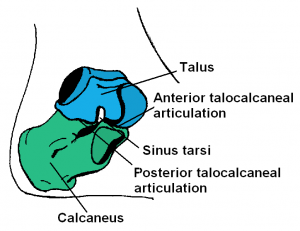
The subtalar joint is multi-articular joint, with three articulated facets that provide a surface for the joint to glide:
- The anterior subtalar joint : Allows joint to glide forward (anterior articulation)
- The medial subtalar joint: Allows joint to glide side-to-side (eversion/inversion)
- The posterior subtalar joint: Allows the to glide backward (posterior articulation)
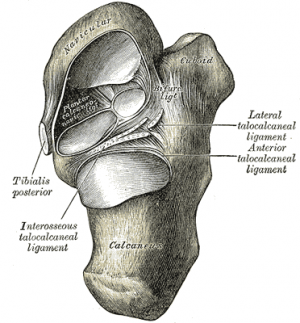
The talus and calcaneous bones are held in place by strong but flexible connective ligaments. The main ligament that attaches these bones is called the interosseous talocalcaneal ligament. Four other weaker ligaments provide the joint with added stability[2].
The interosseous talocalcaneal ligament is composed of two short and broad fibrous bands located in the tarsal sinus. Occupying the central position between the talocalcaneal and talocalcaneonavicular joints, this ligament is associated with the functions of both joints. The primary role of this ligament in the subtalar joint is maintaining stability both at rest and during active movements. Being attached to the talar sulcus and calcaneal sulcus, the interosseous talocalcaneal ligament is taut in pronation of the foot, limiting its movement[3].
In between the calcaneus and talus is the synovial membrane. This tissue secretes fluid to lubricate the joint space, protecting the cartilage and bones from damage.
Sub Heading 3[edit | edit source]
Resources[edit | edit source]
- bulleted list
- x
or
- numbered list
- x
References[edit | edit source]
- ↑ Teach me anatomy The subtalar loint Available:https://teachmeanatomy.info/lower-limb/joints/subtalar/ (accessed 5.6.2022)
- ↑ Very well health Subtalar joint Available: https://www.verywellhealth.com/what-is-the-subtalar-joint-1337686(accessed 5.6.2022)
- ↑ Kenhub Subtalar joint Available: https://www.kenhub.com/en/library/anatomy/subtalar-joint(accessed 5.6.2022)


