Rectus Femoris: Difference between revisions
Abbey Wright (talk | contribs) No edit summary |
Abbey Wright (talk | contribs) No edit summary |
||
| Line 35: | Line 35: | ||
==== Knee extension ==== | ==== Knee extension ==== | ||
* Together with other muscles that are part of the Quadriceps femoris, it facilitates knee extension. | * Together with other muscles that are part of the Quadriceps femoris, it facilitates knee extension. | ||
* In terminal swing phase rectus femoris acts as an extensor of the knee, as a muscle in the quadriceps group, it generate force needed for loading(foot flat phase) in stance phase<ref>https://en.wikipedia.org/wiki/Rectus_femoris_muscle</ref>. | * In terminal swing phase rectus femoris acts as an extensor of the knee, as a muscle in the quadriceps group, it generate force needed for loading(foot flat phase) in stance phase<ref>Rectus femoris muscle [Internet]. En.wikipedia.org. 2018 [cited 15 October 2018]. Available from: https://en.wikipedia.org/wiki/Rectus_femoris_muscle</ref>. | ||
* Rectus femoris is more efficient in movement combining hip hyper-extension and knee flexion or from a position of knee extension and hip flexion. For example kicking a soccer ball<ref name=":1" /><ref name=":2" /> | * Rectus femoris is more efficient in movement combining hip hyper-extension and knee flexion or from a position of knee extension and hip flexion. For example kicking a soccer ball<ref name=":1" /><ref name=":2" /> | ||
== Assessment == | == Assessment == | ||
=== Palpation === | |||
Rectus femoris can be palpated as it is the most superior of the quadriceps muscles. | |||
=== '''Strength'''<ref>Hislop, HJ, Montgomery,J. Daniels and Worthingham's Muscle Testing: Techniques of Manual Examination. 8<sup>th</sup> ed. Missouri: Saunders Elsevier, 2007; p201-204</ref> === | === '''Strength'''<ref>Hislop, HJ, Montgomery,J. Daniels and Worthingham's Muscle Testing: Techniques of Manual Examination. 8<sup>th</sup> ed. Missouri: Saunders Elsevier, 2007; p201-204</ref> === | ||
| Line 48: | Line 51: | ||
* [[FABER Test|FABER]] (Patrick's test) - illicit no pain | * [[FABER Test|FABER]] (Patrick's test) - illicit no pain | ||
* Pain is felt in resisted hip flexion<ref>https://www.ncbi.nlm.nih.gov/pubmed/22864009</ref> | * Pain is felt in resisted hip flexion<ref>Mendiguchia J, Alentorn-Geli E, Idoate F, Myer GD. [https://www.ncbi.nlm.nih.gov/pubmed/22864009 Rectus femoris muscle injuries in football: a clinically relevant review of mechanisms of injury, risk factors and preventive strategies]. Br J Sports Med. 2013 Apr 1;47(6):359-66.</ref> | ||
* [[Ely's test|Ely’ test]] - illicit pain on [[Quadricpes Muscle Contusion|tightness]] | * [[Ely's test|Ely’ test]] - illicit pain on [[Quadricpes Muscle Contusion|tightness]] | ||
Revision as of 22:34, 15 October 2018
Description[edit | edit source]
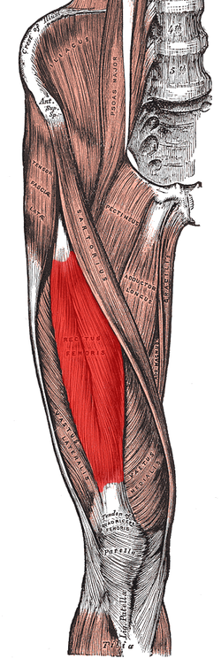
Rectus femoris is a bulk of muscle located in the superior, anterior middle compartment of the thigh and is the only muscle in the quadriceps group that crosses the hip[1].
It is superior and overlying of the vastus intermedius muscle and superior-medial part of Vastus lateralis and Vastus medialis.
The word rectus is a latin word connoting “straight”. Thus the rectus femoris received its name because it runs straight down the thigh[2].
It is a two way acting muscle as it crosses over the hip and knee joint; therefore, it contributes to 90° of knee flexion and assists iliopsoas in hip flexion[3][1].
A short rectus femoris may contribute to a higher positioned patella in relation to the contralateral side. A markedly shortened rectus femoris is suggested by knee flexion of less than 80°or by marked prominence of superior patellar groove[4]
Anatomy[edit | edit source]
Origin[edit | edit source]
Rectus Femoris originates from anterior inferior iliac spine(AIIS) and the part of alar of ilium superior to the acetabulum[1]
Insertion[edit | edit source]
Rectus Femoris together with vastus medialis, vastus lateralis and vastus intermedius joins the quadriceps tendon to insert at the patella and tibial tuberosity (via patellar ligament)[4].
Nerve supply[edit | edit source]
Rectus Femoris is innervated by the femoral nerve, originating from lumbar nerve 2, 3, and 4 nerve roots
Bloody supply[edit | edit source]
Blood is supplied to the Rectus Femoris via descending branch of the lateral circumflex femoral (LCF) artery.
Function[edit | edit source]
Actions[edit | edit source]
Hip flexion[edit | edit source]
- Rectus Femoris acts with iliopsoas to produce hip flexion especially if the knee is flexed[2].
- During gait, as a hip flexor, it acts with the iliopsoas in "Toe off" phase,.
Knee extension[edit | edit source]
- Together with other muscles that are part of the Quadriceps femoris, it facilitates knee extension.
- In terminal swing phase rectus femoris acts as an extensor of the knee, as a muscle in the quadriceps group, it generate force needed for loading(foot flat phase) in stance phase[5].
- Rectus femoris is more efficient in movement combining hip hyper-extension and knee flexion or from a position of knee extension and hip flexion. For example kicking a soccer ball[2][3]
Assessment[edit | edit source]
Palpation[edit | edit source]
Rectus femoris can be palpated as it is the most superior of the quadriceps muscles.
Strength[6][edit | edit source]
To assess muscle strength for the Rectus Femoris (including rest of the quadriceps group) position the patient in sitting with the hip and knee flexed to 90° for grade 5, 4 and 3 while grade 2, is assessed in side-lying with test limb uppermost and knee flexed to 90° position.
Other Tests[8][edit | edit source]
In Rectus femoris injury :
- FABER (Patrick's test) - illicit no pain
- Pain is felt in resisted hip flexion[9]
- Ely’ test - illicit pain on tightness
- Knee ROM is reduced below 80° in shortness of Rectus femoris or prominence of patella grove is noted.[10]
[11]
References[edit | edit source]
|


