Osteogenesis Imperfecta: Difference between revisions
No edit summary |
No edit summary |
||
| Line 5: | Line 5: | ||
</div> | </div> | ||
== Introduction == | == Introduction == | ||
Osteogenesis imperfecta (OI) refers to a heterogeneous group of congenital, non-sex-linked, genetic disorders of collagen type I production, involving connective tissues and bones. | Osteogenesis imperfecta (OI) refers to a heterogeneous group of [[Congenital and Acquired Neuromuscular and Genetic Disorders|congenital]], non-sex-linked, [[Genetic Disorders|genetic disorders]] of [[collagen]] type I production, involving [[Connective Tissue Disorders|connective tissues]] and [[Bone|bones]]. | ||
The hallmark feature of | The hallmark feature of OI is [[osteoporosis]] and fragile bones that [[fracture]] easily, as well as, blue sclera, dental fragility and hearing loss<ref name=":0">Radiopedia [https://radiopaedia.org/articles/osteogenesis-imperfecta-1 Osteogenesisi Imperfecta] Available: https://radiopaedia.org/articles/osteogenesis-imperfecta-1<nowiki/>(accessed 15.10.2021)</ref>. These features result in reduced mobility and function to complete everyday tasks. | ||
OI affects not only the physical but also the social and emotional well-being of children, young people, and their families. The coordinated efforts of a multidisciplinary team can support children with OI to fulfill their potential, maximizing function, independence, and well-being.<ref>Marr C, Seasman A, Bishop N. [https://www.ncbi.nlm.nih.gov/pmc/articles/PMC5388361/ Managing the patient with osteogenesis imperfecta: a multidisciplinary approach]. Journal of multidisciplinary healthcare. 2017;10:145.Available: https://www.ncbi.nlm.nih.gov/pmc/articles/PMC5388361/ (accessed 15.10.2021)</ref> | |||
== Types of OI == | == Types of OI == | ||
Revision as of 01:21, 15 October 2021
Genetic_DisordersOriginal Editors - Barrett Mattingly from Bellarmine University's Pathophysiology of Complex Patient Problems project.
Top Contributors - Barrett Mattingly, Lucinda hampton, Jess Bell, Admin, Uchechukwu Chukwuemeka, Robin Tacchetti, Kim Jackson, Kirenga Bamurange Liliane, Dave Pariser, WikiSysop, Meaghan Rieke, Anna Fuhrmann, 127.0.0.1, Heidi Johnson Eigsti, Elaine Lonnemann and Wendy Walker
Introduction[edit | edit source]
Osteogenesis imperfecta (OI) refers to a heterogeneous group of congenital, non-sex-linked, genetic disorders of collagen type I production, involving connective tissues and bones.
The hallmark feature of OI is osteoporosis and fragile bones that fracture easily, as well as, blue sclera, dental fragility and hearing loss[1]. These features result in reduced mobility and function to complete everyday tasks.
OI affects not only the physical but also the social and emotional well-being of children, young people, and their families. The coordinated efforts of a multidisciplinary team can support children with OI to fulfill their potential, maximizing function, independence, and well-being.[2]
Types of OI[edit | edit source]
Three main types are easily distinguished
Type I. Mildest and most common type. About 50% of all affected children have this type. There are few fractures and deformities
Type II. Most severe type. A baby has very short arms and legs, a small chest, and soft skull. He or she may be born with fractured bones. He or she may also have a low birth weight and lungs that are not well developed. A baby with type II OI usually dies within weeks of birth
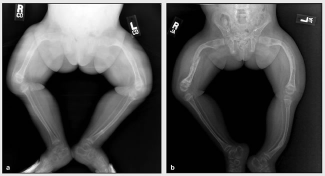
Type III. Most severe type in babies who don’t die as newborns. At birth, a baby may have slightly shorter arms and legs than normal and arm, leg, and rib fractures. A baby may also have a larger than normal head, a triangle-shaped face, a deformed chest and spine, and breathing and swallowing problems. These symptoms are different in each baby[3].
Types IV to VIII are variable in severity and uncommon[1]
Epidemiology[edit | edit source]
The estimated incidence is approximately 1 in every 12,000-15,000 births. OI occurs with equal frequency among males and females and across races and ethnic groups. The lifespan varies with the type. [1]
Etiology[edit | edit source]
OI is a rare genetic disease. In the majority of cases, it occurs secondary to mutations in the COL1A1 and COL1A2 genes. More recently, there has been the identification of diverse mutations related to OI.[4]
Pathology[edit | edit source]
A fundamental pathology in OI is a disturbance in the synthesis of type I collagen, which is the predominant protein of the extracellular matrix of most tissues. In bone, this defect results in osteoporosis, thus increasing the tendency to fracture. Besides bone, type I collagen is also a major constituent of dentine, sclerae, ligaments, blood vessels and skin.[1]
Clinical presentation[edit | edit source]
The clinical presentation of osteogenesis imperfecta is highly variable, ranging from a mild form with no deformity, normal stature and few fractures to a form that is lethal during the perinatal period.
In general, four major clinical features characterise osteogenesis imperfecta:
- Osteoporosis with abnormal bone fragility
- Blue sclera - thinness and transparency of the collagen fibers of the sclera that allow visualization of the underlying uvea. The sclera is the white outer coat of the eye.
- Dentinogenesis imperfecta
- Hearing impairment
Other features include ligamentous laxity and hypermobility of joints, short stature and easy bruising.
Diagnosis[edit | edit source]
The baby's healthcare provider or the specialists may recommend the following diagnostic tests:
- X-rays. These may show many changes such as weak or deformed bones and fractures.
- Lab tests. Blood, saliva, and skin may be checked. The tests may include gene testing.
- Dual Energy X-ray Absorptiometry scan (DXA or DEXA scan). To check for softening.
- Bone biopsy. A sample of the hipbone is checked[3].
Treatment[edit | edit source]
The main goal of treatment is to prevent deformities and fractures. And, once your child gets older, to allow him or her to function as independently as possible.
Management options include:
- Surgical correction of deformities and the prevention of fractures
- intramedullary rods with osteotomy are used to correct severe bowing of the long bones
- intramedullary rods are also recommended for children who repeatedly fracture long bones
- different types of rods (surgical nails) are available to address issues related to surgery, bone size, and the prospect for growth; the two major categories of rods are telescopic and non-telescopic.
- Care of fractures. The lightest possible materials are used to cast fractured bones. To prevent further problems, it is recommended that a child begin moving or using the affected area as soon as possible.
- Bisphosphonates
- Growth hormone therapy[1]
- Dental procedures. Treatments, including capping teeth, braces, and surgery may be needed.
- Physical and occupational therapy. Both are very important in babies and children with OI.
- Assistive devices. Wheelchairs and other custom-made equipment may be needed as babies get older[3].
Below is a documentary from the Discovery Channel titled "Children of Glass" courtesy of Youtube.com.
Characteristics/Clinical Presentation[edit | edit source]
Type III[edit | edit source]
Diagno[edit | edit source]
- Yochum TR, Kulbaba S, Seibert RE. Osteogenesis Imperfecta in a Weightlifter. Journal of Manipulative and Physiological Therapeutics; 25: 334-339. 2002.
- Strevel EL, Adachi JD, Papaioannou A, McNamara M. Case Report: Osteogenesis Imperfecta Elusive Cause of Fractures. Canadian Family Physician; 51: 1655-1657.2005.
- Iwamoto J, Takeda T, Sato Y. Effect of Treatment With Alendronate in Osteogenesis Imperfecta Type I: A Case Report. The Keio Journal of Medicine; 53 (4): 251–255. 2004.
- Aoki T, Kuraoka S, Ohtani S, Kuroda Y. Aortic Valve Replacement in a Woman with Osteogenesis Imperfecta. Annals of Thoracic and Cardiovascular Surgery; 8(1): 51-53. 2002.
Resources[edit | edit source]
- http://www.oif.org
- http://www.genome.gov/25521839
- http://www.osteogenesisimperfecta.org
- http://www.nlm.nih.gov/medlineplus/osteogenesisimperfecta.html
- http://ghr.nlm.nih.gov/condition=osteogenesisimperfecta
- http://www.brittlebone.org
References[edit | edit source]
- ↑ 1.0 1.1 1.2 1.3 1.4 Radiopedia Osteogenesisi Imperfecta Available: https://radiopaedia.org/articles/osteogenesis-imperfecta-1(accessed 15.10.2021)
- ↑ Marr C, Seasman A, Bishop N. Managing the patient with osteogenesis imperfecta: a multidisciplinary approach. Journal of multidisciplinary healthcare. 2017;10:145.Available: https://www.ncbi.nlm.nih.gov/pmc/articles/PMC5388361/ (accessed 15.10.2021)
- ↑ 3.0 3.1 3.2 John Hopkins OI Available: https://www.hopkinsmedicine.org/health/conditions-and-diseases/osteogenesis-imperfecta (accessed 15.10.2021)
- ↑ Subramanian S. StatPearls Publishing LLC.; Treasure Island, FL, USA: 2021. Osteogenesis Imperfecta.Available:https://www.ncbi.nlm.nih.gov/books/NBK536957/ (accessed 15.10.2021)
- ↑ Bublitz Videos. Children of Glass - (Part 1 of 4). Available from: http://www.youtube.com/watch?v=TpAMTOud3bw [last accessed 27/8/2020]
- ↑ Bublitz Videos. Children of Glass - (Part 2 of 4). Available from: http://www.youtube.com/watch?v=GTpSxlPzC8k [last accessed 37/8/2020]
- ↑ Bublitz Videos. Children of Glass - (Part 3 of 4). Available from: http://www.youtube.com/watch?v=L2f8fz6vzoI [last accessed 27/8/2020]
- ↑ Bublitz Videos. Children of Glass - (Part 4 of 4). Available from: http://www.youtube.com/watch?v=QvbY7XqyMz8 [last accessed 27/8/2020]


