Metacarpal Fractures: Difference between revisions

No edit summary
No edit summary
 
(35 intermediate revisions by 9 users not shown)
Line 1: Line 1:
<div class="editorbox">
'''Original Editor '''-&nbsp;--[[User:Marie Avau|Marie Avau]]&nbsp; , Debby Decock, Farrie Bakalli, Margaux Jacobs<br>  
'''Original Editor '''-[[User:Marie Avau|Marie Avau]] '''Top Contributors''' - {{Special:Contributors/{{FULLPAGENAME}}}}</div>
</div>  
== Introduction  ==
= Search strategy<br> =
Hand [[Fracture|fractures]] are common in the general population with relative propensity seen in contact-sport athletes (For example, boxers, football players) and manual laborers<ref name=":0">Moore A, Varacallo M. [https://www.ncbi.nlm.nih.gov/books/NBK536960/ Metacarpal hand fracture.] InStatPearls [Internet] 2019 Jan 16. StatPearls Publishing.Available from:https://www.ncbi.nlm.nih.gov/books/NBK536960/ (last accessed 6.4.2020)</ref>
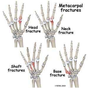
[[File:Types of metacarpal fractures.jpg|right|frameless]]
A metacarpal fracture
* Is a break in one of the five metacarpal bones of either hand.
* Are categorized as being fractures of the head, neck, shaft, and base (from distal at the metacarpal phalangeal joint to proximal 
* at the wrist).<ref name="p1">http://www.physioadvisor.com.au/14681850/metacarpal-fracture-physioadvisor.htm  (level of evidence 5)</ref>&nbsp;  
* [[Boxer's fracture|Boxer fracture]] is another name for a fracture of the fourth or fifth metacarpal, one of the most common metacarpal fractures. <ref name="p2">Blomberg J, Metacarpal fracture, Orthobullets &amp; oral boards, 2014 (level of evidence 5)</ref>
* The mechanisms of these injuries vary from axial loading forces to direct blows to the dorsal hand<ref>Thomas B. McNemar MD, Julianne Wright Howell PT, MS, CHT, Eric Chang MD.Management of metacarpal fractures.Journal of Hand therapy.Volume 16, Issue 2, Pages 143-151</ref><br>


To get a first global view on our topic, we inserted words like metacarpal fracture, anatomy of the hand, fractures of the metacarpals, … in Pedro and PubMed. Then, to complete every part of our subject, we continued with terms like: diagnosis of metacarpal fracture, treatment, epidemiology and characteristics and physical therapy after metacarpal fractures. With the articles and links we found with this keywords, we completed a big part of our task. After this, some of the items were still incomplete or empty, so we did a more thorough search for medical and physical therapy, examination, outcome measures and recent related research. We putted words like treatment of metacarpal fractures, outcome measures of hand fractures and physical therapy metacarpal fractures in the available databases (Pedro, PubMed, research gate, springer link, Medscape, …)  
=== Clinically Relevant Anatomy  ===
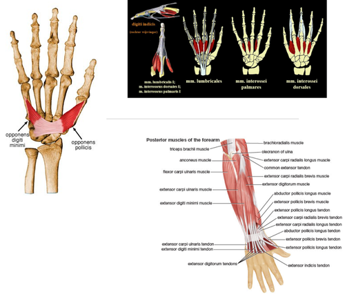
The [[Wrist & Hand|metacarpals]] are long, thin bones that are located between the carpal bones in the wrist and the phalanges in the digits.<ref name="p4">Rafael D. Et al., Current management of metacarpal fractures, hand the clinics, 2013 (level of evidence 5)</ref><ref name="p4" />[[File:Hand_muscles.png|right|frameless|500x500px]]
* Each is comprised of a base, shaft, and head.
* The proximal bases of the metacarpals articulate with the [[Wrist & Hand|carpal bones]],
* Distal heads of the metacarpals articulate with the proximal phalanges and form the knuckles.
* The 1st metacarpal is the thickest and shortest of these bones.
* The 3rd metacarpal is distinguished by a styloid process on the lateral side of its base.  
* Soft tissues generally involved with fractures include cartilage, joint capsule, ligaments, [[fascia]], and the [[Extensor Hood Mechanism Hand|dorsal hood]] fibers.
* With severe polytrauma cases, the [[Tendon Anatomy|tendon]]<nowiki/>s and [[Nerve Injury Rehabilitation|nerves]] adjacent to the fracture can also be injured.&nbsp;<ref name="p8">Hardy MA. Principles of Metacarpal and Phalangeal Fracture Management: A Review of Rehabilitation Concepts. Journal of Orthopedic and Sports Physical Therapy. 2004; 34:781-791.(level of evidence 5)</ref>


<br>  
==== Etiology  ====
Metacarpal fractures typically occur secondary to a direct blow or fall directly onto the hand. 
* These fractures commonly occur during athletic activities, particularly in contact sports. Almost one-fourth of cases occur during athletic events.
* Sporting injury is frequently the cause among younger patients
* Work-related injuries are often the cause in middle-aged patients
* [[Falls in elderly|Falls]] are typically the cause of the elderly.
* Fifth metacarpal fractures often occur secondary to punching a wall or other solid object (hence the eponym, "boxer's fracture")<ref name=":0" />


= Definition/ Description<br>  =
===== Hand Fractures =====
* Makeup about 40% of all acute hand injuries
* Constitute about 20% of all fractures occurring below the elbow


A metacarpal fracture is a break in one of the five metacarpal bones of either hand. Metacarpal fractures are categorized as being fractures of the head, neck, shaft, and base (from distal at the metacarpal phalangeal joint to proximal at the wrist).<ref name="1">http://www.physioadvisor.com.au/14681850/metacarpal-fracture-physioadvisor.htm  (level of evidence 5)</ref>&nbsp;<ref name="2">Blomberg J, Metacarpal fracture, Orthobullets &amp;amp;amp; oral boards, 2014 (level of evidence 5)</ref> (level 5)<br>Thereby we also have the Boxer fracture, this is another name for a fracture of the fourth or fifth metacarpal. This is one of the most common metacarpal fractures, in contrast with the fractures of the thumb (Bennett’s and Rolando’s fracture). <ref name="2" />(Blomberg et al, level 5)<br>  
===== Metacarpal Fractures =====
* Typically occur in patients aged 10-40 years
* Men are more likely to be affected than women. 
* Young men sustain metacarpal fractures secondary to a punching mechanism or a direct blow to the hand
* Geriatric females sustain these injuries secondary to a low energy fall. 
* The incidence rate of fracture seen in association with each digit's metacarpal bone increases from the radial to the ulnar side.
* The incidence rate of 2nd metacarpal fractures is lower than the incidence rate of 5th metacarpal fractures.<ref name=":0" />
*[[Bennett's fracture|Bennett fracture]] is the most common fracture involving the base of the thumb. This fracture refers to an intra-articular fracture that separates the palmar ulnar aspect of the first metacarpal base from the remaining first metacarpal. <ref name="p1" />
The fractures of the metacarpals can be divided into three parts.
# The first, neck fractures, occurs often when a person punches another person or object. In the majority of cases, surgical intervention is not essential to treat this condition.
# The metacarpal shaft fractures are often produced by longitudinal compression, torsion, or direct impact. They are described by the appearance of their respective fracture patterns and can be divided by transverse, oblique, spiral, and comminuted.
# Metacarpal base fractures are rare and have a minimal consequence because the motion of the joint is small. More common are the fractures of the base of the fifth digit and are the result of a longitudinally directed force&nbsp;<ref name="p0">Kathleen M. Kollitz et. Al., Metacarpal fractures: treatment and complication, American Association for Hand Surgery 2013, Springer, Published online: 16 October 2013, HAND (2014) 9:16–23</ref>


= Clinically Relevant Anatomy<br> =
=== Characteristics/Clinical Presentation ===
Patients with metacarpal fractures generally present with <ref name="p8" /><ref name="p9">Michael DelCore ,Metacarpal fractures, orthopaedicsone ,2015.</ref>
* Pain
* Swelling
* Ecchymosis (bruise)
* Limitation of movement
* Deformity - Knuckle asymmetry may be observed, and the knuckle may appear to be missing.
* Finger misalignment may also be noted.
* A metacarpal head fracture is associated with axial compression of the extended digit which causes severe discomfort.
* In a metacarpal base fracture, movement of the wrist or longitudinal compression exacerbates the pain.
* Any metacarpal fracture angulation can produce a pseudo-claw deformity.


The hand is composed of 19 bones (5 metacarpals and 14 phalanges), more than 30 tendinous insertions and numerous complex structures. The metacarpals are long, thin bones which are located between the carpal bones in the wrist and the phalanges in the digits.<ref name="4">Rafael D. Et al., Current management of metacarpal fractures, hand the clinics, 2013 (level of evidence 5)</ref><ref name="14">LIVERNEAUX, PA. et al, Fractures and dislocation of the base of the thumb metacarpal. The Journal of Hand Surgery, 2001.</ref>
==== Differential Diagnosis  ====


[[Image:Hand and wirst bones.png]]<br>
Injuries to neighboring bones (carpal bones, phalanges) and associated soft tissues (ligaments, tendons) need to be excluded.


Each are comprised of a base, shaft, and head. The proximal bases of the metacarpals articulate with the carpal bones, and the distal heads of the metacarpals articulate with the proximal phalanges and form the knuckles. The 1st metacarpal (of the thumb) is the thickest and shortest of these bones. The 3rd metacarpal is distinguished by a styloid process on the lateral side of its base. Soft tissues generally involved with fractures include cartilage, joint capsule, ligaments, fascia, and the dorsal hood fibers. With severe polytrauma cases, the tendons and nerves adjacent to the fracture can also be injured.&nbsp;<ref name="48">Hardy MA. Principles of Metacarpal and Phalangeal Fracture Management: A Review of Rehabilitation Concepts. Journal of Orthopedic and Sports Physical Therapy. 2004; 34:781-791.(level of evidence 5)</ref>
===== Evaluation =====
The evaluation includes:
* Standard radiographs of the hand (antero-posterior, lateral, and oblique). In the vast majority of cases, this will be enough to confirm the diagnosis and form a management plan. Confirmation of more subtle injuries can be obtained using special views such as Brewerton (metacarpal heads), Roberts, and Betts (thumb) views.
* [[CT Scans|CT]] is sometimes necessary for the base of metacarpal fractures to check for any intra-articular displacement and determine if there is a need for surgery


<br>  
=== Outcome Measures  ===
* [[Grip Strength]]: measured with a dynamometer
* Range of motion
* [[Patient Specific Functional Scale]]
* [[DASH Outcome Measure|DASH]]
* [[Michigan Hand Outcomes Questionnaire|Michigan Hand Outcome Questionnaire]] (MHO): In this questionnaire, they assess 6 criteria for people with a hand disorder: overall hand function, activities of daily living (ADL), pain, work performance, aesthetics, and patient satisfaction with hand function. <ref name="p2" /><ref name="p3">Karriem-Norwood, Boxers fracture, webmd , 2014</ref>


Muscles in the metacarpal region: <ref name="3">Karriem-Norwood, Boxers fracture, webmd , 2014</ref><ref name="14" /><br>There are three palmar and four dorsal interossei muscles that arise from metacarpal shafts. We can also find the insertion of extensor carpi radialis longus and brevis at the base of the second and third metacarpal. This muscles assist with wrist extension and radial flexion.<br>In the wrist we can thereby also find the extensor carpi ulnaris muscle, which inserts on the base of metacarpal five. His function is to extend and fixe the wrist when the digits are being flexed and assists with ulnar flexion of wrist.<br>The abductor pollicis longus is situated in the area of the thumb. It inserts on the trapezium and base of metacarpal I and is responsible for abduction of the thumb in the frontal plane and extends the thumb at the carpometacarpal joint. At this site we can also find the opponens pollicis, which inserts on metacarpal I and functions as a flexor of metacarpal I to oppose the thumb to the fingertips.<br>The final muscle at this site is the opponens digiti minimi, it inserts on the medial surface of metacarpal V, flexes metacarpal V at carpometacarpal joint.<br>
==== Medical Management ====
The goal of treatment is a restoration of anatomy and function.
* [[Antibiotics]] and [[tetanus]] prophylaxis are options for open fractures as per standardized guidelines.
* The modality of treatment will vary depending on skin integrity (open versus closed fracture), the number of digits/metacarpals fractured, the stability of the specific, degree of comminution, displacement, and/or rotational malalignment
* In general, increasing degrees of displacement, comminution, and rotational malalignment are critical factors in assessing the fracture patterns potential for stability and reduction maintenance with non-operative management.<ref name=":0" />
* The GP/Specialist after assessing the fracture will perform gentle tests and imaging to work out if surgery is needed.  
* If surgery is not needed a physiotherapist will make a custom splint, which will support the healing fracture.


<br>
=====  Physical Therapy Management  =====
Full strength and range of motion is the goal of rehabilitation.


[[Image:Hand muscles.png]]  
Under the physiotherapist’s instructions
* Hand exercises with light resistance such as rubber bands or squeeze ball can help if there is scarring or extensor lag develops. 
* Soft tissue recovery may be more of a problem than the bony one. 
* Rest and elevation are important, and so is the quality of splinting - poor splinting can cause stiffness, [[Pressure Ulcers|pressure sores]], or even [[Compartment Syndrome of the Forearm|compartment syndrome]]
Physiotherapists use a number of techniques to regain movement in the hand, wrist, and fingers, including:
* Swelling management with [[massage]] and compression garments
* Soft tissue massage to help with muscle tension and pain
* Providing clients with a home exercise program of specific movements and strengthening exercises.
Most hand fractures can be treated non-operatively <ref name="p4" />&nbsp;
This very informative 4 minute video gives a basic run done on Physio treatment
{{#ev:youtube|https://www.youtube.com/watch?v=1xrlrp8Ooa0&feature=youtu.be|width}}<ref>Medicine in a nutshell Physio excercises for patients with metacarpal fractures Available from:https://www.youtube.com/watch?v=1xrlrp8Ooa0&feature=youtu.be (last accessed 6.4.2020)</ref>
'''More specific Advice.'''


= Epidemiology/ Etiology  =
'''These are the steps to be followed in a stable fracture''': <ref name="p5">T. GRANT PHILLIPS, M.D. et al, Diagnosis and Management of Scaphoid Fractures, Washington Hospital Family Practice Residency, Washington, Pennsylvania, 2004.http://coruraltrack.org/wp-content/uploads/2013/01/Scaphoid-Fractures-AFP.pdf</ref>


Hand injuries constitute 5-10% of emergency department visits. The most common mechanisms are road traffic accidents, blunt trauma (e.g. crush injury, contusions), and assault. Fifth metacarpal fractures occur more frequently and are responsible between 16% to 34% of the hand fractures. <ref name="12">Thomas B. McNemar, MD et. al., Management of Metacarpal Fractures, J HAND THER. 2003;16:143–151 (level of evidence 5)</ref>(McNar, level 5) ,<ref name="17">J. J. de Jongel et al, Fractures of the metacarpals. A retrospective analysis of incidence and aetiology and a review of the English-language literature, ‘Department of Traumatology, and ‘Department of Plastic and Reconstructive Surgery, University Hospital Groningen, The Netherlands. Injury, 1994, Vol. 25, 365-369, August.</ref>  
One or other of the below stabilizing techniques could be used :-
* Buddy strapping the injured digit to another digit is used as a non-operative technique. This is used with or without the application of varying degrees of splint. The ‘buddy’ reduces the risk of rotational deformity.  
* The splinting of the fracture should be: 20 degrees wrist extension; MCP joint 60-70 degree flexion and IP joint extension <ref name="p6">Tiel-van Buul MM et al, The value of radiographs and bone scintigraphy in suspected scaphoid fracture. A statistical analysis. J Hand Surg [Br] 1993;18:403-6.</ref>


<br>Men, aged 10-29, have the highest incidence rates for metacarpal fracture (2.5 per cent). Accidental fall is seen as the dominant cause of these fractures this fall is mainly caused by transport accidents and in particular bicycle transport.The main cause of most metacarpal fractures of the fingers is a direct forceful blow on the fingers. For example, punching a solid object with a closed fist, or having a heavy object land on the hand. Occasionally they may also occur due to a direct impact to the hand, from an object travelling at high speed or a fall onto the fingers or thumb. <ref name="10">Kathleen M. Kollitz et. Al., Metacarpal fractures: treatment and complication, American Association for Hand Surgery 2013, Springer, Published online: 16 October 2013, HAND (2014) 9:16–23</ref><ref name="11">Bahubali Aski et. Al., Metacarpal fractures treated by percutaneous Kirschner wire, International Journal of Physical Education, Sports and Health 2015; 1(3): 10-13 (level of evidence 4)</ref>(Bahubali Aski et. Al, level 4)
* Early motion is generally considered appropriate when there are stable fractures or rigid fractures.<ref name="p7">J. J. de Jongel et al, Fractures of the metacarpals. A retrospective analysis of incidence and aetiology and a review of the English-language literature, ‘Department of Traumatology, and ‘Department of Plastic and Reconstructive Surgery, University Hospital Groningen, The Netherlands. Injury, 1994, Vol. 25, 365-369, August.</ref>
* Generally, AROM (active ROM) exercises without resistance can begin 2 to 3 weeks after operative treatment in uninvolved or bordering/adjacent joints. <ref name="p9" /> <ref name="p8" /> <ref name="p0" />&nbsp;<ref name="p1" />
* Active Motion: If the fracture is internally fixed, the active range of motion can start early. Most fractures are treated by immobilization, but the active motion can begin after three weeks of therapy, starting with the joints not splintered during the initial immobilization. This phase usually lasts 3-6 weeks. <ref name="p1" /> <ref name="p2" />&nbsp; <ref name="p5" /><ref name="p6" />
* Specific tendon gliding should be included in the active motion.  
* Tendon gliding is important to prevent adhesions, increased circulation about the fracture site, decreased edema and compression at the fracture site.<br>
[[File:Handen.png|right|frameless]]


The thumb metacarpal fractures, which is the most common fracture type, are usually caused by an axial blow directed against the partially flexed metacarpal. (It is estimated to occur in 4% of hand fractures. (Stanton et al., 2007)) These fractures are called Boxer's fractures and received their name from one of their most common causes of punching an object with a closed fist. This occurs commonly during fist fights or from punching a hard object such as a wall or filing cabinet. Although these breaks usually occur when the hand is closed into a fist, they can also occur when the hand is not clenched and strikes a hard object.<ref name="11" /> (Bahubali Aski et. Al, level 4)
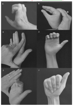
===== Exercises For Tendon Gliding&nbsp;=====
* Claw posture to achieve [[Extensor Digitorum Longus|extensor digitorum communis]] tendon glide over the metacarpal bone
* Intrinsic plus posture to achieve central slip. Lateral bands glide over proximal phalanx 1
* [[Flexor Digitorum Profundus]] (FDP) blocking exercises to glide FDP tendon over the phalanx
* Hook fist posture to promote selective FDP tendon glide
* [[Flexor Digitorum Superficialis]] (Sublimis) blocking exercise to glide FDS tendon over middle phalanx
* Sublimis fist posture to promote selective FDS tendon glide<br><ref name="p5" />


The fractures of the metacarpals can be divided in three parts. The first, neck fractures, occurs often when a person punches another person or object. In the majority of cases, surgical intervention is not essential to treat this condition. The metacarpal shaft fractures are often produced by longitudinal compression, torsion or direct impact. They are described by the appearance of their respective fracture patterns and can be divided by transverse, oblique, spiral and comminuted. The last one is the metacarpal base fractures. They are rare and have minimal consequence because the motion of the joint is small. More common are the fractures of the base of the fifth digit and are the result of a longitudinally directed force&nbsp;<ref name="10" />  
===== Passive Motion =====
* Passive motion can be initiated after sufficient clinical healing at approximately 5-6 weeks of therapy. <ref name="p1" />  <ref name="p3" />&nbsp;<ref name="p2" />&nbsp;
* The timing of initiation of joint mobilization depends on the structures involved in the injury. If the structures resisting the force are not involved in the injury, joint mobilization can be initiated at the same time as active motion. Compression on the fracture can result in shortening, angulation or rotational mal-alignment of the bone.
* Traditional PROM aims to assist in articular cartilage healing, reduce swelling, and stiffness. <ref name="p4" />&nbsp;
* Resistive Motion: Four weeks after the injury light resistance can be performed in most metacarpal fractures which are treated by immobilization. Active motion should only be continued if healing has not started.
* Resistive exercise should also be delayed when a fracture is fixed by pinning until these pins are removed, to ensure the stability of the fracture. Light resistive exercise helps with scar remodeling and improved motion. There are several types of resistive exercises such as the weight-well exercises. This kind of exercise strengthens the finger flexors (FDP and FDS muscles).
* Functional activities and work simulation should be included in the resistive exercises as soon as possible.<ref name="p5" />


<br>  
===== Conclusion =====
[[File:Types of metacarpal fractures.jpg|right|frameless]]
Main points on metacarpal fractures:
* Common hand injury
* Require thorough assessment consisting of the history, examination, and radiological investigations
* They mostly divide into open or closed, based on the digit they affect, intra-articular or extra-articular status, and based on the location on the bone itself (head, neck, shaft, base)
* May have conservative or operative treatment
* Can have long-term sequelae requiring further management
* Rehabilitation goals are return of full strength and range of motion.
* Rest and elevation are important, and so is the quality of splinting - poor splinting can cause stiffness, pressure sores, or even compartment syndrome.
* Physiotherapy is an critical element in the restoration of good [[Hand Function|hand function]]<ref name=":0" />  


= Characteristics/Clinical Presentation =
== References ==


Patients with metacarpal fractures generally present with: <ref name="18">Edward C.  et al, Intra-articular metacarpal head fractures, Vol. 8, No . 4 July 1983.</ref><ref name="19">Michael DelCore ,Metacarpal fractures, orthopaedicsone ,2015.</ref><br>- Pain<br>- Swelling<br>- Ecchymosis (bruise)<br>- Limitation of movement<br>- Deformity. Knuckle asymmetry may be observed, and the knuckle may appear to be missing. <br>- Finger misalignment may also be noted. <br>- A metacarpal head fracture is associated with axial compression of the extended digit which causes severe discomfort. <br>- In a metacarpal base fracture, movement of the wrist or longitudinal compression exacerbates the pain.<br>- Any metacarpal fracture angulation can produce a pseudo-claw deformity. <br>
<references />
 
= Differential diagnosis  =
 
We can distinguish different types of metacarpal fractures: base fractures, shaft fractures, neck and head fractures. These fractures can either be displaced (shifted) or non- displaced They all have a specific cause and clinical presentation. Globally these patients all report a snapping or popping sensation.<ref name="20">Manuel Hernandez, MD, Boxer's Fracture Symptoms, eMedicine Health, 2014.</ref><br>Metacarpal Base Fractures: These fractures are intra-articular and result from high forces (violent accidents), direct blows, or crushing injuries that disrupt the rigid carpal ligaments, or overwhelm the normal flexibility of the ulnar metacarpals. They also occur with an avulsion of the wrist flexors or extensors, that insert on the metacarpal base, resulting from a direct blow or torsional injury. The most common occurrence is at the 5th metacarpal-hamate articulation. The healing rate varies from three to six weeks.<ref name="48" /><ref name="9">Tom Gocke, MS, ATC, PA-C, DFAAPA , Review on the Management of Metacarpal/Hand Fractures, Orthopaedic Educational Services, Inc., 10 April 2013. (level of evidence 5)</ref><br>Metacarpal Shaft Fractures: These fractures are extra-articular and are caused by longitudinal compression, torsion, or direct impact that may result from a fall, blow, or crushing force that usually angulates dorsally. They are described by the fracture configuration as transverse, oblique, spiral, or comminuted. Metacarpal shaft fractures are slower to heal than the more distal or proximal locations because of the predominantly cortical bone found there. The healing rate varies from three to seven weeks<ref name="9" /><ref name="48" /><br>Metacarpal Neck Fractures: Most common metacarpal fracture. The weakest point of the metacarpal bone is the extra-articular neck. These fractures result from a compression force such as a direct blow with a closed fist. The 4th and 5th metacarpals are most often involved and are referred to as a “fighter’s” or “boxer’s” fracture. Trauma causes the fractured metacarpal head to displace with volar angulation. The healing rate is three to five weeks.<ref name="9" /><ref name="48" /><br>Metacarpal Head Fractures: These fractures are intra-articular and are caused by direct impact and high axial loads that can involve avulsion of the collateral ligaments, including a fracture fragment, fracture of one or both condyles, or shattering of the joint surface into many small, comminuted pieces.<ref name="9" /> <ref name="48" />( deze tekst stond op de originele Physiopedia pagina, maar was niet onderveeld in het juiste puntje)<br>Problems in the metacarpal region can also be diagnosed as a skier’s tumb, a metacarpophalangeal joint dislocation or a hand dislocation. These disorders often present with the same symptoms and can be confused with a metacarpal fracture. It is thus important to distinguish both injuries. <ref name="34">David R Steinberg, MD et al, Metacarpal Fracture and Dislocation Differential Diagnoses, Medscape 2015.</ref><br>
 
<br>
 
= Diagnostic Procedures  =
 
To diagnose a metacarpal fracture, it is necessary to submit the patient to a physical examination and radiograph; these can be can be considered as the definitive standard for diagnosis of metacarpal fractures. <ref name="25">T. GRANT PHILLIPS, M.D. et al, Diagnosis and Management of Scaphoid Fractures, Washington Hospital Family Practice Residency, Washington, Pennsylvania, 2004.http://coruraltrack.org/wp-content/uploads/2013/01/Scaphoid-Fractures-AFP.pdf</ref><br>Different types of diagnosis:<br>- Physical examination of a suspected metacarpal fracture should concentrate on hand deformity, tenderness, digit mal rotation, and presence of open wounds. Painful range of motion, point specific bone tenderness and ecchymosis should be considered red flags for fractures. <br>- Routine radiographs include three views: antero-posterior, lateral, and oblique. These views assist in properly assessing angulation of fracture fragments as well as involved joint surfaces. Computed Tomography scans, or CT scans, may be ordered to evaluate complicated fractures.<ref name="50">Freidrich, JB, Vedder, NB. An Evidence-Based Approach to Metacarpal Fractures. Plast. Reconstr. Surg. 126: 2205, 2010.</ref><ref name="52">Kozin SH, Thoder JJ, Leiberman G. Operative Treatment of Metacarpal and Phalangeal Shaft Fractures. J Am Acad Orthop Surg 2000;8:111-121.(level of evidence 5)</ref><br>- Ultrasonography: this type of diagnostic procedure is especially used in scaphoid fractures, as it’s only proven reliable and accurate for this specific type. <br>- Another technique is the bone scintigraphy (this is an imaging technique using radioactive substances that intravenously was injected in small quantities, also called a bone scan)<ref name="26">Tiel-van Buul MM et al, The value of radiographs and bone scintigraphy in suspected scaphoid fracture. A statistical analysis. J Hand Surg [Br] 1993;18:403-6.</ref><br>- An MRI is almost never necessary for an isolated metacarpal fracture, as they normally don’t add any further information beyond a regular x-ray. But if other injuries are suspected, an MRI can be used as a supplementary test.<ref name="27">Bretlau Tet al, Diagnosis of scaphoid fracture and dedicated extremity MRI. Acta Orthop Scand 1999.</ref><ref name="24">Singletary S, Freeland AE, Jarrett CA. Metacarpal fractures in athletes: treatment, rehabilitation, and safe early return to play. J Hand Ther. 2003 Apr-Jun;16(2):171-9.</ref><br>
 
= Outcome Measures  =
 
-Grip Strength: measured with a dynamometer<br>-Range of motion <br>-Disabilities of the Arm, Shoulder, and Hand score (QuickDASH): this questionnaire is scored in two components: the disability/symptom section (30 items, scored 1-5) and the optional high performance Sport/Music or Work section (4 items, scored 1-5).<ref name="32">Dias JJ et al., Which questionnaire is best? The reliability, validity and ease of use of the Patient Evaluation Measure, the Disabilities of the Arm, Shoulder and Hand and the Michigan Hand Outcome Measure., J Hand Surg. 2008 .</ref><br>-Michigan Hand Outcome Questionnaire (MHO): In this questionnaire they asses 6 criteria for people with a hand disorder: overall hand function, activities of daily living (ADL), pain, work performance, aesthetics, and patient satisfaction with hand function. <ref name="32" /><ref name="33">MD Kevin C. Chung et al, Reliability and validity testing of the Michigan Hand Outcomes Questionnaire,  The Journal of Hand Surgery Volume 23, Issue 4, July 1998, Pages 575–587</ref><br>-Patient Evaluation Measure (PEM): this questionnaire is similar to the DASH score and assesses the hand function by asking questions about the patients pain, tenderness, swelling, wrist movement and grip strength.<ref name="31">Hobby J Let al., Validity and responsiveness of the patient evaluation measure as an outcome measure for carpal tunnel syndrome, J Hand Surgery Br,2005</ref><br>- The Hand Clinic Questionnaire: has eight questions addressing pain, stiffness, neurological change, use of the hand and appearance <ref name="32" /><br>- The Patient Evaluation Measure uses visual analogue scales and has three parts: The first part seeks the patient’s opinion on the delivery of care. The second part is entitled ‘‘How your hand is now’’ (Hand Health Profile) and has ten questions concerning feeling, cold sensitivity, pain frequency, use for fiddly (fine dexterous) activities, movement, grip strength, activity, use for work, appearance and general attitude. The third part has three questions, which cover the overall assessment of outcome after the injury.<ref name="28">Kaiser MM et al . , Intramedullary nailing for metacarpal 2-5 fractures, J Pediatr Orthop B. 2009.</ref><br>- The HOSS is a doctor-administered form and combines both subjective and objective measures.<br>The HOSS measures the injury in four tissues: integument, skeletal, motor and neural. These four tissues and their degree of impairment form the basis for measuring the outcome in the HOSS.<br>
 
<u>Reliability and validity of the outcome measures</u>: <ref name="29">R. SHARMA and J. J. DIAS, validity and reliability of three generic outcome measures for hand disorders, the Leicester Hand Surgery Service, Leicester Group of Hospitals, Leicester, UK, Journal of Hand Surgery British and European Volume, 2000,25B: 6: 593–600</ref><ref name="30">J. J. Dias et al., Assessing the outcome of disorders of the hand, Leicester Royal Infirmary, Leicester, England ,2000</ref><br>- Michigan Hand Outcome questionnaire: test-retest reliability was measured using Spearman's correlation and demonstrated substantial agreement, ranging from 0.81 for the aesthetics scale to 0.97 for the ADL scale. In testing for internal consistency, Cronbach's alphas ranged from 0.86 for the pain scale to 0.97 for the ADL scale. Correlation between scales gave evidence of construct validity.<br>- PEM, DASH and MHO all have a good construct validity, but the easiest to use and complete is the PEM questionnaire. They are valid and reliable for wrist and finger disorders. <ref name="31" /><br>
 
= Examination  =
 
The severity of the fracture is evaluated by taking several X-rays of the hand. We can include three views:<br>1. Anteroposterior view: When a x-ray is taken from front to back, with the back against the film plate and the x-ray machine in front of the patient it is called an anteroposterior (AP) view. <ref name="19">Michael DelCore ,Metacarpal fractures, orthopaedicsone ,2015.</ref><br>2. Lateral view: the x-rays beam enters through the lateral side of hand<br>3. Oblique view: the view is obtained from a pronated, palm-down position by rotating the hand 45° laterally. <ref name="18">Edward C.  et al, Intra-articular metacarpal head fractures, Vol. 8, No . 4 July 1983.</ref><br>Special views <br>- Brewerton’s view: a view used to detect occult fractures of the metacarpal head<br>- Skyline view: they are used for the fracture head of the metacarpal bone<br>- 30° pronated and 30° supinated oblique view: it is warranted for the 2nd and 5th metacarpal shaft fracture<br>- Stress view: it may indicate certain fracture dislocations<br>Sophisticated investigations: <br>- CT-scan (computed tomography): it uses computer-processed X-rays to make tomographic images. It evaluate the metacarpal-carpal joints in complicated fractures.<br>- MRI (magnetic resonance imaging): it is used to investigate the physiology and anatomy of the body.<br>The above-mentioned techniques are rarely used in acute skeletal injuries of the hand.<br>
 
= Medical management  =
 
“Evidence for the effective therapy management of these fractures is sparse, varied and of poor quality and does not provide a ‘gold standard’ therapeutic treatment approach.”&nbsp;<ref name="35">Toemen A. et al, Hand therapy management of metacarpal fractures: an evidence-based patient pathway, the royal society of medicine journals,2010</ref> (A. Toemen et al)<br>Knowledge of different therapies, the relevant anatomy, acting muscular forces and the mechanisms of fractures is required to choose the appropriate treatment modality for metacarpal fractures. Thereby, it is important to remember that the medical treatment must not sacrifice the hand functions as they are used in daily life.<ref name="12" /> (McNar et al, level 5)<br>As with any hand fracture, the primary goals are to achieve anatomic and stable reduction, bony<br>union, and early mobilization to minimize disability.<ref name="4" /><br>We can distinguish stable and unstable fractures. A stable fracture sustains an acceptable position at rest when early mobilization are introduced. Unlike unstable fracture, which are likely to displace with early mobilization. The latter is first managed with a period of immobilization or surgical intervention for stabilization. <ref name="23">Geissler WB. Operative fixation of metacarpal and phalangeal fractures in athletes. Hand Clin. 2009 Aug;25(3):409-21.</ref><ref name="13">Benjamin JF Dean et. al., Fractures of the metacarpals and phalanges, ORTHOPAEDICS AND TRAUMA 25:1, 2010 (level of evidence 5)</ref><ref name="15">Mohammad Umar MUMTAZ et al, Unstable metacarpal and phalangeal fractures: treatment by internal fixation using AO mini-fragment plates and screws, Turkish Journal of Trauma &amp;amp; Emergency Surgery, Original Article Klinik Çalışma Ulus Travma Acil Cerrahi Derg 2010;16 (4):334-338. (level of evidence 4)</ref>(level 4)<br>Stable fractures will not lose approximation with muscle tension or controlled motion.
 
First, closed methods of supports are used for 2-3 weeks and then a removable splint for controlled motion is applied. <ref name="12" />(McNar el al, level 5)
 
<br>
 
Potential unstable fractures are those that are aligned, but may get misaligned with certain positions or tension. These are treated with immobilization that maintains reduction or restriction of motion towards position of instability. As healing occurs, immobilization can be modified to allow incremental increases in ROM.<br>
 
Unstable fractures are those that do not maintain reduction. Displacement occurs even with immobilization. Fixation devices are warranted.<ref name="48" /> An unstable fracture can be managed with early mobilization as long as a protective resting splint is used. In such case, a clinical and radiographic follow-up is needed to ensure the undisplaced state of the fracture. Surgical intervention is used when an acceptable displacement occurs.&nbsp;<ref name="12" /> (McNar el al, level 5)&nbsp;<ref name="13" /> <br>Two facts must be provided with therapy referral: date of fracture and method of fixation. Date of fracture provides a timetable on where the bone healing process is. Knowing the method of fixation will determine when motion can be introduced. Open reduction fixations (implementation of implants to guide the healing process of bone) can begin AROM (active range of motion) earlier.<ref name="48" /><br>If there is no intervention, most digit factures will unite. Therefore early mobilization is important to maintain and regain mobility.&nbsp;<ref name="13" />
 
The evaluations of rotational malalignement and stability of these unstable fractures are the most critical elements to choose between operative and non-operative treatment. The latter is cheaper and avoids complications of surgical treatment. There is a chance that it may not restore skeletal position and maintain stability. Open surgery can accomplish the most stable anatomic reduction. Important to notice is that if there has been an aggressive surgeon’s intervention, a more aggressive post-operative rehabilitation is required.<ref name="13" /><br>In recent literature, studies were executed to evaluate the outcome measures in patients with a metacarpal fracture, who received different medical treatments.
 
<br>1. Transverse and short oblique closed metacarpal and proximal phalangeal fractures treated with intramedullary -wire and cross -wires had produced favorable and comparable outcomes in long term. <ref name="21">Singh Ashutosh K et al, Outcome of Displaced Metacarpal and Proximal Phalangeal  Fractures Treated with Intramedullary K-Wire and Cross K-wires: A Prospective Study of 105 Patients, International Journal of Orthopaedics,  June 23 2014. (level of evidence 4)</ref>(Singh Ashutosh K et al, level 4)<br>2. When immobilization was discontinued by five weeks, the position of the metacarpophalangeal joints and the absence or presence of interphalangeal joint motion during the immobilization had little effect on motion, grip strength, or fracture alignment. This finding contradicts the conventional teaching that the metacarpophalangeal joint must be immobilized in flexion to prevent long-term loss of joint extension.<ref name="22">Tavassoli J1 et al, Three cast techniques for the treatment of extra-articular metacarpal fractures. Comparison of short-term outcomes and final fracture alignments, J Bone Joint Surg Am. 2005 Oct;87(10):2196-201. (level of evidence 2C)</ref> (Tavassoli J1 et al, level 2C)
 
= <br>Physical therapy management  =
 
Such as Giddins says “most hand fractures can be treated non-operatively” [39] (Giddins et al, level 2A), which accentuates the important role of the physical therapist in the healing process. But some hand fractures, such as open injuries or displaced intra-articular fractures, are almost always treated operatively. In this section, we will discuss de the modalities and role of the physical therapist after a metacarpal fracture.
 
In early phases of therapy, patient should be educated on edema control. Rest, ice, compression, and elevation should be emphasized.The intervention of the physical therapist consist of applying powertraining modalities or guide the healing process by using a splint. Alongside of improving the functionality, active, passive and resistive exercises also affect the recovery process of bone and cartilage.<br>Before the physical therapist can start the interventions or treatment, certain considerations about the fracture should be made. These are the steps to be followed in a stable fracture: <br>
 
<br>
 
[43] Maureen A. et al (level of evidence 5)
 
<br>
 
Buddy strapping the injured digit to another digit is used as a non-operative technique. This is used with or without the application of varying degrees of splint. The ‘buddy’ reduces the risk of rotational deformity. The splinting of the fracture should be with an aim if 20 degrees wrist extension, the metacarpophalangeal joint in 60-70 degree flexion and interphalangeal joint extension [47] (HAUGHTON et al, Level of evidence 5).
 
Early motion is generally considered appropriate when there are stable fractures or rigid fractures. It is hypothysed that early motion has the potential for improved outcomes.[40] (LM Feehan et. Al, level of evidence 3A)
 
Generally AROM (active ROM) exercises without resistance can begin 2 to 3 weeks after operative treatment in uninvolved or bordering/adjacent joints. [36] (Stern PJ., level of evidence 5), [37] ( Feehan LM. ,level of evidence 5), [38] (Rafael D. Et al., level of evidence 5) , [1]
 
Active Motion: If the fracture is internally fixed, the active range of motion can start early. Most fractures are treated by immobilization, but the active motion can begin after three weeks of therapy, starting with the joints not splintered during the initial immobilization. This phase usually lasts 3-6 weeks. [41] (Tim L et al., level 5), [42] (Sadler JA, Koepfer JM et al, level 5), [43] (Maureen A. Hardy, level 5), [47] (Heiser, R., et al. level 2A)
 
Specific tendon gliding should be included in the active motion. The following muscles should be involved: m. flexor digitorum profundus, m. flexor digitorum superficialis, m., extensor digitorum communis, m. extensor indicis propius and m. extensor digiti minimi<br>
 
Tendon gliding is important to prevent adhesions,increased circulation about the fracture site, decreased edema and compression at the fracture site.<br>FDP tendons and selected tendon glide should be performed by actively flexing the injured PIP joint while positioning the other DIP joints in extension. The EI and EDQ tendons should glide on the adjacent EDC tendon and the EDC tendons on the underlying bone. By extending the MPC joints while IP joints are flexed, tendon gliding of the EDC tendon should be performed.
 
[[Image:Handen.png]]
 
Exercises for tendon gliding&nbsp;:
 
A&nbsp;: Claw posture to achieve extensor digitorum communis tendon glide over metacarpal bone<br>B&nbsp;: Intrinsic plus posture to achieve central slip. Lateral bands glide over proximal phalanx 1<br>C&nbsp;: Flexor digitorum profundus (FDP) blocking exercises to glide FDP tendon over phalanx 1<br>D&nbsp;: Hook fist posture to promote selective FDP tendon glide<br>E&nbsp;: Flexor digitorum sublimis blocking exercise to glide FDS tendon over middle phalanx<br>F&nbsp;: Sublimis fist posture to promote selective FDS tendon glide<br>[43] (Maureen A. Hardy, level 5)<br>
 
<br>
 
Passive Motion: Passive motion can be initiated during the repair phase, when callus is starting to form. This motion will stimulate bone and cartilage healing. The passive range of motion (PROM) can be divided in physiological motion or arthrokinematic motion. Passive motion can be initiated after sufficient clinical healing at approximately 5-6 weeks of therapy. [41] (Tim L et al., level 5) , [44] (Calandruccio JH. et al, level 5), [42] (Sadler JA, Koepfer JM et al, level 5)
 
Once the therapist knows the location of the fracture, joint mobilization can be started. It is preferable to applicate arthrokinematic motion before traditional PROM. The force in physiological PROM is applied at a distance from the joint axis of motion, which can be produced extra load around the fracture site. For arthrokinematic motion, the force is directed perpendicular to the joint surface, which is not stressing the fracture site. <br>The timing of initiation of joint mobilization depends on the structures involved in the injury. If the structures resisting the force are not involved in the injury, joint mobilization can be initiated at the same time as active motion. Compression on the fracture can result in shortening, angulation or rotational malalignment of the bone.<br>Traditional PROM aims to assist in articular cartilage healing, reduce swelling and stiffness. Continuous passive motion (CPM) is a form of PROM. It applies force through the phalanx, thus applies a torque to the fracture site. CPM has the potential to decrease edema and improve synovial fluid production. CPM can be applied as soon as traditional PROM is allowed. However, Continuous passive motion can never replace active and resistive exercise and the application of CPM should not be painful.<br>Important to know is that a full PROM is necessary before full AROM and ultimate function of the hand can be achieved. [46] (Jagannath B et al. level of evidence 5)
 
Resistive Motion:
 
Four weeks after the injury light resistance can be performed in most metacarpal fractures which are treated by immobilization. Active motion should only be continued if healing has not started. Resistive exercise should also be delayed when a fracture is fixed by pinning until these pins are removed, to ensure stability of the fracture.<br>Light resistive exercise helps with scar remodeling and improved motion. There are several types pf resistive exercises such as the weight-well exercises. This kind of exercise strengthens the finger flexors (FDP and FDS muscles). Functional activities and work simulation should be included in the resistive exercises as soon as possible. [45] (Sander Richards Saunders, level of evidence 5)<br>
 
= Key research  =
 
Hardy MA. Principles of Metacarpal and Phalangeal Fracture Management: A Review of Rehabilitation Concepts. Journal of Orthopedic and Sports Physical Therapy. 2004; 34:781-791.
 
<br>  


= Resources  =
[[Category:Hand]]
 
[[Category:Conditions]]
Hand and Metacarpal Fractures<br>Bennett's fracture<br>http://www.sciencedirect.com.ezproxy.vub.ac.be:2048/science/article/pii/S089411300380011X<br> https://www.youtube.com/watch?v=uWQtpqXRx-w<br>http://www.physioadvisor.com.au/14681850/metacarpal-fracture-physioadvisor.htm <br>http://www.orthobullets.com/hand/6037/metacarpal-fractures <br>http://medical-dictionary.thefreedictionary.com/fracture+of+fifth+metacarpal
[[Category:Hand - Conditions]]
 
[[Category:Hand - Conditions]]
http://www.journalagent.com/pubmed/linkout.asp?ISSN=1306-696X&amp;PMID=20849050 <br>http://www.webmd.com/a-to-z-guides/boxers-fracture?print=true<br>
[[Category:Fractures]]
 
= Clinical Bottom Line  =
 
Bottom line (=The main or essential point)<br>A metacarpal fracture is a break in one of the five metacarpal bones of either hand. It is categorized as being head fractures, neck fractures, shaft fractures and base fractures. These fractures can either be displaced or non- displaced. The main cause of most metacarpal fractures of the fingers is a direct forceful blow on the fingers. Each fracture-type has specific clinical presentation. <br>In some cases, there may be a wrong diagnosis for a metacarpal fracture because other disorders often present the same symptoms. Because of that, it is necessary to submit the patient to a physical examination and radiograph. By taking several X-rays of the hand, the severity of the fracture can be evaluated.https://www.youtube.com/watch?v=uWQtpqXRx-w
 
<br>Medical treatment must not sacrifice the hand functions. The primary goals of medical management are to achieve anatomic and stable reduction, bony union, and early mobilization to minimize disability. Knowledge of the relevant anatomy, acting muscular forces and the mechanisms of fractures is required to choose the appropriate treatment.
 
The intervention of the physical therapist consist of applying strength exercises or guide the healing process by using a splint. Alongside of the improved functionality, active, passive and resistive exercises also affect the recovery process.
 
<br>
 
= References  =
 
<references />

Latest revision as of 13:16, 9 January 2023

Introduction[edit | edit source]

Hand fractures are common in the general population with relative propensity seen in contact-sport athletes (For example, boxers, football players) and manual laborers[1]

Types of metacarpal fractures.jpg

A metacarpal fracture

  • Is a break in one of the five metacarpal bones of either hand.
  • Are categorized as being fractures of the head, neck, shaft, and base (from distal at the metacarpal phalangeal joint to proximal
  • at the wrist).[2] 
  • Boxer fracture is another name for a fracture of the fourth or fifth metacarpal, one of the most common metacarpal fractures. [3]
  • The mechanisms of these injuries vary from axial loading forces to direct blows to the dorsal hand[4]

Clinically Relevant Anatomy[edit | edit source]

The metacarpals are long, thin bones that are located between the carpal bones in the wrist and the phalanges in the digits.[5][5]

Hand muscles.png
  • Each is comprised of a base, shaft, and head.
  • The proximal bases of the metacarpals articulate with the carpal bones,
  • Distal heads of the metacarpals articulate with the proximal phalanges and form the knuckles.
  • The 1st metacarpal is the thickest and shortest of these bones.
  • The 3rd metacarpal is distinguished by a styloid process on the lateral side of its base.
  • Soft tissues generally involved with fractures include cartilage, joint capsule, ligaments, fascia, and the dorsal hood fibers.
  • With severe polytrauma cases, the tendons and nerves adjacent to the fracture can also be injured. [6]

Etiology[edit | edit source]

Metacarpal fractures typically occur secondary to a direct blow or fall directly onto the hand. 

  • These fractures commonly occur during athletic activities, particularly in contact sports. Almost one-fourth of cases occur during athletic events.
  • Sporting injury is frequently the cause among younger patients
  • Work-related injuries are often the cause in middle-aged patients
  • Falls are typically the cause of the elderly.
  • Fifth metacarpal fractures often occur secondary to punching a wall or other solid object (hence the eponym, "boxer's fracture")[1]
Hand Fractures[edit | edit source]
  • Makeup about 40% of all acute hand injuries
  • Constitute about 20% of all fractures occurring below the elbow
Metacarpal Fractures[edit | edit source]
  • Typically occur in patients aged 10-40 years
  • Men are more likely to be affected than women. 
  • Young men sustain metacarpal fractures secondary to a punching mechanism or a direct blow to the hand
  • Geriatric females sustain these injuries secondary to a low energy fall. 
  • The incidence rate of fracture seen in association with each digit's metacarpal bone increases from the radial to the ulnar side.
  • The incidence rate of 2nd metacarpal fractures is lower than the incidence rate of 5th metacarpal fractures.[1]
  • Bennett fracture is the most common fracture involving the base of the thumb. This fracture refers to an intra-articular fracture that separates the palmar ulnar aspect of the first metacarpal base from the remaining first metacarpal. [2]

The fractures of the metacarpals can be divided into three parts.

  1. The first, neck fractures, occurs often when a person punches another person or object. In the majority of cases, surgical intervention is not essential to treat this condition.
  2. The metacarpal shaft fractures are often produced by longitudinal compression, torsion, or direct impact. They are described by the appearance of their respective fracture patterns and can be divided by transverse, oblique, spiral, and comminuted.
  3. Metacarpal base fractures are rare and have a minimal consequence because the motion of the joint is small. More common are the fractures of the base of the fifth digit and are the result of a longitudinally directed force [7]

Characteristics/Clinical Presentation[edit | edit source]

Patients with metacarpal fractures generally present with [6][8]

  • Pain
  • Swelling
  • Ecchymosis (bruise)
  • Limitation of movement
  • Deformity - Knuckle asymmetry may be observed, and the knuckle may appear to be missing.
  • Finger misalignment may also be noted.
  • A metacarpal head fracture is associated with axial compression of the extended digit which causes severe discomfort.
  • In a metacarpal base fracture, movement of the wrist or longitudinal compression exacerbates the pain.
  • Any metacarpal fracture angulation can produce a pseudo-claw deformity.

Differential Diagnosis[edit | edit source]

Injuries to neighboring bones (carpal bones, phalanges) and associated soft tissues (ligaments, tendons) need to be excluded.

Evaluation[edit | edit source]

The evaluation includes:

  • Standard radiographs of the hand (antero-posterior, lateral, and oblique). In the vast majority of cases, this will be enough to confirm the diagnosis and form a management plan. Confirmation of more subtle injuries can be obtained using special views such as Brewerton (metacarpal heads), Roberts, and Betts (thumb) views.
  • CT is sometimes necessary for the base of metacarpal fractures to check for any intra-articular displacement and determine if there is a need for surgery

Outcome Measures[edit | edit source]

Medical Management[edit | edit source]

The goal of treatment is a restoration of anatomy and function.

  • Antibiotics and tetanus prophylaxis are options for open fractures as per standardized guidelines.
  • The modality of treatment will vary depending on skin integrity (open versus closed fracture), the number of digits/metacarpals fractured, the stability of the specific, degree of comminution, displacement, and/or rotational malalignment
  • In general, increasing degrees of displacement, comminution, and rotational malalignment are critical factors in assessing the fracture patterns potential for stability and reduction maintenance with non-operative management.[1]
  • The GP/Specialist after assessing the fracture will perform gentle tests and imaging to work out if surgery is needed.
  • If surgery is not needed a physiotherapist will make a custom splint, which will support the healing fracture.
Physical Therapy Management[edit | edit source]

Full strength and range of motion is the goal of rehabilitation.

Under the physiotherapist’s instructions

  • Hand exercises with light resistance such as rubber bands or squeeze ball can help if there is scarring or extensor lag develops.
  • Soft tissue recovery may be more of a problem than the bony one.
  • Rest and elevation are important, and so is the quality of splinting - poor splinting can cause stiffness, pressure sores, or even compartment syndrome

Physiotherapists use a number of techniques to regain movement in the hand, wrist, and fingers, including:

  • Swelling management with massage and compression garments
  • Soft tissue massage to help with muscle tension and pain
  • Providing clients with a home exercise program of specific movements and strengthening exercises.

Most hand fractures can be treated non-operatively [5]  This very informative 4 minute video gives a basic run done on Physio treatment

[10]

More specific Advice.

These are the steps to be followed in a stable fracture: [11]

One or other of the below stabilizing techniques could be used :-

  • Buddy strapping the injured digit to another digit is used as a non-operative technique. This is used with or without the application of varying degrees of splint. The ‘buddy’ reduces the risk of rotational deformity.
  • The splinting of the fracture should be: 20 degrees wrist extension; MCP joint 60-70 degree flexion and IP joint extension [12]
  • Early motion is generally considered appropriate when there are stable fractures or rigid fractures.[13]
  • Generally, AROM (active ROM) exercises without resistance can begin 2 to 3 weeks after operative treatment in uninvolved or bordering/adjacent joints. [8] [6] [7] [2]
  • Active Motion: If the fracture is internally fixed, the active range of motion can start early. Most fractures are treated by immobilization, but the active motion can begin after three weeks of therapy, starting with the joints not splintered during the initial immobilization. This phase usually lasts 3-6 weeks. [2] [3]  [11][12]
  • Specific tendon gliding should be included in the active motion.
  • Tendon gliding is important to prevent adhesions, increased circulation about the fracture site, decreased edema and compression at the fracture site.
Handen.png
Exercises For Tendon Gliding [edit | edit source]
  • Claw posture to achieve extensor digitorum communis tendon glide over the metacarpal bone
  • Intrinsic plus posture to achieve central slip. Lateral bands glide over proximal phalanx 1
  • Flexor Digitorum Profundus (FDP) blocking exercises to glide FDP tendon over the phalanx
  • Hook fist posture to promote selective FDP tendon glide
  • Flexor Digitorum Superficialis (Sublimis) blocking exercise to glide FDS tendon over middle phalanx
  • Sublimis fist posture to promote selective FDS tendon glide
    [11]
Passive Motion[edit | edit source]
  • Passive motion can be initiated after sufficient clinical healing at approximately 5-6 weeks of therapy. [2] [9] [3] 
  • The timing of initiation of joint mobilization depends on the structures involved in the injury. If the structures resisting the force are not involved in the injury, joint mobilization can be initiated at the same time as active motion. Compression on the fracture can result in shortening, angulation or rotational mal-alignment of the bone.
  • Traditional PROM aims to assist in articular cartilage healing, reduce swelling, and stiffness. [5] 
  • Resistive Motion: Four weeks after the injury light resistance can be performed in most metacarpal fractures which are treated by immobilization. Active motion should only be continued if healing has not started.
  • Resistive exercise should also be delayed when a fracture is fixed by pinning until these pins are removed, to ensure the stability of the fracture. Light resistive exercise helps with scar remodeling and improved motion. There are several types of resistive exercises such as the weight-well exercises. This kind of exercise strengthens the finger flexors (FDP and FDS muscles).
  • Functional activities and work simulation should be included in the resistive exercises as soon as possible.[11]
Conclusion[edit | edit source]
Types of metacarpal fractures.jpg

Main points on metacarpal fractures:

  • Common hand injury
  • Require thorough assessment consisting of the history, examination, and radiological investigations
  • They mostly divide into open or closed, based on the digit they affect, intra-articular or extra-articular status, and based on the location on the bone itself (head, neck, shaft, base)
  • May have conservative or operative treatment
  • Can have long-term sequelae requiring further management
  • Rehabilitation goals are return of full strength and range of motion.
  • Rest and elevation are important, and so is the quality of splinting - poor splinting can cause stiffness, pressure sores, or even compartment syndrome.
  • Physiotherapy is an critical element in the restoration of good hand function[1]

References[edit | edit source]

  1. 1.0 1.1 1.2 1.3 1.4 Moore A, Varacallo M. Metacarpal hand fracture. InStatPearls [Internet] 2019 Jan 16. StatPearls Publishing.Available from:https://www.ncbi.nlm.nih.gov/books/NBK536960/ (last accessed 6.4.2020)
  2. 2.0 2.1 2.2 2.3 2.4 http://www.physioadvisor.com.au/14681850/metacarpal-fracture-physioadvisor.htm (level of evidence 5)
  3. 3.0 3.1 3.2 3.3 Blomberg J, Metacarpal fracture, Orthobullets & oral boards, 2014 (level of evidence 5)
  4. Thomas B. McNemar MD, Julianne Wright Howell PT, MS, CHT, Eric Chang MD.Management of metacarpal fractures.Journal of Hand therapy.Volume 16, Issue 2, Pages 143-151
  5. 5.0 5.1 5.2 5.3 Rafael D. Et al., Current management of metacarpal fractures, hand the clinics, 2013 (level of evidence 5)
  6. 6.0 6.1 6.2 Hardy MA. Principles of Metacarpal and Phalangeal Fracture Management: A Review of Rehabilitation Concepts. Journal of Orthopedic and Sports Physical Therapy. 2004; 34:781-791.(level of evidence 5)
  7. 7.0 7.1 Kathleen M. Kollitz et. Al., Metacarpal fractures: treatment and complication, American Association for Hand Surgery 2013, Springer, Published online: 16 October 2013, HAND (2014) 9:16–23
  8. 8.0 8.1 Michael DelCore ,Metacarpal fractures, orthopaedicsone ,2015.
  9. 9.0 9.1 Karriem-Norwood, Boxers fracture, webmd , 2014
  10. Medicine in a nutshell Physio excercises for patients with metacarpal fractures Available from:https://www.youtube.com/watch?v=1xrlrp8Ooa0&feature=youtu.be (last accessed 6.4.2020)
  11. 11.0 11.1 11.2 11.3 T. GRANT PHILLIPS, M.D. et al, Diagnosis and Management of Scaphoid Fractures, Washington Hospital Family Practice Residency, Washington, Pennsylvania, 2004.http://coruraltrack.org/wp-content/uploads/2013/01/Scaphoid-Fractures-AFP.pdf
  12. 12.0 12.1 Tiel-van Buul MM et al, The value of radiographs and bone scintigraphy in suspected scaphoid fracture. A statistical analysis. J Hand Surg [Br] 1993;18:403-6.
  13. J. J. de Jongel et al, Fractures of the metacarpals. A retrospective analysis of incidence and aetiology and a review of the English-language literature, ‘Department of Traumatology, and ‘Department of Plastic and Reconstructive Surgery, University Hospital Groningen, The Netherlands. Injury, 1994, Vol. 25, 365-369, August.