Lung Cancer: Difference between revisions
Kim Jackson (talk | contribs) m (Categorisation) |
Kapil Narale (talk | contribs) No edit summary |
||
| (26 intermediate revisions by 5 users not shown) | |||
| Line 1: | Line 1: | ||
<div class="editorbox"> '''Original Editor '''- [[User:User Name|Sarah Hansford]] and [[User:User Name|Kaden Johnson]] '''Top Contributors''' - {{Special:Contributors/{{FULLPAGENAME}}}}</div> | <div class="editorbox"> | ||
== | '''Original Editor '''- [[User:User Name|Sarah Hansford]] and [[User:User Name|Kaden Johnson]] | ||
[[Image:Lung anatomy nci 407.jpg | '''Top Contributors''' - {{Special:Contributors/{{FULLPAGENAME}}}} | ||
</div> | |||
== Introduction == | |||
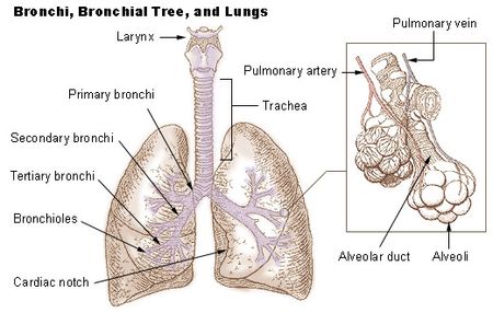
[[Image:Lung anatomy nci 407.jpg|right|Lung Anatomy|frameless|450x450px]]Lung [[Oncology|cancer]] refers to tumors originating in the [[Lung Anatomy|lung]] parenchyma or within bronchi. It is: | |||
* A broad term referring to the main histological subtypes of primary lung malignancies that are mainly linked with inhaled carcinogens, with [[Smoking Cessation and Brief Intervention|cigarette smoke]] being a key culprit.<ref name=":0">Radiopedia [https://radiopaedia.org/articles/lung-cancer-3 Lung cancer] </ref> | |||
* | * The most common cancer worldwide, with 2 million new cases in 2018<ref>World cancer research fund [https://www.wcrf.org/dietandcancer/cancer-trends/lung-cancer-statistics Lung cancer] Available from:https://www.wcrf.org/dietandcancer/cancer-trends/lung-cancer-statistics (last accessed 7.9.2020)</ref>. | ||
* One of the leading causes of cancer-related deaths in the United States. | |||
Due to the aetiology of lung cancer, the older age of patients, and presence of multi-morbidities, people with lung cancer constitute a complex patient population to manage.<ref name=":2" /> | |||
* At the beginning of the 20th century, lung cancer was a relatively rare disease. Its dramatic rise in later decades is mostly attributable to the increase in smoking among both males and females<ref name=":1">Siddiqui F, Siddiqui AH. [https://www.ncbi.nlm.nih.gov/books/NBK482357/ Cancer, lung]. InStatPearls [Internet] 2020 Apr 12. StatPearls Publishing.</ref> | |||
== Epidemiology == | == Epidemiology == | ||
* Is a leading type of cancer, equal in prevalence with [[Breast Cancer|breast cancer.]] | |||
* Is the leading cause of cancer mortality worldwide; accounting for ~20% of all cancer deaths<ref name=":0" /> | |||
* Is dramatically rising with nearly half of new cases, 49.9%, diagnosed in the underdeveloped world. | |||
* In the United States, mortality is high in men compared to women. | |||
* Overall, there is no difference between blacks and whites, but age-adjusted mortality is higher in black males than their white counterparts<ref name=":1" />. | |||
== Aetiology == | == Aetiology == | ||
Smoking is the most common cause of lung cancer. | |||
* It is estimated that 90% of the cases of lung cancer are attributable to smoking. | |||
* The risk is highest in males who smoke. | |||
* The risk is further compounded with exposure to other carcinogens, such as asbestos<ref name=":1" />. | |||
== | == Risk Factors == | ||
* There is no correlation between lung cancer and the number of packs smoked per year due to the complex interplay between smoking and environmental and genetic factors. | |||
* The risk of lung cancer by passive smoking increases by 20% to 30%. | |||
* asbestos: 5x increased risk | |||
* occupational exposure: uranium, radon, arsenic, chromium | |||
* diffuse [[Pulmonary Fibrosis|lung fibrosis]]: 10x increased risk | |||
* [[Chronic Obstructive Pulmonary Disease Rehabilitation Class|chronic obstructive pulmonary disease]]<ref name=":0" /> | |||
== | == Lung Metastases == | ||
The lungs are one of the most common metastatic disease target organs. Lung metastases are most commonly caused by cancers of the head and neck, breast, stomach, pancreas, kidney, bladder, male and female genitourinary tract, and sarcomas. The presence of pulmonary nodules, lymphangitic carcinomatosis, endobronchial tumours, and pleural involvement indicates pulmonary metastatic disease. Nonetheless, the differential diagnosis is critical, especially in patients with solitary pulmonary nodules and systemic disease. <ref name=":3">Herold CJ, Bankier AA, Fleischmann D. [http://dx.doi.org/10.1007/bf00187656 Lung metastases]. Eur Radiol. 1996;6(5). </ref> | |||
Plain [[X-Rays|chest radiography]] is typically used for detection and therapeutic monitoring; however, [[CT Scans|CT scans]] is increasingly being used for these purposes. Spiral CT scans appears to be the most sensitive imaging technique for detecting metastases at the moment, as it detects a greater number of pulmonary nodules than other techniques. <ref name=":3" /> | |||
== Investigations == | == Investigations == | ||
The overall goal is a timely diagnosis and accurate staging. | |||
Only 26% and 8% of cancers are diagnosed at stages I and II, whereas 28% and 38% are diagnosed at stages III and IV respectively. Hence curative surgery is an option for a minority of patients<ref name=":1" />. | |||
'''Lung cancer evaluation can be divided in 2 ways:''' | |||
# [[X-Rays|Radiological]] staging | |||
# Invasive staging | |||
=== | === Goals of Initial Evaluation === | ||
* Clinical extent and stage of the disease | |||
* Optimal target site and modality of 1st tissue biopsy | |||
* Specific histologic subtypes | |||
* Presence of co-morbidities, para-neoplastic syndromes | |||
* Patient values and preferences regarding therapy | |||
=== Radiologic Staging === | |||
Every patient suspected of having lung cancer should undergo the following tests: | |||
* | * Contrast-enhanced [[CT Scans|CT]] chest with extension to upper abdomen up to the level of adrenal glands. | ||
* | * Imaging with PET or PET-CT directed at sites of potential metastasis when symptoms or focal findings are present or when chest CT shows evidence of advanced disease.<ref name=":1" /><br> | ||
== Clinical Manifestations == | == Clinical Manifestations == | ||
Patients with lung cancer may be asymptomatic in up to 50% of cases. | |||
* Cough and [[dyspnoea]] are rather non-specific symptoms that are common amongst those with lung cancer. | |||
* Central tumours may result in haemoptysis | |||
* Peripheral lesions may result in pleuritic chest pain. | |||
* [[Pneumonia]], [[Pleural Effusion|pleural effusion]], wheeze, [[Lymphatic System|lymphadenopathy]] may be present. | |||
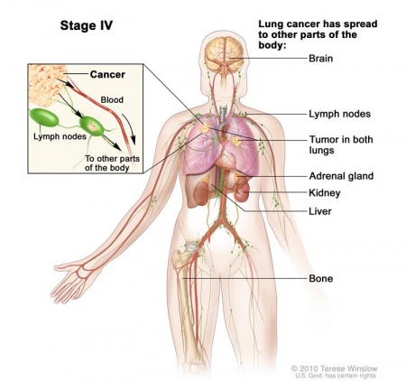
* Other symptoms may be secondary to metastases (bone, contralateral lung, brain, adrenal glands, and liver particularly) or [[Paraneoplastic Syndrome|paraneoplastic syndromes]]. | |||
== Treatment/Prognosis == | |||
[[File:Stage 4.jpg|right|frameless|450x450px]] | |||
Treatment and prognosis vary not only with stage but also with cell type.In general, surgery, [[Chemotherapy Side Effects and Syndromes|chemotherapy]], and [[Radiation Side Effects and Syndromes|radiotherapy]] are offered according to the stage, resectability, operability, and functional status.<ref name=":0" /> | |||
* Despite all the advances, the outcomes for lung cancer remain abysmal. The key reason is that most patients are diagnosed with advanced-stage disease. To improve outcomes, an interprofessional team approach with close communication between the members may perhaps lead to earlier diagnosis and treatment. | |||
* The definitive diagnosis and management of lung cancer is done by the thoracic surgeon with collaboration with the radiologist and pathologist. | |||
* After surgery, the patients are usually monitored by nurses for [[Oxygen Therapy|oxygenation]], [[Ventilation and Weaning|ventilation]], and [[Cancer Pain|pain]]. Since many of these patients are smokers, they also have other comorbidities like [[Coronary Artery Disease (CAD)|heart disease]] and [[Peripheral Arterial Disease|peripheral vascular disease]], which often presents with symptoms in the post-operative period. | |||
* After surgery, patients need prolonged rehabilitation. Some may need chemotherapy and radiation. | |||
* Lung cancer is not curable and all clinicians should urge patients to quit smoking; screening may be useful in selective patients. | |||
== Physiotherapy == | |||
Physiotherapy interventions vary depending on the stage in disease trajectory and timing relative to treatment. | |||
* The cornerstone of physiotherapy management in lung cancer should be prescription and delivery of [[Therapeutic Exercise|exercise]] intervention. | |||
* | * [[Physical Activity|Physical activity]] and exercise are vital components targeting three main aspects of the cancer continuum: prevention, mortality and morbidity. | ||
* The American Cancer Society recommends that adults with cancer engage in at least 150 minutes of moderate-intensity aerobic exercise and two sessions of resistance exercise per week, which is the same as the guidelines for the general adult<ref name=":2">Granger CL. [https://www.researchgate.net/publication/298433271_Physiotherapy_management_of_lung_cancer/fulltext/56e9aa3b08ae3a5b48cc8aac/Physiotherapy-management-of-lung-cancer.pdf?origin=publication_detail Physiotherapy management of lung cancer.] Journal of physiotherapy. 2016 Apr 1;62(2):60-7.</ref>. | |||
* | |||
* | |||
==== Exercise ==== | ==== Exercise ==== | ||
Aerobic exercise and resistance training have a positive impact on lung functioning including a reduction in airflow obstruction and clearing of airways therefore the improved functional capabilities increase energy levels and the release of sputum.<ref name="p7" / | * Aerobic exercise and resistance training have a positive impact on lung functioning including a reduction in airflow obstruction and clearing of airways therefore the improved functional capabilities increase energy levels and the release of sputum.<ref name="p7">Ozalevli S. [http://www.ncbi.nlm.nih.gov/pubmed/24177683 Impact of physiotherapy on patients with advanced lung cancer.] Chron Respir Dis [Internet]. 2013;10(4):223–32. </ref> | ||
* [[Pulmonary Rehabilitation|Pulmonary Rehabilitation programs]] is tailored to the individual who has recently had eg lung surgery, with the aim of optimizing their respiratory function and therefore their [[Quality of Life|quality of life]] (QOL) and participation in their everyday lives. | |||
* Exercise following surgery or treatment aims to restore physical status (addressing loss of functional capacity and muscle strength, which may occur during treatment) and to maximise function, physical activity, psychological status and health-related quality of life in the long term<ref name=":2" /> | |||
* Research shows positive results of physical exercises on cancer-related fatigue, physical function, symptom distress, sarcopenia and health-related quality of life (HRQoL)<ref>Edbrooke L, Granger CL, Denehy L. [https://pubmed.ncbi.nlm.nih.gov/32233342/ Physical activity for people with lung cancer.] Australian Journal of General Practice. 2020 Apr;49(4):175.</ref>. | |||
== Prevention == | == Prevention == | ||
* Smoking cessation: The best way to prevent lung cancer is to quit smoking. The risk of getting diagnosed with lung cancer will decrease the sooner an individual quits smoking. After 10 years of not smoking, the chance of developing lung cancer decreases to half that of someone who smokes.<ref name="p5">Dela Cruz CS, Tanoue LT, Matthay R a. Lung Cancer: epidemiology, etiology and prevention. Clin Chest Med. 2011;32(4):1–61.</ref> | |||
The best way to prevent lung cancer is to quit smoking. The risk of getting diagnosed with lung cancer will decrease the sooner an individual quits smoking. After 10 years of not smoking, the chance of developing lung cancer decreases to half that of someone who smokes.<ref name="p5" | |||
* Diet: Research suggests that eating a low-fat, high-fibre diet, including at least five portions of fresh fruit and vegetables and whole grains every day, can help reduce the risk of developing lung cancer, as well as other types of cancer and heart disease.<ref name="p8" /> | |||
* [[Physical Activity]]/Exercise: Studies show that higher levels of physical activity may lower lung cancer risk. <ref>Lee IM. Physical activity and cancer prevention--data from epidemiologic studies. Medicine and science in sports and exercise. 2003 Nov;35(11):1823-7.</ref> It is important to exercise regularly, attempting to perform at least 150 minutes of moderate intensity [[Aerobic Exercise|aerobic activity]] each week and incorporate muscle strengthening activities two days per week.<ref name="p8">[https://www.nhs.uk/conditions/lung-cancer/prevention/ Lung cancer - Prevention] [Internet]. nhs.uk. </ref> | |||
== References == | == References == | ||
<br> | |||
[[Category:Glasgow_Caledonian_University_Project]] | [[Category:Glasgow_Caledonian_University_Project]] | ||
[[Category:Cardiopulmonary]] | [[Category:Cardiopulmonary]] | ||
<references /> | <references /> | ||
[[Category: | [[Category:Chronic Respiratory Disease - Conditions]] | ||
[[Category:Oncology]] | |||
Latest revision as of 01:09, 2 December 2023
Original Editor - Sarah Hansford and Kaden Johnson Top Contributors - Sarah Hansford, Kaden Johnson, Lucinda hampton, Kim Jackson, Vidya Acharya, Admin, WikiSysop, 127.0.0.1, Mariam Hashem, Michelle Lee, Adam Vallely Farrell, Kapil Narale and Aya Alhindi
Introduction[edit | edit source]
Lung cancer refers to tumors originating in the lung parenchyma or within bronchi. It is:
- A broad term referring to the main histological subtypes of primary lung malignancies that are mainly linked with inhaled carcinogens, with cigarette smoke being a key culprit.[1]
- The most common cancer worldwide, with 2 million new cases in 2018[2].
- One of the leading causes of cancer-related deaths in the United States.
Due to the aetiology of lung cancer, the older age of patients, and presence of multi-morbidities, people with lung cancer constitute a complex patient population to manage.[3]
- At the beginning of the 20th century, lung cancer was a relatively rare disease. Its dramatic rise in later decades is mostly attributable to the increase in smoking among both males and females[4]
Epidemiology[edit | edit source]
- Is a leading type of cancer, equal in prevalence with breast cancer.
- Is the leading cause of cancer mortality worldwide; accounting for ~20% of all cancer deaths[1]
- Is dramatically rising with nearly half of new cases, 49.9%, diagnosed in the underdeveloped world.
- In the United States, mortality is high in men compared to women.
- Overall, there is no difference between blacks and whites, but age-adjusted mortality is higher in black males than their white counterparts[4].
Aetiology[edit | edit source]
Smoking is the most common cause of lung cancer.
- It is estimated that 90% of the cases of lung cancer are attributable to smoking.
- The risk is highest in males who smoke.
- The risk is further compounded with exposure to other carcinogens, such as asbestos[4].
Risk Factors[edit | edit source]
- There is no correlation between lung cancer and the number of packs smoked per year due to the complex interplay between smoking and environmental and genetic factors.
- The risk of lung cancer by passive smoking increases by 20% to 30%.
- asbestos: 5x increased risk
- occupational exposure: uranium, radon, arsenic, chromium
- diffuse lung fibrosis: 10x increased risk
- chronic obstructive pulmonary disease[1]
Lung Metastases[edit | edit source]
The lungs are one of the most common metastatic disease target organs. Lung metastases are most commonly caused by cancers of the head and neck, breast, stomach, pancreas, kidney, bladder, male and female genitourinary tract, and sarcomas. The presence of pulmonary nodules, lymphangitic carcinomatosis, endobronchial tumours, and pleural involvement indicates pulmonary metastatic disease. Nonetheless, the differential diagnosis is critical, especially in patients with solitary pulmonary nodules and systemic disease. [5]
Plain chest radiography is typically used for detection and therapeutic monitoring; however, CT scans is increasingly being used for these purposes. Spiral CT scans appears to be the most sensitive imaging technique for detecting metastases at the moment, as it detects a greater number of pulmonary nodules than other techniques. [5]
Investigations[edit | edit source]
The overall goal is a timely diagnosis and accurate staging.
Only 26% and 8% of cancers are diagnosed at stages I and II, whereas 28% and 38% are diagnosed at stages III and IV respectively. Hence curative surgery is an option for a minority of patients[4].
Lung cancer evaluation can be divided in 2 ways:
- Radiological staging
- Invasive staging
Goals of Initial Evaluation[edit | edit source]
- Clinical extent and stage of the disease
- Optimal target site and modality of 1st tissue biopsy
- Specific histologic subtypes
- Presence of co-morbidities, para-neoplastic syndromes
- Patient values and preferences regarding therapy
Radiologic Staging[edit | edit source]
Every patient suspected of having lung cancer should undergo the following tests:
- Contrast-enhanced CT chest with extension to upper abdomen up to the level of adrenal glands.
- Imaging with PET or PET-CT directed at sites of potential metastasis when symptoms or focal findings are present or when chest CT shows evidence of advanced disease.[4]
Clinical Manifestations[edit | edit source]
Patients with lung cancer may be asymptomatic in up to 50% of cases.
- Cough and dyspnoea are rather non-specific symptoms that are common amongst those with lung cancer.
- Central tumours may result in haemoptysis
- Peripheral lesions may result in pleuritic chest pain.
- Pneumonia, pleural effusion, wheeze, lymphadenopathy may be present.
- Other symptoms may be secondary to metastases (bone, contralateral lung, brain, adrenal glands, and liver particularly) or paraneoplastic syndromes.
Treatment/Prognosis[edit | edit source]
Treatment and prognosis vary not only with stage but also with cell type.In general, surgery, chemotherapy, and radiotherapy are offered according to the stage, resectability, operability, and functional status.[1]
- Despite all the advances, the outcomes for lung cancer remain abysmal. The key reason is that most patients are diagnosed with advanced-stage disease. To improve outcomes, an interprofessional team approach with close communication between the members may perhaps lead to earlier diagnosis and treatment.
- The definitive diagnosis and management of lung cancer is done by the thoracic surgeon with collaboration with the radiologist and pathologist.
- After surgery, the patients are usually monitored by nurses for oxygenation, ventilation, and pain. Since many of these patients are smokers, they also have other comorbidities like heart disease and peripheral vascular disease, which often presents with symptoms in the post-operative period.
- After surgery, patients need prolonged rehabilitation. Some may need chemotherapy and radiation.
- Lung cancer is not curable and all clinicians should urge patients to quit smoking; screening may be useful in selective patients.
Physiotherapy[edit | edit source]
Physiotherapy interventions vary depending on the stage in disease trajectory and timing relative to treatment.
- The cornerstone of physiotherapy management in lung cancer should be prescription and delivery of exercise intervention.
- Physical activity and exercise are vital components targeting three main aspects of the cancer continuum: prevention, mortality and morbidity.
- The American Cancer Society recommends that adults with cancer engage in at least 150 minutes of moderate-intensity aerobic exercise and two sessions of resistance exercise per week, which is the same as the guidelines for the general adult[3].
Exercise[edit | edit source]
- Aerobic exercise and resistance training have a positive impact on lung functioning including a reduction in airflow obstruction and clearing of airways therefore the improved functional capabilities increase energy levels and the release of sputum.[6]
- Pulmonary Rehabilitation programs is tailored to the individual who has recently had eg lung surgery, with the aim of optimizing their respiratory function and therefore their quality of life (QOL) and participation in their everyday lives.
- Exercise following surgery or treatment aims to restore physical status (addressing loss of functional capacity and muscle strength, which may occur during treatment) and to maximise function, physical activity, psychological status and health-related quality of life in the long term[3]
- Research shows positive results of physical exercises on cancer-related fatigue, physical function, symptom distress, sarcopenia and health-related quality of life (HRQoL)[7].
Prevention[edit | edit source]
- Smoking cessation: The best way to prevent lung cancer is to quit smoking. The risk of getting diagnosed with lung cancer will decrease the sooner an individual quits smoking. After 10 years of not smoking, the chance of developing lung cancer decreases to half that of someone who smokes.[8]
- Diet: Research suggests that eating a low-fat, high-fibre diet, including at least five portions of fresh fruit and vegetables and whole grains every day, can help reduce the risk of developing lung cancer, as well as other types of cancer and heart disease.[9]
- Physical Activity/Exercise: Studies show that higher levels of physical activity may lower lung cancer risk. [10] It is important to exercise regularly, attempting to perform at least 150 minutes of moderate intensity aerobic activity each week and incorporate muscle strengthening activities two days per week.[9]
References[edit | edit source]
- ↑ 1.0 1.1 1.2 1.3 Radiopedia Lung cancer
- ↑ World cancer research fund Lung cancer Available from:https://www.wcrf.org/dietandcancer/cancer-trends/lung-cancer-statistics (last accessed 7.9.2020)
- ↑ 3.0 3.1 3.2 Granger CL. Physiotherapy management of lung cancer. Journal of physiotherapy. 2016 Apr 1;62(2):60-7.
- ↑ 4.0 4.1 4.2 4.3 4.4 Siddiqui F, Siddiqui AH. Cancer, lung. InStatPearls [Internet] 2020 Apr 12. StatPearls Publishing.
- ↑ 5.0 5.1 Herold CJ, Bankier AA, Fleischmann D. Lung metastases. Eur Radiol. 1996;6(5).
- ↑ Ozalevli S. Impact of physiotherapy on patients with advanced lung cancer. Chron Respir Dis [Internet]. 2013;10(4):223–32.
- ↑ Edbrooke L, Granger CL, Denehy L. Physical activity for people with lung cancer. Australian Journal of General Practice. 2020 Apr;49(4):175.
- ↑ Dela Cruz CS, Tanoue LT, Matthay R a. Lung Cancer: epidemiology, etiology and prevention. Clin Chest Med. 2011;32(4):1–61.
- ↑ 9.0 9.1 Lung cancer - Prevention [Internet]. nhs.uk.
- ↑ Lee IM. Physical activity and cancer prevention--data from epidemiologic studies. Medicine and science in sports and exercise. 2003 Nov;35(11):1823-7.


