Iliotibial Tract: Difference between revisions
Eman Ammar (talk | contribs) No edit summary |
No edit summary |
||
| (42 intermediate revisions by 2 users not shown) | |||
| Line 1: | Line 1: | ||
<div class="editorbox"> | <div class="editorbox">'''Original Editor '''- [[User:Eman Ammar|Eman Ammar]] | ||
'''Top Contributors''' - {{Special:Contributors/{{FULLPAGENAME}}}} | '''Top Contributors''' - {{Special:Contributors/{{FULLPAGENAME}}}} | ||
</div> | </div> | ||
== Description | == Description == | ||
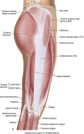
The | [[File:Iliotibial tract.jpg|560x560px|alt=|right|frameless]]The iliotibial band (ITB) is a thick band of fascia formed proximally at the [[hip]] by the [[fascia]] of the [[Gluteus Maximus|gluteus maximus]], [[Gluteus Medius|gluteus medius]] and [[Tensor Fascia Lata|tensor fasciae latae]] muscles<ref name=":0">Radiopedia ITB Available: https://radiopaedia.org/articles/iliotibial-band?lang=gb (accessed 27.12.2021)</ref>. Its main functions are pelvic stabilisation and posture control.<ref>Musculoskeletal Key Deep Dry Needling of the Hip and Pelvic Muscles Available:https://musculoskeletalkey.com/deep-dry-needling-of-the-hip-and-pelvic-muscles/ (accessed 28.12.2021)</ref> | ||
The | The ITB runs along the lateral thigh and serves as an important structure involved in lower extremity motion. | ||
There are multiple clinical conditions that can present secondary to a spectrum of ITB dysfunction eg [[Snapping Hip Syndrome|external snapping hip syndrome]], [[Iliotibial Band Syndrome|ITB syndrome]]<ref name=":1">Hyland S, Graefe S, Varacallo M. [https://www.ncbi.nlm.nih.gov/books/NBK537097/ Anatomy, bony pelvis and lower limb, iliotibial band (tract).] StatPearls [Internet]. 2020 Aug 10.Available: https://www.ncbi.nlm.nih.gov/books/NBK537097/<nowiki/>(accessed 27.12.2021)</ref>. | |||
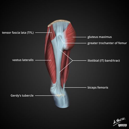
* Due to the ITBand’s insertion on Gerdy’s tubercle, it actually has no bony attachment along the femur. Therefore, it has the tendency to shift anterior/posterior (front to back) as your knee flexes and extends.<ref name=":2">Boulder sports Physio Iliotibial Band (ITBand) Syndrome Available:https://www.bouldersportsphysio.com/blog/blog-post-title-two-5k22t (accessed 27.12.2021)</ref> | |||
'''Image 1''': The iliotibial band (ITB). is a thick band of fascia formed proximally at the hip by the fascia of the gluteus maximus, gluteus medius and tensor fasciae latae muscles. | |||
=== | === Anatomy === | ||
[[File:Iliotibial-band-itb-anatomy-diagrams.jpeg|right|frameless|495x495px|alt=]] | |||
The iliotibial band (ITB) is a thick band of [[fascia]] formed proximally at the [[hip]] by the fascia of the [[Gluteus Maximus|gluteus maximus]], [[Gluteus Medius|gluteus medius]] and [[Tensor Fascia Lata|tensor fasciae latae]] muscles. | |||
It traverses superficial to the [[Vastus Lateralis|vastus lateralis]] and inserts on the Gerdy tubercle of the lateral [[Tibial Plateau Fractures|tibial plateau]] and partially to the supracondylar ridge of the lateral [[Femur|femur.]] There is also an anterior extension called the iliopatella band that connects the lateral [[patella]] and prevents medial translation of the patella.<ref>Hadeed A, Tapscott DC. [https://www.ncbi.nlm.nih.gov/books/NBK542185/ Iliotibial band friction syndrome]. 2019 Available: https://www.ncbi.nlm.nih.gov/books/NBK542185/<nowiki/>(accessed 28.12.2021)</ref> | |||
the | |||
= | A small recess exists between the lateral femoral epicondyle and the ITB, which contains a [[Synovium & Synovial Fluid|synovial]] extension of the [[knee]] joint capsule (lateral synovial recess)<ref name=":0" /> | ||
The ITB | The ITB shares the innervation of the TFL and [https://physio-pedia.com/Gluteus_Maximus?utm_source=physiopedia&utm_medium=search&utm_campaign=ongoing_internal gluteus maximus] via the superior gluteal nerve and inferior gluteal nerve. | ||
Composition: The Iliotibial Band is made up of mostly collagen fibers. [[Collagen]] is the strongest [[Muscle Proteins|protein]] found in nature. The collagen fibres are aligned in a very organized, vertical fashion as this allows for better force absorption with weight bearing activities. There is a small amount of elastin fibers amongst the layers of collagen, which allow it to be slightly elastic and pliable helping it act as a spring. However, this does not give it the ability to [[Stretching|stretch]] like a muscle<ref name=":2" /> | |||
The | |||
== | == Function. == | ||
[[File:ITB.png|right|frameless|702x702px]] | |||
Proximal ITB function includes: | |||
# Hip extension | |||
# Hip abduction | |||
# Lateral hip rotation | |||
Distally, ITB function depends on the position of the knee joint | |||
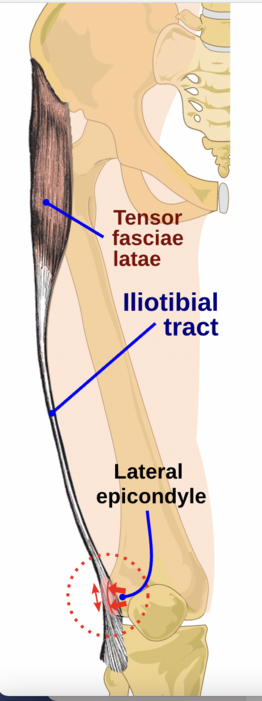
# Full extension to 20 to 30 degrees of flexion: Active knee extensor, ITB lying anterior to the lateral femoral epicondyle | |||
# 20 to 30 degrees of flexion to full flexion ROM: Active knee flexor, ITB lies posterior relative to the lateral femoral epicondyle<ref name=":1" /> | |||
== | == Physiotherapy == | ||
The iliotibial band is one of the most common [[Assessment of Running Biomechanics|running]] injuries we see as physiotherapists. It is considered a non-traumatic overuse injury and is often concomitant with underlying weakness of hip abductor muscles. For more see [[Iliotibial Band Syndrome|Iliotibial Band Syndrome.]] Clinical examination testing for [[Iliotibial Band Syndrome|ITB dysfunction]] is best elicited utilizing the [https://physio-pedia.com/Ober%27s_Test Ober Test, see here] | |||
< | External [[Snapping Hip Syndrome|snapping hip syndrome]] is another ITB pathology you may encounter<ref>Winston P, Awan R, Cassidy JD, et al. Clinical examination and ultrasound of self-reported snapping hip syndrome in elite ballet dancers. Am J Sports Med. 2007 Jan;35(1):118–126. [PubMed] </ref>. Snapping Hip Syndrome is a condition that is characterized by a snapping sensation, and/or audible “snap” or “click” noise, in or around the [[hip]] when it is in motion. | ||
It has been proposed that a tight iliotibial band (ITB) through its attachment of the lateral retinaculum into the patella could cause lateral patella tracking, patella tilt and compression. <ref>Hudson Z, Darthuy E. Iliotibial band tightness and patellofemoral pain syndrome: a case-control study. Manual therapy. 2009 Apr 1;14(2):147-51. Available: https://pubmed.ncbi.nlm.nih.gov/18313972/ (accessed 28.12.2021)</ref>This has implications in subjects presenting with [[Patellofemoral Pain Syndrome|patellofemoral pain syndrome]] (PFPS) | |||
Image 3: Iliotibial band syndrome | |||
== References == | |||
[[Category:Anatomy]] | [[Category:Anatomy]] | ||
[[Category:Muscles]] | [[Category:Muscles]] | ||
<references /> | |||
Latest revision as of 02:00, 28 December 2021
Top Contributors - Eman Ammar and Lucinda hampton
Description[edit | edit source]
The iliotibial band (ITB) is a thick band of fascia formed proximally at the hip by the fascia of the gluteus maximus, gluteus medius and tensor fasciae latae muscles[1]. Its main functions are pelvic stabilisation and posture control.[2]
The ITB runs along the lateral thigh and serves as an important structure involved in lower extremity motion.
There are multiple clinical conditions that can present secondary to a spectrum of ITB dysfunction eg external snapping hip syndrome, ITB syndrome[3].
- Due to the ITBand’s insertion on Gerdy’s tubercle, it actually has no bony attachment along the femur. Therefore, it has the tendency to shift anterior/posterior (front to back) as your knee flexes and extends.[4]
Image 1: The iliotibial band (ITB). is a thick band of fascia formed proximally at the hip by the fascia of the gluteus maximus, gluteus medius and tensor fasciae latae muscles.
Anatomy[edit | edit source]
The iliotibial band (ITB) is a thick band of fascia formed proximally at the hip by the fascia of the gluteus maximus, gluteus medius and tensor fasciae latae muscles.
It traverses superficial to the vastus lateralis and inserts on the Gerdy tubercle of the lateral tibial plateau and partially to the supracondylar ridge of the lateral femur. There is also an anterior extension called the iliopatella band that connects the lateral patella and prevents medial translation of the patella.[5]
A small recess exists between the lateral femoral epicondyle and the ITB, which contains a synovial extension of the knee joint capsule (lateral synovial recess)[1]
The ITB shares the innervation of the TFL and gluteus maximus via the superior gluteal nerve and inferior gluteal nerve.
Composition: The Iliotibial Band is made up of mostly collagen fibers. Collagen is the strongest protein found in nature. The collagen fibres are aligned in a very organized, vertical fashion as this allows for better force absorption with weight bearing activities. There is a small amount of elastin fibers amongst the layers of collagen, which allow it to be slightly elastic and pliable helping it act as a spring. However, this does not give it the ability to stretch like a muscle[4]
Function.[edit | edit source]
Proximal ITB function includes:
- Hip extension
- Hip abduction
- Lateral hip rotation
Distally, ITB function depends on the position of the knee joint
- Full extension to 20 to 30 degrees of flexion: Active knee extensor, ITB lying anterior to the lateral femoral epicondyle
- 20 to 30 degrees of flexion to full flexion ROM: Active knee flexor, ITB lies posterior relative to the lateral femoral epicondyle[3]
Physiotherapy[edit | edit source]
The iliotibial band is one of the most common running injuries we see as physiotherapists. It is considered a non-traumatic overuse injury and is often concomitant with underlying weakness of hip abductor muscles. For more see Iliotibial Band Syndrome. Clinical examination testing for ITB dysfunction is best elicited utilizing the Ober Test, see here
External snapping hip syndrome is another ITB pathology you may encounter[6]. Snapping Hip Syndrome is a condition that is characterized by a snapping sensation, and/or audible “snap” or “click” noise, in or around the hip when it is in motion.
It has been proposed that a tight iliotibial band (ITB) through its attachment of the lateral retinaculum into the patella could cause lateral patella tracking, patella tilt and compression. [7]This has implications in subjects presenting with patellofemoral pain syndrome (PFPS)
Image 3: Iliotibial band syndrome
References[edit | edit source]
- ↑ 1.0 1.1 Radiopedia ITB Available: https://radiopaedia.org/articles/iliotibial-band?lang=gb (accessed 27.12.2021)
- ↑ Musculoskeletal Key Deep Dry Needling of the Hip and Pelvic Muscles Available:https://musculoskeletalkey.com/deep-dry-needling-of-the-hip-and-pelvic-muscles/ (accessed 28.12.2021)
- ↑ 3.0 3.1 Hyland S, Graefe S, Varacallo M. Anatomy, bony pelvis and lower limb, iliotibial band (tract). StatPearls [Internet]. 2020 Aug 10.Available: https://www.ncbi.nlm.nih.gov/books/NBK537097/(accessed 27.12.2021)
- ↑ 4.0 4.1 Boulder sports Physio Iliotibial Band (ITBand) Syndrome Available:https://www.bouldersportsphysio.com/blog/blog-post-title-two-5k22t (accessed 27.12.2021)
- ↑ Hadeed A, Tapscott DC. Iliotibial band friction syndrome. 2019 Available: https://www.ncbi.nlm.nih.gov/books/NBK542185/(accessed 28.12.2021)
- ↑ Winston P, Awan R, Cassidy JD, et al. Clinical examination and ultrasound of self-reported snapping hip syndrome in elite ballet dancers. Am J Sports Med. 2007 Jan;35(1):118–126. [PubMed]
- ↑ Hudson Z, Darthuy E. Iliotibial band tightness and patellofemoral pain syndrome: a case-control study. Manual therapy. 2009 Apr 1;14(2):147-51. Available: https://pubmed.ncbi.nlm.nih.gov/18313972/ (accessed 28.12.2021)


