Aorta: Difference between revisions
No edit summary |
Sona Eyobe (talk | contribs) No edit summary |
||
| Line 6: | Line 6: | ||
== Introduction == | == Introduction == | ||
[[File:Human-heart-chambers.jpg|right|frameless|399x399px]] | [[File:Human-heart-chambers.jpg|right|frameless|399x399px]] | ||
The aorta is the large [[Arteries|artery]] that carries oxygen-rich [[Blood Physiology|blood]] from | The aorta is the large [[Arteries|artery]] that carries oxygen-rich [[Blood Physiology|blood]] from the [[Left Ventricle Heart|left ventricle]] of the heart to other parts of the body. The aorta begins at the top of the left ventricle, the [[Anatomy of the Human Heart|heart]]'s muscular pumping chamber. The heart pumps blood from the left ventricle into the aorta through the aortic valve. Three leaflets on the [[Aortic Valve Disease|aortic]] valve open and close with each heartbeat to allow a one-way flow of blood. | ||
* The aorta is important because it gives the body access to the oxygen-rich blood it needs to survive. | * The aorta is important because it gives the body access to the oxygen-rich blood it needs to survive. | ||
| Line 16: | Line 16: | ||
The aorta is a tube about a foot long and just over an inch in diameter. The aorta is divided into four sections: | The aorta is a tube about a foot long and just over an inch in diameter. The aorta is divided into four sections: | ||
# The ascending aorta rises | # The ascending aorta rises from the heart and is about 2 inches long. The [[Coronary Artery|coronary]] [[arteries]] branch off the ascending aorta to supply the heart with blood. | ||
# The aortic arch curves over the heart, giving rise to branches that bring blood to the head, neck, and arms. | # The aortic arch curves over the heart, giving rise to branches that bring blood to the head, neck, and arms. | ||
# The descending thoracic aorta travels down through the [[Thoracic Anatomy|chest]]. Its small branches supply blood to the [[ribs]] and some chest structures. | # The descending thoracic aorta travels down through the [[Thoracic Anatomy|chest]]. Its small branches supply blood to the [[ribs]] and some chest structures. | ||
| Line 25: | Line 25: | ||
Aortic [[atherosclerosis]]: Plaque collects and hardens inside the aorta blocking the free flow of blood through it and weakening the aortic walls. It can lead to aortic aneurysms, arterial thrombosis, strokes, and anginas. | Aortic [[atherosclerosis]]: Plaque collects and hardens inside the aorta blocking the free flow of blood through it and weakening the aortic walls. It can lead to aortic aneurysms, arterial thrombosis, strokes, and anginas. | ||
[[Aortic Valve Disease|Aortic stenosis]]: With this condition, the aortic valve doesn’t open completely when it should, making the heart have to pump harder to get blood through the valve and into the aorta. It can lead to complications like left ventricular hypertrophy (LVH), diastolic dysfunction, and diastolic heart failure. Image: Aortic Stenosis | [[Aortic Valve Disease|Aortic stenosis]]: With this condition, the aortic valve doesn’t open completely when it should, making the heart have to pump harder to get blood through the valve and into the aorta. It can lead to complications like left ventricular hypertrophy (LVH), diastolic dysfunction, and diastolic heart failure. Image: Aortic Stenosis | ||
Aortic regurgitation: | Aortic regurgitation: It is the inadequate closure of the aortic valve during diastole that results in reverse blood flow through the aortic valve. Its acute form is caused by infective endocarditis and aortic dissection in the ascending part. The chronic form is caused by the deterioration of the aortic valve, aneurysm in the thoracic aorta, rheumatic fever, infective endocarditis, and trauma, which typically doesn't show any symptoms for a long time. It can lead to [[Pulmonary Oedema|pulmonary oedema]], left ventricular hypertrophy (LVH), [[Heart Arrhythmias: Assessment|arrhythmias]], and [[Heart Failure|heart failure]]. It is also known as aortic insufficiency.<ref>Very Well health Aorta Available:https://www.verywellhealth.com/aorta-anatomy-4588336 (accessed 16.7.2021)</ref> | ||
[[File:Aortic aneurysm.jpg|right|frameless|alt=|408x408px]] | [[File:Aortic aneurysm.jpg|right|frameless|alt=|408x408px]] | ||
An [[Aortic Aneurysm|aortic aneurysm]] is an abnormal enlargement or bulging of the wall of the aorta. An aneurysm can occur anywhere in the vascular tree. Eg [[Abdominal Aortic Aneurysm|Abdominal]] Aorta. Once an aneurysm is diagnosed, treatment may be needed, depending on the size of the aneurysm. Ruptured aneurysms require emergency [[Surgery and General Anaesthetic|surgery]] to stop the bleeding. | An [[Aortic Aneurysm|aortic aneurysm]] is an abnormal enlargement or bulging of the wall of the aorta. An aneurysm can occur anywhere in the vascular tree. Eg [[Abdominal Aortic Aneurysm|Abdominal]] Aorta. Once an aneurysm is diagnosed, treatment may be needed, depending on the size of the aneurysm. Ruptured aneurysms require emergency [[Surgery and General Anaesthetic|surgery]] to stop the bleeding. | ||
| Line 33: | Line 33: | ||
The aorta has 3 layers. Aortic dissection is a tear that develops in the inner layer of the aorta, causing blood to flow between the layers. The layers then separate, interrupting the blood flow and possibly causing the arterial wall to burst. Aortic dissection can be a life-threatening emergency, in some situations requiring emergency surgery to repair or replace the damaged segment of the aorta.<ref name=":0">Cleveland Clinic Aorta Available:https://my.clevelandclinic.org/health/articles/17058-aorta-anatomy (accessed 16.7.2021)</ref> | The aorta has 3 layers. Aortic dissection is a tear that develops in the inner layer of the aorta, causing blood to flow between the layers. The layers then separate, interrupting the blood flow and possibly causing the arterial wall to burst. Aortic dissection can be a life-threatening emergency, in some situations requiring emergency surgery to repair or replace the damaged segment of the aorta.<ref name=":0">Cleveland Clinic Aorta Available:https://my.clevelandclinic.org/health/articles/17058-aorta-anatomy (accessed 16.7.2021)</ref> | ||
== | == Education == | ||
[[File:Healthy food 2.jpg|right|frameless]] | [[File:Healthy food 2.jpg|right|frameless]] | ||
Educate clients on the ways to keep their aorta healthy: | Educate clients on the ways to keep their aorta healthy: | ||
Revision as of 00:10, 20 November 2023
Original Editor - Lucinda hampton
Top Contributors - Lucinda hampton, Sona Eyobe and Rucha Gadgil
Introduction[edit | edit source]
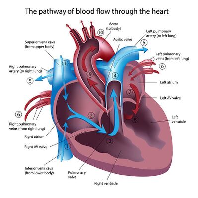
The aorta is the large artery that carries oxygen-rich blood from the left ventricle of the heart to other parts of the body. The aorta begins at the top of the left ventricle, the heart's muscular pumping chamber. The heart pumps blood from the left ventricle into the aorta through the aortic valve. Three leaflets on the aortic valve open and close with each heartbeat to allow a one-way flow of blood.
- The aorta is important because it gives the body access to the oxygen-rich blood it needs to survive.
- The heart itself gets oxygen from arteries that come off the ascending aorta.
- The head (including the brain), neck and arms get oxygen from arteries that come off the aortic arch.
- The stomach, intestines, kidneys and other vital organs get oxygen from arteries that come off the abdominal aorta.[1]
Sections of the Aorta[edit | edit source]
The aorta is a tube about a foot long and just over an inch in diameter. The aorta is divided into four sections:
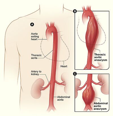
- The ascending aorta rises from the heart and is about 2 inches long. The coronary arteries branch off the ascending aorta to supply the heart with blood.
- The aortic arch curves over the heart, giving rise to branches that bring blood to the head, neck, and arms.
- The descending thoracic aorta travels down through the chest. Its small branches supply blood to the ribs and some chest structures.
- The abdominal aorta begins at the diaphragm, splitting to become the paired iliac arteries in the lower abdomen. Most of the major organs receive blood from branches of the abdominal aorta[2]
Clinical Significance[edit | edit source]
Aortic atherosclerosis: Plaque collects and hardens inside the aorta blocking the free flow of blood through it and weakening the aortic walls. It can lead to aortic aneurysms, arterial thrombosis, strokes, and anginas.
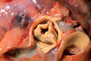
Aortic stenosis: With this condition, the aortic valve doesn’t open completely when it should, making the heart have to pump harder to get blood through the valve and into the aorta. It can lead to complications like left ventricular hypertrophy (LVH), diastolic dysfunction, and diastolic heart failure. Image: Aortic Stenosis
Aortic regurgitation: It is the inadequate closure of the aortic valve during diastole that results in reverse blood flow through the aortic valve. Its acute form is caused by infective endocarditis and aortic dissection in the ascending part. The chronic form is caused by the deterioration of the aortic valve, aneurysm in the thoracic aorta, rheumatic fever, infective endocarditis, and trauma, which typically doesn't show any symptoms for a long time. It can lead to pulmonary oedema, left ventricular hypertrophy (LVH), arrhythmias, and heart failure. It is also known as aortic insufficiency.[3]
An aortic aneurysm is an abnormal enlargement or bulging of the wall of the aorta. An aneurysm can occur anywhere in the vascular tree. Eg Abdominal Aorta. Once an aneurysm is diagnosed, treatment may be needed, depending on the size of the aneurysm. Ruptured aneurysms require emergency surgery to stop the bleeding.
The aorta has 3 layers. Aortic dissection is a tear that develops in the inner layer of the aorta, causing blood to flow between the layers. The layers then separate, interrupting the blood flow and possibly causing the arterial wall to burst. Aortic dissection can be a life-threatening emergency, in some situations requiring emergency surgery to repair or replace the damaged segment of the aorta.[4]
Education[edit | edit source]
Educate clients on the ways to keep their aorta healthy:
- The aorta can be damaged by high cholesterol and high blood pressure, so anything you do to keep those risk factors under control will also help you maintain a healthy aorta.
- This includes eating a balanced diet, getting regular exercise and avoiding cigarettes.[4]
References[edit | edit source]
- ↑ Columbia Surgery Aorta Available:https://columbiasurgery.org/aortic/all-about-aorta (accessed 16.7.2021)
- ↑ WebMD Aorta Available:https://www.webmd.com/heart/picture-of-the-aorta#1 (accessed 16.7.2021)
- ↑ Very Well health Aorta Available:https://www.verywellhealth.com/aorta-anatomy-4588336 (accessed 16.7.2021)
- ↑ 4.0 4.1 Cleveland Clinic Aorta Available:https://my.clevelandclinic.org/health/articles/17058-aorta-anatomy (accessed 16.7.2021)


