Talus: Difference between revisions
No edit summary |
No edit summary |
||
| (2 intermediate revisions by the same user not shown) | |||
| Line 2: | Line 2: | ||
<div class="editorbox"> '''Original Editor '''- [[User:Lucinda hampton|Lucinda hampton]] '''Top Contributors''' - {{Special:Contributors/{{FULLPAGENAME}}}}</div> | <div class="editorbox"> '''Original Editor '''- [[User:Lucinda hampton|Lucinda hampton]] '''Top Contributors''' - {{Special:Contributors/{{FULLPAGENAME}}}}</div> | ||
== Introduction == | == Introduction == | ||
The talus is the second largest bone in the hindfoot region of the human body. Responsible for transmitting body weight and forces passing between the lower leg and the foot. | <blockquote>Nearly 70% of ankle injuries can cause varying degrees of [[Cartilage|chondral]] and osteochondral injuries to the talus. </blockquote>The talus is the second largest bone in the hindfoot region of the human body. Responsible for transmitting body weight and forces passing between the lower leg and the foot. | ||
[[File:Talus bone - animation03.gif|right|frameless]] | [[File:Talus bone - animation03.gif|right|frameless]] | ||
The Talus (left talus shown in image) | The Talus (left talus shown in image) | ||
* Is a component of many multiple joints, including the [[Ankle and Foot|talocrural]] (ankle), subtalar, and transverse tarsal joints. | * Is a component of many multiple joints, including the [[Ankle and Foot|talocrural]] (ankle), subtalar, and transverse tarsal joints. | ||
* Does not have any direct muscular attachments and has a tenuous and limited blood supply, | * Does not have any direct muscular attachments and has a tenuous and limited blood supply, | ||
* | * The attachment site for many [[Ligament|ligaments]], including the lateral ankle ligaments and medial deltoid ligament complex. | ||
Talus fractures comprise about 1% of all [[Foot Anatomy|foot]] and ankle [[fracture]]<nowiki/>s. | Talus fractures comprise about 1% of all [[Foot Anatomy|foot]] and ankle [[fracture]]<nowiki/>s. | ||
It is important to be aware of the occurrence of both isolated and associated talar injuries and their extent of clinical and potentially long-term implications.<ref name=":0">Khan IA, Varacallo M. Anatomy, Bony Pelvis and Lower Limb, Foot Talus. [Updated 2023 Aug 8]. In: StatPearls [Internet]. Treasure Island (FL): StatPearls Publishing; 2023 Jan-. Available from: https://www.ncbi.nlm.nih.gov/books/NBK541086/ [last access 29.10.2023] | |||
It is important to be aware of the occurrence of both isolated and associated talar injuries and their extent of clinical and potentially long-term implications.<ref name=":0"> | |||
</ref> | </ref> | ||
== Structure == | == Structure == | ||
[[File:Talus .png|right|frameless|400x400px]] | [[File:Talus .png|right|frameless|400x400px]] | ||
The talus is an incredible bone; despite its small size, it transmits considerable force during the normal gait cycle and even more | The talus is an incredible bone; despite its small size, it transmits considerable force during the normal gait cycle and even more significantly during impact activities. | ||
* The talus is shaped like a truncated cone and is wider anteriorly than posteriorly (it is an irregular saddle-shaped bone). | * The talus is shaped like a truncated cone and is wider anteriorly than posteriorly (it is an irregular saddle-shaped bone). | ||
* Approximately two-thirds of its surface area is covered with articular [[cartilage]] and it has a tenuous blood supply, similar to the [[scaphoid]]. | * Approximately two-thirds of its surface area is covered with articular [[cartilage]], and it has a tenuous blood supply, similar to the [[scaphoid]]. | ||
* The talus has seven articular surfaces and is divided into the head, neck and body, and two processes, the posterior process and the lateral process | * The talus has seven articular surfaces and is divided into the head, neck and body, and two processes, the posterior process and the lateral process | ||
* The talar body has a curved smooth trochlear surface also termed the talar dome, | * The talar body has a curved smooth trochlear surface, also termed the talar dome, covered with hyaline cartilage and convex from front to back. The medial and lateral surfaces articulate with the medial malleolus (of the tibia) and lateral malleolus (of the fibula), respectively. The lateral articular surface is large and projects more inferiorly. The lower part of the lateral surface forms a bony projection called the lateral process, which supports the lower portion of the lateral articular facet. The posterior aspect has a backward and medially facing posterior process, with a lateral and medial tubercle separated by a groove for the tendon of [[flexor hallucis longus]]. | ||
*The talar head is the part that articulates with the navicular bone. On its inferior aspect, this is continuous with three articular facets | *The talar head is the part that articulates with the navicular bone. On its inferior aspect, this is continuous, with three articular facets separated by smooth ridges. There are anterior and middle facets which articulate with corresponding facets on the calcaneus. There is another facet, medial to the above facets, for articulation with the [[Spring Ligament|spring]] ligament. The talar head and body are connected by the talar neck, which is inclined downwards distally and medially.<ref>Majeed H, McBride DJ. [https://www.ncbi.nlm.nih.gov/pmc/articles/PMC5890124/pdf/eor-3-85.pdf Talar process fractures: An overview and update of the literature]. EFORT Open Rev. 2018 Mar 29;3(3):85-92. </ref> | ||
* The inferior surface of the talar neck has a deep groove, the sulcus tali, that passes obliquely forward and expands from medial to lateral. It forms the tarsal sinus with the calcaneal sulcus of the [[Calcaneal Fractures|calcaneum]]. <ref name=":1"> | * The inferior surface of the talar neck has a deep groove, the sulcus tali, that passes obliquely forward and expands from medial to lateral. It forms the tarsal sinus with the calcaneal sulcus of the [[Calcaneal Fractures|calcaneum]]. <ref name=":1">Morgan M, Worsley C, Yu Y, et al. Talus. Reference article, Radiopaedia.org. Available from https://radiopaedia.org/articles/talus [last access 29.10.2023] </ref> | ||
[[File:Talus bone - animation01.gif|right|frameless|350x350px]] | [[File:Talus bone - animation01.gif|right|frameless|350x350px]] | ||
| Line 35: | Line 32: | ||
== Muscle Attachments == | == Muscle Attachments == | ||
The talus does not serve as the site of any muscle attachment, which is significant because that means the talus does not have secondary sources of blood supply. | The talus does not serve as the site of any muscle attachment, which is significant because that means the talus does not have secondary sources of blood supply. | ||
* If there is vascular compromise to the talus through traumatic injuries such as in the setting of displaced fractures, there is a relatively high risk of developing post-traumatic, avascular necrosis (AVN)<ref name=":0" /> | * If there is a vascular compromise to the talus through traumatic injuries such as in the setting of displaced fractures, there is a relatively high risk of developing post-traumatic, avascular necrosis (AVN)<ref name=":0" /> | ||
== Ligaments == | == Ligaments == | ||
| Line 51: | Line 48: | ||
== Blood Supply == | == Blood Supply == | ||
* Posterior tibial artery into the medial side of body and sinus | * Posterior tibial artery into the medial side of the body and sinus | ||
* Anterior tibial artery/dorsalis pedis artery into head and neck | * Anterior tibial artery/dorsalis pedis artery into head and neck | ||
* Peroneal artery into the lateral side of the body and sinus<ref name=":1" /> | * Peroneal artery into the lateral side of the body and sinus<ref name=":1" /> | ||
| Line 66: | Line 63: | ||
=== Avascular Necrosis (AVN) === | === Avascular Necrosis (AVN) === | ||
Fractures of the talus have a high risk of developing AVN due to its inherently tenuous and limited blood supply. | Fractures of the talus have a high risk of developing AVN due to its inherently tenuous and limited blood supply. | ||
* Talar fractures | * Talar fractures comprise one percent of all foot and ankle fractures, and the most common (around 50%) talar fractures occur in the talar neck, the weakest part of the talus.<ref name=":0" /> | ||
* Talar body fractures make up 20% of all talar fractures, and if a fracture extends through an articulating surface on the talar body, sequelae of osteoarthritis can occur. | * Talar body fractures make up 20% of all talar fractures, and if a fracture extends through an articulating surface on the talar body, sequelae of osteoarthritis can occur. | ||
=== Avulsion Fractures of the Talus === | === Avulsion Fractures of the Talus === | ||
Lateral ankle sprains are the most common athletic injury, constituting 15 to 20% of all athletic injuries | Lateral ankle sprains are the most common athletic injury, constituting 15 to 20% of all athletic injuries and 85% of all ankle sprains. | ||
* Due to the attachment of the ATFL and PTFL on the talus, avulsion fractures | * Due to the attachment of the ATFL and PTFL on the talus, avulsion fractures may occur when those ligaments suffer injury in lateral ankle sprains. | ||
* One percent of all ankle sprains cause avulsion fractures of the talus, so it’s important to consider them in the differential diagnosis when patients present with symptoms a lateral ankle sprain, especially if patients have marked swelling, bruising, or difficulty weight-bearing. | * One percent of all ankle sprains cause avulsion fractures of the talus, so it’s important to consider them in the differential diagnosis when patients present with symptoms of a lateral ankle sprain, especially if patients have marked swelling, bruising, or difficulty weight-bearing. | ||
[[File:Ankle Post Med View Google.jpg|right|frameless]] | [[File:Ankle Post Med View Google.jpg|right|frameless]] | ||
=== Tarsal Tunnel Syndrome === | === Tarsal Tunnel Syndrome === | ||
[[Tarsal Tunnel Syndrome]] occurs on the ankle's medial aspect, the tarsal tunnel forms a bony floor consisting of the tibia, talus, and calcaneus, and a fibrous roof consisting of the fibrous flexor retinaculum. The posterior tibial nerve, artery, vein, | [[Tarsal Tunnel Syndrome]] occurs on the ankle's medial aspect, the tarsal tunnel forms a bony floor consisting of the tibia, talus, and calcaneus, and a fibrous roof consisting of the fibrous flexor retinaculum. The posterior tibial nerve, artery, vein, posterior tibialis, flexor hallucis longus, and flexor digitorum longus tendons are inside the tarsal tunnel, and the posterior tibial nerve has the potential to become entrapped within the space. | ||
* Characterized by pain, paresthesia, loss of sensation over the medial foot and ankle, patients may also experience weakness and muscle atrophy in severe cases. | * Characterized by pain, paresthesia, and loss of sensation over the medial foot and ankle, patients may also experience weakness and muscle atrophy in severe cases. | ||
* Bony lesions on the talus, such as osteophytes or osteosarcoma, may cause the posterior tibial nerve to get compressed in the tarsal tunnel, leading to tarsal tunnel syndrome. | * Bony lesions on the talus, such as osteophytes or osteosarcoma, may cause the posterior tibial nerve to get compressed in the tarsal tunnel, leading to tarsal tunnel syndrome. | ||
=== Pes Planus === | === Pes Planus === | ||
Through the subtalar joint, the talus maintains the medial and lateral longitudinal arches. [[Pes Planus|pes planus]] occurs when there is an alteration of talus morphology of the articular facets | Through the subtalar joint, the talus maintains the medial and lateral longitudinal arches. [[Pes Planus|pes planus]] occurs when there is an alteration of talus morphology of the articular facets and the biomechanics of the subtalar joint change, which can lead to pes planus. <ref name=":0" /> | ||
=== Os Trigonum Syndrome === | === Os Trigonum Syndrome === | ||
'''[[The Os Trigonum Syndrome|Os trigonum syndrome]] | '''[[The Os Trigonum Syndrome|Os trigonum syndrome]]'' refers to pain posterior to the ankle and reduced plantarflexion caused by “the nutcracker-phenomenon”. When an os trigonum is present, this accessory ossicle and surrounding soft tissues can wedge between the tibia, talus and calcaneus. This can lead to inflammation of the involved structures. <ref>Russell JA, Kruse DW, Koutedakis Y, McEwan IM, Wyon MA. Pathoanatomy of posterior ankle impingement in ballet dancers. Clin Anat. 2010 Sep;23(6):613-21. </ref><ref>M. Nyska, G. Mann Unstable Ankle. Leeds: Human Kinetics Publishers, 2002</ref> | ||
== References == | == References == | ||
Latest revision as of 15:11, 29 October 2023
Introduction[edit | edit source]
Nearly 70% of ankle injuries can cause varying degrees of chondral and osteochondral injuries to the talus.
The talus is the second largest bone in the hindfoot region of the human body. Responsible for transmitting body weight and forces passing between the lower leg and the foot.

The Talus (left talus shown in image)
- Is a component of many multiple joints, including the talocrural (ankle), subtalar, and transverse tarsal joints.
- Does not have any direct muscular attachments and has a tenuous and limited blood supply,
- The attachment site for many ligaments, including the lateral ankle ligaments and medial deltoid ligament complex.
Talus fractures comprise about 1% of all foot and ankle fractures.
It is important to be aware of the occurrence of both isolated and associated talar injuries and their extent of clinical and potentially long-term implications.[1]
Structure[edit | edit source]

The talus is an incredible bone; despite its small size, it transmits considerable force during the normal gait cycle and even more significantly during impact activities.
- The talus is shaped like a truncated cone and is wider anteriorly than posteriorly (it is an irregular saddle-shaped bone).
- Approximately two-thirds of its surface area is covered with articular cartilage, and it has a tenuous blood supply, similar to the scaphoid.
- The talus has seven articular surfaces and is divided into the head, neck and body, and two processes, the posterior process and the lateral process
- The talar body has a curved smooth trochlear surface, also termed the talar dome, covered with hyaline cartilage and convex from front to back. The medial and lateral surfaces articulate with the medial malleolus (of the tibia) and lateral malleolus (of the fibula), respectively. The lateral articular surface is large and projects more inferiorly. The lower part of the lateral surface forms a bony projection called the lateral process, which supports the lower portion of the lateral articular facet. The posterior aspect has a backward and medially facing posterior process, with a lateral and medial tubercle separated by a groove for the tendon of flexor hallucis longus.
- The talar head is the part that articulates with the navicular bone. On its inferior aspect, this is continuous, with three articular facets separated by smooth ridges. There are anterior and middle facets which articulate with corresponding facets on the calcaneus. There is another facet, medial to the above facets, for articulation with the spring ligament. The talar head and body are connected by the talar neck, which is inclined downwards distally and medially.[2]
- The inferior surface of the talar neck has a deep groove, the sulcus tali, that passes obliquely forward and expands from medial to lateral. It forms the tarsal sinus with the calcaneal sulcus of the calcaneum. [3]

Articulations[edit | edit source]
- superiorly through the talar dome to form the mortise joint of the ankle with the tibia and fibula
- inferoposteriorly: large oblique facet that is concave articulates with the calcaneus to form the talocalcaneal joint
- anteroinferiorly: two facets for articulation with the calcaneus to form part of the talocalcaneonavicular joint
- talar head (domed articular surface) with the navicular bone (circular depression on the posterior surface)[3]
Muscle Attachments[edit | edit source]
The talus does not serve as the site of any muscle attachment, which is significant because that means the talus does not have secondary sources of blood supply.
- If there is a vascular compromise to the talus through traumatic injuries such as in the setting of displaced fractures, there is a relatively high risk of developing post-traumatic, avascular necrosis (AVN)[1]
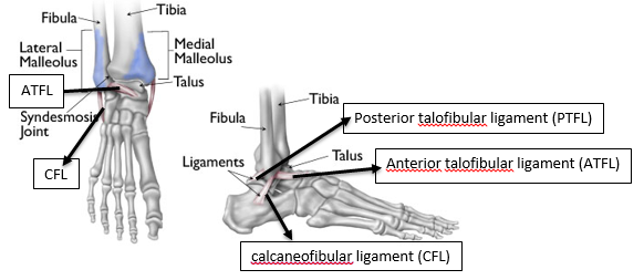
Ligaments[edit | edit source]
- Anterior talofibular ligament
- Posterior talofibular ligament
- Talocalcaneal ligaments
- Tarsal sinus ligaments
- Cervical ligament
- Talocalcaneal interosseous ligament
- Deltoid ligament
- Anterior tibiotalar ligament
- Posterior superficial tibiotalar ligament
- Posterior deep tibiotalar ligament
- Dorsal talonavicular ligament[3]

Blood Supply[edit | edit source]
- Posterior tibial artery into the medial side of the body and sinus
- Anterior tibial artery/dorsalis pedis artery into head and neck
- Peroneal artery into the lateral side of the body and sinus[3]

Innervation[edit | edit source]
- Deep peroneal nerve
- Tibial nerve
- Saphenous nerve
- Sural nerves[3]
Clinical Significance[edit | edit source]
Avascular Necrosis (AVN)[edit | edit source]
Fractures of the talus have a high risk of developing AVN due to its inherently tenuous and limited blood supply.
- Talar fractures comprise one percent of all foot and ankle fractures, and the most common (around 50%) talar fractures occur in the talar neck, the weakest part of the talus.[1]
- Talar body fractures make up 20% of all talar fractures, and if a fracture extends through an articulating surface on the talar body, sequelae of osteoarthritis can occur.
Avulsion Fractures of the Talus[edit | edit source]
Lateral ankle sprains are the most common athletic injury, constituting 15 to 20% of all athletic injuries and 85% of all ankle sprains.
- Due to the attachment of the ATFL and PTFL on the talus, avulsion fractures may occur when those ligaments suffer injury in lateral ankle sprains.
- One percent of all ankle sprains cause avulsion fractures of the talus, so it’s important to consider them in the differential diagnosis when patients present with symptoms of a lateral ankle sprain, especially if patients have marked swelling, bruising, or difficulty weight-bearing.

Tarsal Tunnel Syndrome[edit | edit source]
Tarsal Tunnel Syndrome occurs on the ankle's medial aspect, the tarsal tunnel forms a bony floor consisting of the tibia, talus, and calcaneus, and a fibrous roof consisting of the fibrous flexor retinaculum. The posterior tibial nerve, artery, vein, posterior tibialis, flexor hallucis longus, and flexor digitorum longus tendons are inside the tarsal tunnel, and the posterior tibial nerve has the potential to become entrapped within the space.
- Characterized by pain, paresthesia, and loss of sensation over the medial foot and ankle, patients may also experience weakness and muscle atrophy in severe cases.
- Bony lesions on the talus, such as osteophytes or osteosarcoma, may cause the posterior tibial nerve to get compressed in the tarsal tunnel, leading to tarsal tunnel syndrome.
Pes Planus[edit | edit source]
Through the subtalar joint, the talus maintains the medial and lateral longitudinal arches. pes planus occurs when there is an alteration of talus morphology of the articular facets and the biomechanics of the subtalar joint change, which can lead to pes planus. [1]
Os Trigonum Syndrome[edit | edit source]
'Os trigonum syndrome refers to pain posterior to the ankle and reduced plantarflexion caused by “the nutcracker-phenomenon”. When an os trigonum is present, this accessory ossicle and surrounding soft tissues can wedge between the tibia, talus and calcaneus. This can lead to inflammation of the involved structures. [4][5]
References[edit | edit source]
- ↑ 1.0 1.1 1.2 1.3 Khan IA, Varacallo M. Anatomy, Bony Pelvis and Lower Limb, Foot Talus. [Updated 2023 Aug 8]. In: StatPearls [Internet]. Treasure Island (FL): StatPearls Publishing; 2023 Jan-. Available from: https://www.ncbi.nlm.nih.gov/books/NBK541086/ [last access 29.10.2023]
- ↑ Majeed H, McBride DJ. Talar process fractures: An overview and update of the literature. EFORT Open Rev. 2018 Mar 29;3(3):85-92.
- ↑ 3.0 3.1 3.2 3.3 3.4 Morgan M, Worsley C, Yu Y, et al. Talus. Reference article, Radiopaedia.org. Available from https://radiopaedia.org/articles/talus [last access 29.10.2023]
- ↑ Russell JA, Kruse DW, Koutedakis Y, McEwan IM, Wyon MA. Pathoanatomy of posterior ankle impingement in ballet dancers. Clin Anat. 2010 Sep;23(6):613-21.
- ↑ M. Nyska, G. Mann Unstable Ankle. Leeds: Human Kinetics Publishers, 2002

