When refering to evidence in academic writing, you should always try to reference the primary (original) source. That is usually the journal article where the information was first stated. In most cases Physiopedia articles are a secondary source and so should not be used as references. Physiopedia articles are best used to find the original sources of information (see the references list at the bottom of the article).
If you believe that this Physiopedia article is the primary source for the information you are refering to, you can use the button below to access a related citation statement.
Post traumatic vision Syndrome can occur after a traumatic brain injury(TBI), Cerebrovascular insult or a neurovascular incident( Cerebral palsy, multiple sclerosis, concussion, whiplash)[1]. The visual system is significant vulnerable to any brain insult due the vast of anatomy and physiology throughout the brain. For visual and sensory processing a person needs approximately 70% of the brain.[2] Trauma influence vision depending on parts of brain and external eye structures involved. Muscle imbalances in extra ocular eye muscles affect visual function as it depends on both motor and sensory components of vision. Sensory component of vision is dependent on proper motor function. Extraocular muscles align our eyes allowing to use both eyes together to perceive and process information. Affected visual processing mechanisms cause altered ability to comprehend and process visual and sensory feedback causing dysfunction in vision. Following a neurological insult various symptoms can related to vision: frontal headaches, blurred or double vision, ocular strain or ocular fatigue, light sensitivity, difficulty in taking notes in class, reading difficulties and impaired visual based concentration.[2]
It is estimated that upwards of 76.2% of acute concussion patients will present with visual oculomotor dysfunction.
Clinical Signs and Symptoms
Image of Diplopia.
Binocular vision and accommodative disturbances in vision are common after mild Traumatic brain injuries. Oculomotor deficits in accommodative and binocular vision dysfunctions may occur from damage to the cranial nerves( Oculomotor, trochlear and abducens nerve). Exotropia and Exophoria are common after a neurological event. There are also difficulty in convergence and difficulty in accommodation and increased myopia (Blurred near vision). Patient also suffer from oculomotor dysfunction leading to vision problems. Patient report to have symptoms like double vision(Diplopia), dizziness, nausea, eye strain (asthenopia), sensitivity to light(Photo phobia) and perceived movement of print or stationary objects.
In addition to visual motor function there could be balance and posture can be affected as there can be mid line shift. In case of homonymous hemianopsia (loss of vision on one side) there is a mid line shift affecting balance and posture leading to instability and frequent falls. Due to mismatch between visual processes and sensorimotor information there is spatial disruption affecting binocular vision and patient reporting vertigo, dizziness, and balance problems[3][1].
Symptoms generally are better in the mornings but get worse with activity and in busy high visual sensory environment. Sleep disturbances are also noticed in Post traumatic vision syndrome[4] .
Tests for Diagnosis
Cranial nerve testing – Motor weakness in Cranial nerve 3, 4, or 6 may be contributing to diplopia, or vertical imbalances[5]. Most commonly patients with post-traumatic vision syndrome experience difficulties with eye tracking, eye teaming/convergence, and eye focusing problems.
Nuclear eye movements (Ductions)- Ductions compromise of each eye separately with one eye covered. Limited eye movement or Ductions informs us about neurological involvement of eye.
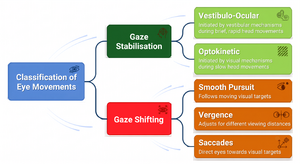
Supranuclear eye movements –Supranuclear eye movement involves versions, pursuits, saccades, convergence and vestibulo-occular reflex. Supranuclear eye movements inform us about upper or lower motor neuron lesion caused by trauma.
Visual field testing – Can give information on any type of vision loss if the patient is experiencing any. If a visual field is affected it can indicate towards conditions affecting visual pathways from optical nerve to occipital lobe, tumours, injury, ot poor circulation in the brain.
Vision Test– Using letters and shapes can help identifiy possible neglect or vision loss that could indicate a neurological condition. Visual acuity test generally utilises reading from a standardised chart (Snellen chart) or a card held 20 feet (6 meters) away.
Visual neglect testing – Patient demonstrate neglect of one side of body[6].
Aneisokonia testing – Testing show discrepancies in object size between eyes when you are looking at same object in same direction.
Hirschberg test & Cover Test- Test for ocular alignment . where we try to use a light source in midline from 2 feet to reflect on patient eyes while instructing them to focus on light source. Reflection of light should appear as a pin point while light near the center of pupil in each eye.
Management
Management strategies involves remediation of dysfunctions that were discovered during the assessment.Early treatment in post traumatic vision syndrome is advised to avoid compensatory changes.
If vestibular related oculomotor issues arises the treating therapist should refer the patient to a Neuro-ophtalmalogy/neuro-optometrist for further treatment. Investigation to find out if prism lenses with or with out occlusion are indicated should be done to start rehabilitation early.Optical devices like lenses and prisms can be used to help improve spatial orientation, posture and balance, and address issues regarding convergence, focus and binocular dysfunction. Presence of symptoms such as double vision, headaches, blurry vision, dizziness or nausea of post traumatic vision syndrome warrants referral to a specialist.[3]
Vision exercises with optical devices help effective management of the symptoms[7][8]. Different diagnostic methods like testing of saccades, convergence and Vestibulo-ocuar reflex can also be used for training vision. If Oculomotor dysfunctions are present oculomotor training through vision exercises has been shown to be beneficial. Computer based programs and video games have been shown to be beneficial in vision rehabilitation[9].
Vestibular physiotherapy rehabilitation programs are tailored according to the patients symptoms but typically the physiotherapist works with the patient to improve their balance, gaze stability, canalith repositioning maneuvers (if Benign Paroxysmal Positional Vertigo is present) and habituation exercises for specific head and body movements associated with the patients sport challenge or daily activities.[2]