When refering to evidence in academic writing, you should always try to reference the primary (original) source. That is usually the journal article where the information was first stated. In most cases Physiopedia articles are a secondary source and so should not be used as references. Physiopedia articles are best used to find the original sources of information (see the references list at the bottom of the article).
If you believe that this Physiopedia article is the primary source for the information you are refering to, you can use the button below to access a related citation statement.
Osteoporotic (fragility) vertebral fractures (VFs) are fractures that result from mechanical forces that would not ordinarily result in a fracture. [1] These types of fractures are insufficiency fractures, caused by normal stresses on abnormal bone, often indicating frailty.
Classification of Vertebral Fractures
Classification of VFs has been described by type of deformity (wedge, biconcavity, or crush compression) and further by the degree of deformity (grades I, II). [2][3]
X-Ray of an anterior wedge fracture.
Wedge VFs are the most common, occuring in the midthoracic region with compression of the anterior segment of the vertebral body (VB) and the anterior longitudinal ligament (ALL), and reports of more than 50% of all VFs. [4] Wedge fractures do not involve the posterior half of the VB and do not involve the posterior osseous ligamentous complex. The former distinguishes a compression fracture from a burst fracture.
Biconcave compression VFs are the second-most common (approximately 17% of all VFs); here, only the middle portion of the vertebral body is collapsed, with no compromise of the anterior and posterior walls. [3]
Crush compression VFs are the least common (13%), and are characterised by the collaplse of the entire anterior column, including anterior and posterior margins. [3]
The remaining 20% of VFs may involve complex fractures of the spine. [3]
Grade I refers to 3 SD < deformity < 4 SD and grade 2 deformity > 4 SD from the normal range for each vertebra. [2]
Etiology
The most common etiology of VFs is osteoporosis, making these fractures the most common fragility fracture. [5][6]
In contrast, vertebral compression fractures (VCFs) demonstrate a bimodal distribution with younger patients sustaining these injuries secondary to high energy mechanisms (fall from a height, MVA, etc.).
VFs occur secondary to an axial/compressive (and to a lesser extent, flexion) load with a resultant biomechanical failure of the bone resulting in a fracture.
Watch this video analysing causes of VFs (including osteoporotic):
European population studies have shown that the annual incidence of VCFs is 10.7 per 1000 women and 5.7 per 1000 men. [8]
VCFs are the most common fragility fractures reported in the literature. [9]
VCFs are common in Asian and Caucasian women, and less common in African-American women. [3]
Approximately 1.5 million VCFs occur annually in the United States (US) alone in the general population. [10]
Prevalence increases with age, and it is estimated that 40% to 50% of patients over age 80 years have sustained a VCF either acutely, or recognised incidentally during clinical workup for a separate condition.[11]
Thoracolumbar junction (i.e., the segment from T12 to L2) location most often affected and the second most common site is the L2 to L5 region. [3]
Estimated annual cost of VCFs in the US is $746 million. [12]
In the elderly patients,
30% of VCFs in severe cases of osteoporosis occur while the patient is in bed [3][13]
As the population continues to age, the population at risk of sustaining low energy fragility fractures will continue to increase as well.
Largely unreported and are probably more common radiographically (present up to 14% of women older than 60 years in one study)[14]
Currently, 10 million Americans are already diagnosed with osteoporosis, and another 34 million have osteopenia.
Back pain is common in elderly patients. Decreases when lying on the back (reduced weight baring conditions). [15][16]
Most patients with vertebral fractures do not seek medical attention and up to 70% of vertebral fractures may not result in notably severe symptoms.[6]
Loss of height. Height loss of the osteoporotic fractured vertebra may be mild (20-25%), moderate (25-40%) or severe (> 40%). It commonly affects the thoracolumbar region, though any vertebra may be disturbed.
The pain of acute fracture usually lasts 4 to 6 weeks with intense pain at the site of fracture.
The pain resolves over a period of six to 12 weeks.
Chronic pain may also occur in patients with multiple compression fractures, height loss and low bone density (also due to structural changes or osteoarthritis).[3]
Radiographic VCF may not be symptomatic. The greater the deformity, the greater the likelihood of pain and disability.
As height is lost, patients experience discomfort from the rib cage pressing downward on the pelvis.
Patients develop an exaggerated thoracic kyphosis and/or an exaggerated lumbar lordosis[6] - due to multiple adjacent VCFs - [3]which may result in decreased exercise tolerance and reduced abdominal space giving rise to early satiety and weight loss.
Sleep disorders may occur and decreased self esteem +/- depression.
Associated with an increased morbidity and increased mortality[6][20]
Fracture Risk Assessment Tool
A Fracture Risk Assessment Tool (FRAX) has been developed with the aim to predict osteoporotic fractures in general. It is based on cohort data generated from European, North American, Australian and Asian men and women. Although it does not refer to vertebral fractures exclusively, the FRAX algorithm indicates the 10-year chance of a major osteoporotic fracture taking place. [21] Clinicians may use of the FRAX tool in combination with indications for assessment of osteoporotic vertebral fractures.
Indications for Vertebral Fracture Assessment
A Vertebral Fracture Assessment (VFA) is indicated: [22]
In postmenopausal women, and men aged ≥ 50 years,
In the presence of a history of ≥ 4 cm height loss, or kyphosis,
If there is a history of recent or current long-term oral glucocorticoid therapy, or other therapy that compromises the bone
In BMD T-scores ≤ − 2.5 at either the spine or hip
If an acute onset of back pain with risk factors for osteoporosis at an older age is reported. An updated Cochrane review assessing the diagnostic accuracy of red flags for vertebral fracture in patients complaining for low back pain [23] concluded that appropriate sets of vertebral fracture indicators of various aetiologies are still lacking, so this finding may be interpreted with caution.
Diagnosed on Xrays when there is a loss of height in the anterior, middle, or posterior dimension of the vertebral body that exceeds 20%.
Osteoporotic spine fractures can be graded based on vertebral height loss as:
Mild: up to 20-25%
Moderate: 25-40%
Severe: >40%
Chronicity of the fracture indicates its temporal relationship with symptoms and hence is an important determination when deciding on conservative or surgical treatment. [24]
On conventional imaging, acute fracture signs include cortical breaking or impaction of trabeculae; in the absence of these signs fractures are chronic.
In uncertain cases, MRI signs of oedema (acute) and presence of radiotracer uptake on bone scintigraphy (acute) help decide the age of the fracture.
Quality of Life questionnaire: this can be measured with the use of the Quality of Life Questionnaire of the European Foundation for Osteoporosis (QUALEFFO). An other possibility is to use the Assessment of Quality of Life (AQoL) questionnaire or the European Quality of Life–5 Dimensions (EQ–5D) scale.
Physical functioning: measured by a modified 23-item version of the Roland–Morris Disability Questionnaire.
Research suggests that the four-factor Mini-BESTest model can be effective in evaluating multiple dynamic balance aspects in older adults with femoral or vertebral fractures and may help therapists in making clinical decisions, considering factors that indicated a decline in function. [26]
Examination
Clinical assessment of vertebral fractures is generally poor and reliance is made on imaging studies for diagnosis. [18]
Two tests that help the practitioner to predict more accurately which patients have an acute vertebral fracture: [27]
Closed-fist percussion sign:[27]This test has a sensitivity of 87,5% and a specificity of 90%, which is good. Stand behind the patiënt and the patiënt stand in front of a mirror allowing you to see the reaction of the patiënt. Firm closed-fist percussion is used to examine the entire length of the spine. The clinical sign is positive when the patiënt complains of a sharp, sudden pain.
The supine sign:[27] This test has a sensitivity of 81,25% and a specificity of 93,33%, which is also pretty good. The patiënt is asked to lie supine with only one pillow for the head. The clinical sign is positive when the patiënt is unable to lie supine due to severe pain.
Medical Management
VFs are usually considered stable and do not require surgical instrumentation.[28]
Conservative management of acute osteoporotic compression fractures may involve the following goals: reducing pain and improving functional status. This can be accomplished with the following:[29]
Acetaminophen, ibuprofen, opioids
Nutritional support
Bed rest (but not continuous or prolonged)
Modifications in daily activities
Pacing or graded activity
Physiotherapy e.g. exercises after 12 weeks post fracture
Rehabilitation programmes
Taping and bracing for compression fractures is often done for patient comfort and is unlikely to influence spinal stability. A small study did support the use of semirigid thoracolumbar orthosis for gait improvement.[30]
Treatment of the underlying disease (osteoporosis) is the recommended approach.
In 2023, an international consensus was reached on the non-pharmacological and non-surgical management of osteoporotic vertebral fractures. [29] Multidisciplinary biopsychosocial recommendations were made on pain, nutrition, safe movement, and exercise for individuals with acute and chronic vertebral fractures.[29]
For persistent pain and the failure of conservative treatments, surgical intervention may be indicated.[31]
Vertebroplasty is usually an outpatient procedure that takes one to two hours where under guided imaging, a needle get inserted into the vertebral body and medical-grade cement is injected. This cement hardens quickly and stabilises the fracture. [32]
Kyphoplasty is a very similar procedure, but in this case, a balloon is used to expand the vertebral body before injecting the cement. [33]
After a short period of bed rest, patients begin with a rehabilitation exercise programme. A "how-to" language is preferred rather than only suggesting activity restrictions. [29] The goals of rehabilitation are the prevention of falls, providing correct spine alignment (reduction of the Thoracic Hyperkyphosis), improving respiratory function in severe cases, and enhancing axial muscle strength. [1]
Exercises such as walking help to maintain or improve bone density in an osteoporotic population.
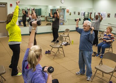
Strengthening exercises, using weights or resistance bands help maintain or improve bone density at the location of the targeted muscle attachments. [37] Maintaining bone health is extremely important, especially in the elderly as there typically is a decline in bone mass with age.
Strengthening and flexibility exercises improve overall physical function and postural control (important to reduce risk for falls).[38]
Combined balance and progressive strength training programme produced the best results in terms of maintaining leg strength, balance, bone mineral density and physical function compared to balance or strength training alone. [39][40][41]
Spinal extensor strengthening programme and a dynamic proprioceptive programme increase bone density and reduced the risk of VCFs.[42]
Back extensor exercises improve muscle strength, providing a better dynamic-static posture and reduction of the kyphotic deformity. Correction of the kyphosis also results in pain relief, increased mobility and an improvement in the quality of life.[43]
Postural taping can help in postural alignment. Tape is applied to the skin to provide increased proprioceptive feedback about postural alignment, improve thoracic extension, reduce pain and facilitate postural muscle activity and balance. [44] For example;
Osteoporotic vertebral fractures are fractures of one or more of the vertebrae due to osteoporosis.Osteoporosis causes the bones to be more fragile and more likely to fracture. Osteoporotic vertebral fractures classically occur during normal day-to-day activities such as bending, twisting, walking or lifting relatively light objects.
The pain of acute fracture usually lasts 4 to 6 weeks with intense pain at the site of fracture. Chronic pain may also occur in patients with multiple compression fractures, height loss and low bone density. Although this is probably due to structural changes or osteoarthritis.
Vertebral fractures don't only occur due to osteoporosis. Also a trauma or metastasis can cause a vertebral fracture. The diagnosis of osteoporosis can be confirmed by Dual Energy X-ray Absorptiometry (DXA).
Often used outcome measures to evaluate the progression of a patient are: Visual analogue scale for overall pain and pain during day and night (VAS), a Quality of Life questionnaire and a Physical functioning questionaire ( modified 23-item version of the Roland–Morris Disability Questionnaire).
Conservative treatment (bed rest, analgesic medication, physiotherapy and bracing) still is the method of choice as medical treatment. Vertebral fractures can also be treated surgically using vertebroplasty and kyphoplasty.
As physical therapy management there are a number of treatment approaches, including strengthening exercise, stabilisation exercise, balance training, stretching, relaxation techniques, manual therapy and taping.
Key Research
The following articles are key evidence pieces for physical therapy interventions:
International consensus on the non-pharmacological and non-surgical management of osteoporotic vertebral fractures. [29]
Exercise interventions to reduce fall-related fractures and their risk factors in individuals with low bone density: a systematic review of randomized controlled trials [46]
A multi-component exercise regimen to prevent functional decline and bone fragility in home-dwelling elderly women: a randomized, controlled trial [47]
Positive effects of exercise on falls and fracture risk in osteopenic women [48]
Reducing the risk of falls through proprioceptive dynamic posture training in osteoporotic women with kyphotic posturing: a randomized pilot study
↑Centre for Metabolic Bone Diseases, University of Sheffield. UK. Welcome to FRAX. Available from: https://frax.shef.ac.uk/FRAX/ [accessed 17-11-2023]
↑Gregson CL, Armstrong DJ, Bowden J, Cooper C, Edwards J, Gittoes NJL, Harvey N, Kanis J, Leyland S, Low R, McCloskey E, Moss K, Parker J, Paskins Z, Poole K, Reid DM, Stone M, Thomson J, Vine N, Compston J. UK clinical guideline for the prevention and treatment of osteoporosis. Arch Osteoporos. 2022 Apr 5;17(1):58.
↑Liu JT et al. Balloon kyphoplasty versus vertebroplasty for treatment of osteoporotic vertebral compression fracture: a prospective, comparative, and randomized clinical study. Osteoporos Int. 2010 Feb;21(2):359-64. Level of Evidence: 1B.
↑Gauthier A, Kanis JA, Jiang Y, Martin M, Compston JE, Borgström F, Cooper C, McCloskey EV. Epidemiological burden of postmenopausal osteoporosis in the UK from 2010 to 2021: estimations from a disease model. Arch Osteoporos 2011 6:179–188.
↑Kim DH, Vaccaro AR. Contemporary Concepts in Spine Care: Osteoporotic compression fractures of the spine; current options and considerations for treatment. The Spine Journal. 2006 6 479–487.
↑ 49.049.1NICE. Alendronate, etidronate, risedronate, raloxifene and strontium ranelate for the primary prevention of osteoporotic fragility fractures in postmenopausal women (amended): NICE technology appraisal guidance 160. Manchester: National Institute for Health and Care Excellence, 2011.