Lumbar Conditions in Ballet Dancers
Top Contributors - Diana Yang, Jessica Bedwell, Rebecca McFarlane, Kimberley Anteh, Denise Payet, Carina Therese Magtibay, Cindy John-Chu, Bruno Serra and Kim Jackson
This article or area is currently under construction and may only be partially complete. Please come back soon to see the finished work! (11/10/2023)
Introduction[edit | edit source]
Ballet dancers are both highly skilled artists and elite athletes[1]. Due to their intense and highly repetitive nature of movement patterns, ballet dancers are often at significant risk for injury[2], especially overuse injuries (as opposed to traumatic injuries) in the lower extremities and the lumbar region. Many of the injuries trace back to technique error, or external factors such as the footwear and the stage[2][3][4][5][6].
The number of overuse injuries are seen more in female dancers than their male counterpart, this is mostly due to the higher technical requirements, leading to higher repetition of physically demanding movements in rehearsals and performances, making the female dancers more prone to overuse injuries[7][8].
Dance medicine literature is small and very heterogeneous with a mostly ballet focus[6]. Overuse injuries in the spine are generally regarded as the second most effected region with the lower limbs being the first in ballet dancers[9], these injuries are typically presented as pain to the affected region[8].
Certain movements in ballet are especially prone to put additional strain on the lower back in both female and male dancers. While male dancers are generally expected to meet tougher athletic demands such as lifting their partners, female dancers are usually required to meet stricter technical demands[8]. Excessive stress on the lumbar spine during lifts and hyperlordosis in the back to compensate for hip extension contribute to the strain in the musculoskeletal structures in the lumbar region[1][4][6][10][11].
Lumbar Conditions in Ballet Dancers[edit | edit source]
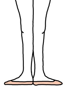
Turnout (Figure 1), the first position in ballet, requires maximal hip external rotation. When the tightness of the psoas muscle limits the degree of external rotation at the hip, dancers will attempt to compensate by increasing hip flexion and thus lumbar lordosis[9][4][11].
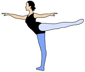
Similarly, in movements such as arabesque (Figure 2) that require extension at the hip. If there is insufficient hip extension or the spine is too straight, compensatory techniques such as increasing lumbar lordosis will be employed, resulting in excessive torsion strain[4][12][10].
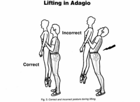
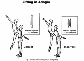
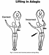
Both mechanisms will lead to the shortening of the erector spinae, and thoracolumbar fascia, contributing to a hyperlordotic posture in the lumbar region [4][12]. In male dancers, excessive lumbar lordosis is seen during lifts of another dancer, especially away from their center of gravity[4][10].
Incorrect technique and posture adding excessive stress to the lumbar spine in ballet dancers can result in a hyperlordotic posture, which can lead to low back pain (LBP)[3], and joint trauma[13] such as spondylolysis, or trauma to the pars interarticularis, the intervertebral discs, and the facet joints[4]. It predisposes dancers to muscle spasm, piriformis syndrome, and other spinal conditions like spondylolisthesis[6].
This page will focus on the following three conditions in the lumbar region:
- Hyperlordosis
- Spondylolysis
- Spondylolisthesis
Clinically Relevant Anatomy[edit | edit source]
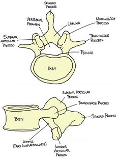
The lumbar spine consists of 5 vertebrae, situated between the thoracic spine and the sacrum. Each vertebra consists of a vertebral body and a neural arch, with the body anterior to the arch (Figure 3). When viewed superiorly, the body is wider transversely, and the neural arch is triangular shaped (especially distinguishable at L5). The facet angles range from 120 – 150 degrees relative to the sagittal plane, and consistently decrease from L1 to L5[14].
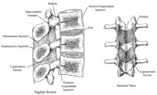
As with the rest of the spinal column, the articulations consist of the intervertebral disc anteriorly, and a pair of facet joints posteriorly. The ligaments (Figure 4) stabilize the spine as one unit, including the following major ligaments:
- Anterior longitudinal ligament: originating from the skull and inserting into the upper part of the sacrum, connecting the whole anterior aspect of the vertebral bodies and intervertebral discs
- Posterior longitudinal ligament: originating from the occipital bone and inserting into the sacrum, connecting the whole posterior aspect of the vertebral bodies and discs
- Ligamentum flava: present in between the laminae of each vertebra
- Supraspinous and Interspinous ligaments: connecting the spinous processes of each vertebra
The lordotic curvature of the lumber spine is formed because the sacrum is tilt forwards and inclined downwards, so to compensate for the inclination angle of the sacrum, the spine must assume a lordotic curve. Moreover, the wedge shaped lumbosacral intervertebral disc and L5 vertebral body contribute to a forward inclination at the L5/S1 joint. As a result of the inclination, the anterior longitudinal ligament is stretched, and the intervertebral discs are compressed slightly[15].
Clinical Presentation[edit | edit source]
| Condition | Subjective Presentation | Objective Presentation | Diagnosis |
|---|---|---|---|
| Spondylolysis and Spondylothesis |
|
|
|
| Hyperlordosis |
|
|
|
Epidemiology[edit | edit source]
Within existing literature there are limited studies regarding the prevalence of spine conditions in ballet. Available research shows the lumbar region is the fifth most prevalent form of injury amongst ballet dancers, accounting for 10%-15% of dance injuries[16][17] [4][12] . Micheli reports spinal injuires being one of the most dangerous injuries across dance injuries as it results in removing dancers from performing for longer durations of time than other injuries[18]. Dancers, specifically ballet dancers, have a higher incidence of spinal deviation compared to the general population[6]. The majority of spinal conditions have been associated with excessive extension of the lumbar region[12], which can result in hyperlordosis and other recognised spinal conditions such as spondylolisthesis and spondylosis[6].
According to a recent study of dance students from Indonesia, hyperlordosis affects 79.5% of the ballet dancers[19]. In another study of 65 ballet dancers, 32% experienced spondylolysis as a result of frequent loading of the spine in rotated and hyperextended positions required for technical positions such as lifts and arabesques[20]. Research shows there is a higher angle of lordosis in females than males[21][22]and young females (15-19 years old) are more prone to lumbar stress fractures such as spondylosis[23].
Aetiology[edit | edit source]
| Condition | Causes/risk factors |
|---|---|
| Hyperlordosis[24] |
|
| Spondylosis[25] |
|
| Sponsylolithesis[26] |
|
Other factors[edit | edit source]
Overuse - Repetitive loading has been shown to contribute in the cause of stress fractures[23].
Incorrect technique - Incorrect technique can lead to weakness and tension in specific muscles, most often the posas muscle, which increases lumbar lordosis this causes a displacement of load on the lumbosacral joints resulting in injuries[27][28][2].
Diet - Although no studies have yet shown a direct correlation between poor diet and back injuries, poor nutrition has been linked to increased osteopenia and stress fractures[29][30]. Females are more vulnerable in acquiring stress fractures due to predisposed factors such as decreased bone density and low calcium intake[31].
External factors - Ballet dancers are known for performing on hard inflexible floors, with inadequate shock absorption footwear and sometimes are known to dance barefoot in modern ballet[9][32]. The impact or contact on this floor repetitively can contribute to the risk of injuries especially in lower limbs and spine[9].
Social Factors[edit | edit source]
Meeting high physical demands - Ballet is extremely physically demanding [33]. Dancers are expected to repeat technical moves until they are executed correctly which in turn results in over use injuries[9]. Young, skeletally immature female dancers that participate in intensive ballet training are the most suseptible to acquiring stress fractures[23]. Injuries are also seen to be more frequent with those who have insufficient rest time and the transition from rest cycles to maximum activity[34].
Psychological Factors[edit | edit source]
External pressure within the dancing industry has caused dancers to push through injuries resulting in further injuries[35]. It has been reported that dancers experience anxiety when approaching teachers about an injury due to the fear of potentially 'making or breaking' their career; with others stating that they felt they had no choice but to continue dancing[36].
Dancing at a high level has been associated with pain, and some dancers believe suffering and pain is part of the process to be successful[35]. This mindset can jeopardise dancers performance as they are unable to recognise when they have sustained a serious injury[37] [38]. Previous research has shown that dancers social identity is associated to their profession and being unable to dance can threaten their identity[36]. Dancers perceived that if they admitted to having an injury that they are viewed as weak and lack commitment and passion for the profession; therefore coverup injuries and dance through pain which in turn prolongs recovery or even worsen the injury[36][38][39].
Hyperlordosis[edit | edit source]
Hyperlordosis is an excessive curve of the lower spine, resulting in a c-shape curve in the lumbar region. The lumbar curvature begins at the first vertebra of the lumbar spine and extends to the top of the sacrum. Lumbar hyperlordosis may contribute to dancers' injuries due to the relationship between repetitive stress on the posterior elements and discs. Common symptoms include back pain and limited movement. In severe cases bilateral weakness in the lower limbs, numbness and tingling.
An increase in lordotic angle increases the strain and stress and shifts the centre of gravity anteriorly. A systematic review discovered a strong relationship between lower back pain and decreased lumbar lordosis[40]. However, a specific study on lumbar lordosis in college dancers showed an increase in the relationship between lower back pain and lumber lordosis[11].
There are various factors influencing lumbar hyperlordosis:
- Congenital spinal deformities
- Anterior tilt of the hip
- Poor posture
- Short back muscles
- Sedentary lifestyle
- Imbalance between muscles surrounding the pelvic bones this may include tights hip flexors, weak core muscles and gluteal muscles (lower crossed syndrome). This could have an effect on the length of hamstrings, causing the muscles to be shortened.
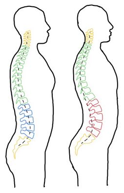
The mean angle of lumbar lordosis is said to be around 48-61 degrees[41]. Lumbar hyperlordosis is indicated when the angle is greater than 68 degrees[15]. Figure 5 demonstrate the comparison between the normal lumbar curvature and an increased lordosis in the lumbar spine. The hyperlordotic and hyperextended positions implicate the lumbar spine is more vulnerable to microtrauma to the pars interarticularis, intervertebral discs, and the facet joints. This can subsequently contribute to overuse or stress fractures in the lumbar spine. In the hyperlordotic position, the erector spinae muscle in the back shortens, contributing to the increasing lumbar lordosis in a standing position[10].
Relevant Literature[edit | edit source]
The extreme ranges of motion required during dancing and gymnastics may contribute to the participants' high lumbar lordosis[42]. Ambegaonkar (2014)[42] examined 17 female dancers using 2-dimensional sagittal plane photographs and the Watson Maconncha Posture Analysis Instrument. A strength of this study are the experimental methods due to be found reliable and accurate in previous studies and are commonly used in a clinical and research setting for assessing lumbar lordosis. Results showed among dancers, 9 had significant lumber lordosis deviations and 5 had moderate deviations. This shows the extreme ranges of motion required during the technique when dancing may contribute to the participants high lumbar lordosis. Additionally, the study only examined lumbar lordosis levels at one point in time, future research could enquire longitudinal studies to determine if there is a relationship between lumbar lordosis levels and lumbar injury incidence in dancers. Alongside this, a study by Byran and Smith (1992)[32] discussed the patterns of weakness and tightness in certain muscle groups in ballet dancers that contribute to hyperlordotic posture such as lumbodorsal fascia and hamstring can occur during the growth spurt phase of development. This could be common as the average starting ages ranging from 5 to 11 years old with full time training typically beginning around 14 to 15 years old [43] [44]. Due to the demands of dance involve extreme and repetitive lumber hyperextension alongside areas of weak and tight muscles during their growth spurt where full time intense training begins, may result in a hyperlordotic posture[32]. However, the limitation of both these study are that it entails female dancers, therefore the findings may not be applicable to other populations as well as not investigating the influence of age, training onset, training regimes, or other factors on lumbar hyperlordosis.
Spondylolysis[edit | edit source]
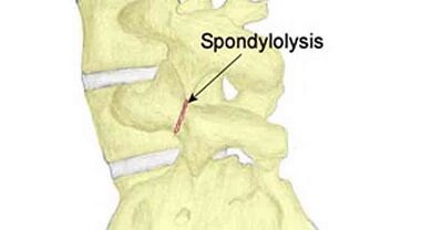
Spondylolysis is an isolated defect in the neural arch of the vertebrae [45]. It refers to a stress fracture of the pars interarticularis of a vertebra in the spine. Lumbar spondylolysis results from repetitive, mechanical stress on the lower back, however, biological factors are not ruled out [45]. There are congenital factors such as spina bifida occulta and exaggerated lumbar lordosis that predispose individuals to pars interarticularis fractures [46]. The defect can either be asymptomatic or associated with significant low back pain [47].
The pars interarticularis is the weakest area of the vertebrae, making it the most susceptible to injury. Spondylolysis can occur in one of the pars interarticularis or in both of them in one vertebra, meaning that is can be unilateral or bilateral [48]. The vast majority of spondylolitic defects occur at the L5 level (85-95%) and L4 being the second most commonly involved level (5-15%)[49]. Spondylolysis is found in 3-10% of the general population [50]. It is one of the most common types of skeletal injury in paediatric dancers and gymnasts.
The most common symptom of spondylolysis is back pain [51], usually with an insidious onset and may be referred to the buttocks or posterior aspect of the thigh. The pain can be brought on by loading the lumbar spine in combination with lumbar extension and rotation movements [52]. The diagnosis of spondylolysis cannot be made with physical examination alone [52]. Two-view plain film x-rays are typically used for initial diagnosis as they are inexpensive and relatively low in radiation [53].
Spondylolisthesis[edit | edit source]
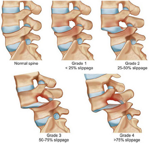
Spondylolisthesis is closely related to Spondylolysis. It refers to the forward slip of the vertebrae [6]. This is due to stress fractures (Spondylolysis) causing a weakening of the vertebral column to the extent that the vertebrae cannot maintain its position, resulting in the vertebrae to move outside of its normal anatomical place [54]. Spondylolisthesis can be defined by four grades, grade I is when there 1 to 25% slippage, grade II up to 50% slippage and grade III is up to 75% in slippage and lastly grade IV is when there is greater than 75% slippage[55].
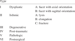
Wiltse and Winter (1983)[56] developed six classifications for Spondylolisthesis and dancers are likely to develop grade II Isthmic spondylolithesis. Grade II is further divided into three parts, type A which is fatigue fractures of the pars, type B which refers to elongated but intact pars and type C which is acute fracture of the pars [56]. Grade II Isthmic spondylolithesis is greatly associated with flexion, twisting and most notably hyperextension activities and movement repetitively performed by ballet dancers[56]. Symptoms of dull aches in the lower back and radicular pain into the glutes and anterior thigh usually accompany Spondylolisthesis and dancers who experience this, usually can not identify an exactly when the initial injury occurred[6]. Additionally, stigma and dance culture means that injured dancers often delay seeking medical help and "dance through" the pain[57].
Spondylolysis and Spondylolisthesis Relevant Literature[edit | edit source]
The literature shows a fourfold increase in spondylolysis and spondylolisthesis in adolescent dancers when compared with the general population[58]. This incidence is mainly caused by the repetitive microtrauma and shear forces of hyperextension into positions and the poor mechanics and alignment often used by young dancers to obtain these hyperextension positions. A review found that 590 out of the 4243 athletes (13.9%) had a radiological diagnosis of spondylolysis [59]. It recommended a radiological examination of the lumbar spine in symptomatic athletes who are considered to be at high risk of developing spondylolysis. The review included both male and female athletes meaning that it has high population validity. However, this review was conducted a significant amount of time ago. Therefore, the prevalence of spondylolysis in elite athletes may have changed in recent years. Another limitation of this review was the age range of patients included in the review was 15-27 years, therefore, it is not representative of elite athletes outside of this age range.
A questionnaire study[60] conducted within the Finnish National Ballet included 25 male and 35 female ballet dancers between the ages of 16- 36. The study aimed to investigate the incidence of spondylolysis in this population due to the frequent rotational, extensional and axial load that ballet dancers put on their spines. They found that in this population, 19 (32%) of the dancers had spondylolysis, 15 of which had spondylolisthesis and 4 without spondylolisthesis. This figure is 6 times greater than prevalence of this condition in the Finnish population at the time of this study[60]. Therefore, supporting the notion that the mechanical load that ballet requires contributes to the aetiology and prevalence of both spondylosis and spondylolithesis in this population. Although this study is valuable for understanding the population as it accounts for both conditions and included both genders, it is an old study (conducted in 1997) which can affect its validity when applying these findings to current ballet dancers. Additionally the study is finnish, limiting its generalisability as there may be differences between populations that are not accounted for. However, this was one of the few studies in the area and therefore necessary to include in the synthesis of the literature.
Management Recommendations[edit | edit source]
Core Strengthening - home exercises and dynamic sling work[edit | edit source]
Within ballet it is important to have flexibility within the lumbar region to execute technical skills therefore it is important to stabilise the abdominal and lumbar extensor muscles to protect the spine.
Lumbar stabilisation exercises have been shown to reduce chronic low back pain through increasing dynamic stability and strengthening the abdominals.[61]
Sling exercises are also used to improve lumbar stabilisation and treat low back pain.[61] Participants are suspended on a sling pulley system in an approach to strength training using isometric, eccentric and concentric movements that focus on activate their core muscles whilst stabilising their spine to reduce pain and disability. [62][63]
In a randomised control trial of pre-professional ballet dancers, participants were assigned to a home exercise programme (HEP) and the others were assigned to the dynamic sling exercises as shown in the table below.[63]
| Home exercise programme | Dynamic sling programme |
|---|---|
20 mins of exercises 3x a week for 6 weeks
|
|
At the end of the study all participants in both groups had a significant increase in strength and reduction in pain.[63]
William Training[edit | edit source]
As shown above, low back pain is common in spinal conditions. Williams back exercises is a home exercise protocol designed to relieve pain and improve lower-body stability by training the abdominal, gluteals, and hamstring muscles[64].
Main exercises:
- Pelvic tilt
- Single knee to chest motion
- Double knee to chest motion
- Partial sit-up
- Hamstring stretch
- Hip flexor stretch
- Squat
It is recommended to complete these exercises 10-20 minutes every day[64].
This protocol aims to reduce lumbar lordosis by minimising the pressure placed on the posterior aspect of the lumbar vertebra which, enhances disc flexion and thus reduces disc herniation and low back pain[64].
In an 8 week randomised controlled trial, 40 females were divided between a control group and an intervention group that followed the William exercises protocol. The Williams exercises group showed a significant reduction in lumbar angle and back pain, increase in hamstring, hip flexor and lumbar extensor muscles and an increase in abdominal strength[65].
Positioning[edit | edit source]
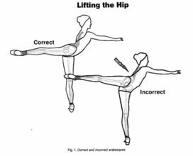
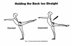
Gelabert (1986) [10] explains that in a ballet dancer, if the iliofemoral ligament in front of the hip joint restricts hypertension of the leg and the external rotation at the hip socket, the dancer will often disguise this limitation by lifting the hip to the side like shown in Figure 9. The repetition of this incorrect movement has a massive effect on low back afflictions and multiplies when the back is kept too straight shown in Figure 10. This is performed instead of the correct position by increasing the inclination of the pelvis forward as the leg raises backwards.
This study shows in 100 cases of low back syndromes in ballet dancers, 45% are related to those who lift at the hip in arabesque and other movements of the leg backwards and 25% of those who forcefully try to raise their leg above 90 degrees while keeping their back too straight. It has been found that these injuries will arise faster with individuals with longer backs and tight hip muscles and ligaments, which is often found in male physiques. Alongside this, spasm of the lumbar muscles occurs usually from faults of technique in hyperextension. Tension within the upper spine and chest muscles will be reflected in the thoracic region and to lumbar, pelvis and to the legs, increasing fatigue which causes inefficient breathing patterns and inclining the spine to strain. The impact of this is instrumental to male dancers when lifting, this is shown in Figure 11, 12, and 13, it can lead to weak muscle pull, inefficient energy use and an increased risk of low back strain.
Stretching[edit | edit source]
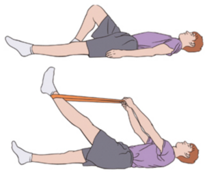
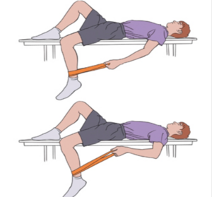
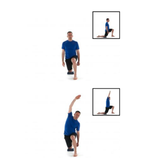
Brennan (2022)[66], Husney (2022)[67] and Walden (2022)[68] show stretches for spondylolysis, spondylolisthesis and hyperlordosis. These exercises specifically focus on hamstring and hip flexor muscles.
Examples[edit | edit source]
Static hamstring stretch (Figure 14)
Static hip flexor stretches (Figure 15 and 16)
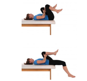
Knee to chest or single knee to chest (Figure 17)
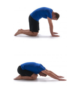
Child pose (Figure 18)
Conclusion[edit | edit source]
Certain movements in ballet are especially prone to put additional strain on the lower back. This results in the prevalence of many lumbar spine conditions in ballet such as hyperlordosis, spondylolysis and spondylolisthesis. Hyperlordosis is the excessive curvature of the lumbar spine and is a desired position in many ballet moves. Spondylolysis refers to a stress fracture in the pars interarticularis caused by repetitive and mechanical stress to the lumbar spine. This can lead to spondylolisthesis which is the forward slip of the vertebra. The available literature shows that the lumbar spine is the fifth most prevalent form of injury amongst ballet dancers, accounting for 10-15% of dance injuries. Therefore, it is important to have a good understanding of these injuries to attempt to prevent them from occurring and manage them properly when the do occur. There is a lack of research surrounding this topic currently, therefore, future research should focus on this due to the high prevalence of lumbar conditions in this population.
References[edit | edit source]
- ↑ 1.0 1.1 Allen, N., Nevill, A., Brooks, J., Koutedakis, Y. and Wyon, M. (2012) Ballet injuries: Injury incidence and severity over 1 year. Journal of Orthopaedic and Sports Physical Therapy. 42(9), 781–790.
- ↑ 2.0 2.1 2.2 Gamboa, J.M., Robert, L.A. and Fergus, A. (2008) Injury patterns in elite preprofessional ballet dancers and the utility of screening programs to identify risk characteristics. The Journal of orthopaedic and sports physical therapy. 38(3), 126–136.
- ↑ 3.0 3.1 Pinnelli, M., Pulcinelli, M., Carnevale, A., Di Tocco, J., Massaroni, C., Schena, E., Longo, U.G. and Denaro, V. (2022) A Wearable System for Detecting Lumbar Hyperlordosis in Ballet Dancers: Design, Development and Feasibility Assessment. 2022 IEEE International Workshop on Metrology for Industry 4.0 and IoT, MetroInd 4.0 and IoT 2022 - Proceedings. 277–282.
- ↑ 4.0 4.1 4.2 4.3 4.4 4.5 4.6 4.7 Milan, K.R. (1994) Injury in Ballet: A Review of Relevant Topics for the Physical Therapist. J Orthop Sports Phys Ther. 19(2), 121–129.
- ↑ Cejudo, A., Gómez-Lozano, S., de Baranda, P.S., Vargas-Macías, A. and Santonja-Medina, F. (2021) Sagittal Integral Morphotype of Female Classical Ballet Dancers and Predictors of Sciatica and Low Back Pain. International Journal of Environmental Research and Public Health. 18(9).
- ↑ 6.0 6.1 6.2 6.3 6.4 6.5 6.6 6.7 Gottschlich, L.M. and Young, C.C. (2011) Spine injuries in dancers. Current Sports Medicine Reports. 10(1), 40–44.
- ↑ Sobrino, F.J., Cuadra, C. de la and Guillén, P. (2015) Overuse Injuries in Professional Ballet Injury-Based Differences Among Ballet Disciplines. The Orthopaedic Journal of Sports Medicine. 3(6), 1–7.
- ↑ 8.0 8.1 8.2 Sobrino, F.J. and Guillén, P. (2017) Overuse Injuries in Professional Ballet: Influence of Age and Years of Professional Practice. Orthopaedic Journal of Sports Medicine. 5(6).
- ↑ 9.0 9.1 9.2 9.3 9.4 Sobrino, F.J. and Guillen, P. (2017) Overuse Injuries in Professional Ballet. Sport and Exercise Science.
- ↑ 10.0 10.1 10.2 10.3 10.4 Gelabert, R. (1986) Dancers’ Spinal Syndromes. The Journal of Orthopaedic and Sports Physical Therapy. 7(4), 180–191.
- ↑ 11.0 11.1 11.2 Skallerud, A., Brumbaugh, A., Fudalla, S., Parker, T., Robertson, K. and Pepin, M.-E. (2022) Comparing Lumbar Lordosis in Functional Dance Positions in Collegiate Dancers with and without Low Back Pain. J Dance Med Sci. 26(3), 191–201.
- ↑ 12.0 12.1 12.2 12.3 Livanelioglu, A., Otman, S., Yakut, Y. and Uygur, F. (1998) The Effects of Classical Ballet Training on the Lumbar Region. Journal of Dance Medicine & Science. 2(2), 52–55.
- ↑ Roussel, N.A., Nijs, J., Mottram, S., Van Moorsel, A., Truijen, S. and Stassijns, G. (2009) Altered lumbopelvic movement control but not generalized joint hypermobility is associated with increased injury in dancers. A prospective study. Manual Therapy. 14(6), 630–635.
- ↑ Ebraheim, N.A., Hassan, A., Lee, M. and Xu, R. (2004) Functional anatomy of the lumbar spine. Seminars in Pain Medicine. 2 (3), 131–137.
- ↑ 15.0 15.1 Bogduk, N. (2005) Clinical Anatomy of the Lumbar Spine and Sacrum. 4th edition. Elsevier: Churchill Livingstone.
- ↑ Ramkumar, P. N., Farber, J., Arnouk, J., Varner, K. E. and McCulloch, P. C. (2016) Injuries in a Professional Ballet Dance Company a 10-year Retrospective Study, Journal of Dance Medicine & Science, 20(1), 30-37.
- ↑ Schneider, H.J., King, A.Y., Bronson, J.L. and Miller, E.H. (1974) Stress injuries and developmental change of lower extremities in ballet dancers. Radiology, 113(3), 627-632.
- ↑ Micheli, L. J. (1983) Back injuries in dancers. Clinics in Sports Medicine, 2(3), 473-484.
- ↑ Wiraputri, A. A. W., Wardana, I. N. G., Widianti, I. G. A. and Muliani, M. (2022) Prevalence of Lumbar Hyperlordosis in Dance Department Students, Faculty of Performing Arts, Indonesian Art Institutes Denpasar Bali Batch of 2018-2020, E-Jurnal Medika Udayana, (5), 101-107.
- ↑ Seppo Seitsalo (1997) Spondylolysis in Ballet Dancers. SAGE Journals, 1(2), 51–54.
- ↑ Bergenudd, H., Nilsson, B., Udén, A. and Willner, S. (1989) Bone mineral content, gender, body posture, and build in relation to back pain in middle age, Spine (Phila Pa 1976), 14(6), 577-9.
- ↑ Lang-Tapia, M., España-Romero, V., Anelo, J. and Castillo, M. J. (2011) Differences on spinal curvature in standing position by gender, age and weight status using a noninvasive method, J Appl Biomech, 27(2), 143-50.
- ↑ 23.0 23.1 23.2 Lundon, K., Melcher, L. and Bray, K. (1999) Stress Fractures in Ballet: A Twenty-Five Year Review, Journal of dance medicine & science, 3(3), 101-107.
- ↑ Lillis, C. (2021) What to know about Hyperlordosis, MedicalNewsToday. Available at: https://www.medicalnewstoday.com/articles/321959 [Accessed: 18 May, 2023].
- ↑ MacGill, M. (2019) Spondylosis: All you need to know, MedicalNewsToday. Available at: [https://www.medicalnewstoday.com/articles/312598] [Accessed: 18 May, 2023].
- ↑ Fletcher, J. (2017) What to know about spondylolisthesis, MedicalNewsToday. Available at: https://www.medicalnewstoday.com/articles/318925 [Accessed: 18 May, 2023].
- ↑ MacSweeney, N., Pedlar, C., Cohen, D., Mahaffey, R. and Price, P. (2022) The Use of Physical Screening Tools to IdentifyInjury Risk Within Pre-Professional Ballet Dancers: An Integrative Review, Revisit de investigación en Ciencias de la Salud, 4(2), 95-120.
- ↑ Coplan, J. A. (2002) Ballet dancer's turnout and its relationship to self reported injury. The Journal of Orthopaedic and Sport Physical Therapy, 32(11), 579-584.
- ↑ Horváth, C. and Holló, I. (1989) Osteopenia as a risk factor for injuries to dancers. BMJ, 298(6681), 1176-1177.
- ↑ Benson, J. E., Geiger, C. J., Eiserman, P. A. and Wardlaw, G. M. (1989) Relationship between nutrient intake, body mass index, menstrual function, and ballet injury. Journal of the American Dietetic Association, 89(1), 58-63.
- ↑ Myburgh, K.H., Hutchins, J., Fatter, A.B., Hough, S.F. and Noakes, T.D. (1990) Low Bone Density Is an Etiologic Factor for Stress Fractures in Athletes. Annals of Internal Medicine, 113(10), 754.
- ↑ 32.0 32.1 32.2 Bryan, N. and Smith, B.M. (1992) The Ballet Dancer. Occupational Medicine: State of the Art Reviews, 7(1), 67–75.
- ↑ Koutedakis, Y. and Jamurtas, T. (2004) The dancer as a performing athlete: Physiological considerations, Sports medicine (Auckland, N.Z.), 34(10), 651-61.
- ↑ Bronner, S., Ojofeitimi, S. and Rose, D. (2003) Injuries in a Modern Dance Company. American Orthopaedic Society for Sports Medicine, 31(3), 365-373.
- ↑ 35.0 35.1 Soundy, A. and Lim, J. Y. (2023) Pain Perceptions, Suffering and Pain Behaviours of Professional and Pre-Professional Dancers towards Pain and Injury: A Qualitative Review, Behav Sci (Basel),13(3).
- ↑ 36.0 36.1 36.2 Wainwright, S.P., Williams, C. and Turner, B.S (2005) Fractured identities: injury and the balletic body. Health An Interdisciplinary Journal for the Social Study of Health, Illness and Medicine, 9(1), 49-66.
- ↑ Alaten, A. (2007) Listening to the Dancer's Body. The Sociological Review, 55, 109-125.
- ↑ 38.0 38.1 Harrison, C. and Ruddock-Hudson,M. (2017) Perceptions of Pain, Injury, and Transition-Retirement: The Experiences of Professional Dancers. Journal of Dance Medicine & Science, 21(2), 43-52.
- ↑ McEwen, K. and Young, K. (2011) Ballet and pain: reflections on a risk-dance culture. Qualitative Research in Sport, Exercise and Health, 3(2),152-173.
- ↑ Chun, S.W., Lim, C.Y., Kim, K., Hwang, J. and Chung, S.G. (2017) The relationships between low back pain and lumbar lordosis: a systematic review and meta-analysis. The Spine Journal. 17(8), 1180–1191.
- ↑ Jentzsch, T., Geiger, J., König, M.A. and Werner, C.M.L. (2017) Hyperlordosis is Associated with Facet Joint Pathology at the Lower Lumbar Spine. Clinical Spine Surgery. 30(3), 129–135.
- ↑ 42.0 42.1 Ambegoankar, J.P., Caswell, A.M., Kenworthy, K.L., Cortes, N. and Caswell, S.V. (2014) Lumbar lordosis in female collegiate dancers and gymnasts. Medical problems of performing artists, 29(4), 189-192.
- ↑ Hutchinson, C. U., Sachs-Ericsson, N. J. and Ericsson, A. K. (2013) Generalizable aspects of the development of expertise in ballet across countries and cultures: a perspective from the expert-performance approach. High Ability Studies, 24(1), 21-47.
- ↑ Bernacki, J.L., Stracciolini, A., Fraser, J., Micheli, L. and Sugimoto, D. (2021) Risk Factors for Lower-Extremity Injuries in Female Ballet Dancers: A Systematic Review. Clinical Journal of Sports Medicine, 31(2), 4-79.
- ↑ 45.0 45.1 Berger, R. G. and Doyle, S. M. (2019) Spondylolysis 2019 update. Current Opinion in Pediatrics, 31(1), 61-68.
- ↑ Watkins, R. G. and Watkins, R. G. (2010) Lumbar Spondylolysis and Spondylolisthesis in Athletes. Seminars in Spine Surgery, 22(4), 210-217.
- ↑ Syrmou, E., Tsitsopoulos, P. P., Marinopoulos, D., Tsonidis, C., Anagnostopoulos, I. and Tsitsopoulos, P. D. (2010) Spondylolysis: A review and reappraisal. Hippokratia Quarterly Medical Journal, 14(1), 17-21.
- ↑ Leone, A., Cianfoni, A., Cerase, A., Magarelli, N. and Bonomo, L. (2011) Lumbar spondylolysis: a review. Skeletal Radiology, 40, 683-700.
- ↑ Standaert, D. C. and Herring, S. (2000) Spondylolysis: a critical review. British Journal of Sports Medicine, 34, 415-422.
- ↑ Lemoine, T., Fournier, J., Odent, T., Sombrely-Taveau, C., Merenda, P., Sirinelli, D. and Morel, B. (2018) The prevalence of lumbar spondylolysis in young children: a retrospective analysis using CT. European Spine Journal, 27, 1067-1072.
- ↑ Porter, R. W. and Hibbert, C. S. (1984) Symptoms associated with lysis of the pars interarticularis. Spine, 9, 755-758.
- ↑ 52.0 52.1 Goetzinger, S., Courtney, S., Yee, K., Welz, M., Kalani, M. and Neal, M. (2020) Spondylolysis in Young Athletes: An Overview Emphasizing Nonoperative Management. Journal of Sports Medicine, 2020, 1-15.
- ↑ Miller, R., Beck, N. A., Sampson, N. R., Zhu, X., Flynn, J. M. and Drummond, D. (2013) A Review of Spondylolysis and Undiagnosed Mechanical Back Pain. Journal of Pediatric Orthopaedics, 33(3), 282-288.
- ↑ NHS (2022) Spondylolisthesis. 1 June 2022. NHS, [online]. Available at: https://www.nhs.uk/conditions/spondylolisthesis/ [Accessed: 19 May, 2023].
- ↑ Hu, S.S., Tribus, C.B., Diab, M. and Ghanayem, A.J. (2008) Spondylolisthesis and Spondylolysis. JBJS, 90(3), 656.
- ↑ 56.0 56.1 56.2 Wiltse, L.L. and Winter, R.B. (1983) Terminology and measurement of spondylolisthesis. The Journal of Bone & Joint Surgery, 65(6), 768–772.
- ↑ Vassallo, A.J., Pappas, E., Stamatakis, E. and Hiller, C.E. (2019) Injury Fear, Stigma, and Reporting in Professional Dancers. Safety and Health at Work, 10(3), 260–264.
- ↑ Tsirikos, A. and Garrido, E. (2010) Spondylolysis and spondylolisthesis in children and adolescents. Journal of Bone Joint Surgery, 92(6), 741-749.
- ↑ Rossi, F. and Dragoni, S. (2001) The prevalence of spondylolysis and spondylolisthesis in symptomatic elite athletes: radiographic findings. Radiography, 7, 37-42.
- ↑ 60.0 60.1 Seppo Seitsalo (1997) Spondylolysis in Ballet Dancers. Journal of Dance Medicine & Science, 1(2), 51–54.
- ↑ 61.0 61.1 Ko, K.J., Ha, G.C., Yook, Y.S. and Kang, S.J. (2018) Effects of 12-week lumbar stabilization exercise and sling exercise on lumbosacral region angle, lumbar muscle strength, and pain scale of patients with chronic low back pain. J Phys Ther Sci, 30(1), 18-22.
- ↑ Lee, J.S., Yang, S.H., Koog, Y.H., Jun, H.J., Kim, S.H. and Kim, K.J. (2014) Effectiveness of sling exercise for chronic low back pain: a systematic review. J Phys Ther Sci, 26(8), 1301-1306.
- ↑ 63.0 63.1 63.2 Kline, J. B., Krauss, J. R., Maher, S. F. and Qu, X. (2013) Core strength training using a combination of home exercises and a dynamic sling system for the management of low back pain in pre-professional ballet dancers: a case series. J Dance Med Sci, 17(1), 24-33.
- ↑ 64.0 64.1 64.2 Dydyk, A. M. and Sapra, A. (2022) Williams Back Exercises. StatPearls.
- ↑ Fatemi, R., Javid, M. and Najafabadi, E.M. (2015) Effects of William training on lumbosacral muscles function, lumbar curve and pain. Journal of Back and Musculoskeletal Rehabilitation, 28(3), 591–597.
- ↑ Brennan, D. (2020) Best Exercises for Spondylolisthesis. [Online]. Available at: https://www.webmd.com/back-pain/best-exercises-spondylolisthesis [Accessed: 18 May, 2023].
- ↑ Husney, A. (2022) Spondylolysis and Spondylolisthesis: Exercises. [Online]. Available at: https://myhealth.alberta.ca/Health/aftercareinformation/pages/conditions.aspx?hwid=bo1583& [Accessed: 18 May, 2023].
- ↑ Walden, M. (2022) Hyperlordosis. [Online]. Available at: https://www.sportsinjuryclinic.net/sport-injuries/back/low-back-pain/hyperlordosis [Accessed: 18 May, 2023].
