
General Assessment of a Patient with Burns
Top Contributors - Jess Bell, Carin Hunter, Kim Jackson, Tarina van der Stockt, Vidya Acharya, Rucha Gadgil and Ahmed M Diab
Introduction
Burn injuries vary widely in their severity, distribution and impact, so management should always be tailored to the individual, their injury and their context. This requires a detailed and accurate initial assessment. Investing time in the initial evaluation helps ensure the best possible immediate care, reduces the risk of long-term complications, and maximises a patient's functional recovery. By conducting a comprehensive assessment, the multidisciplinary team can become familiar with the patient's long-term goals and align therapy to these objectives. This, in turn, enhances patient engagement with the treatment plan.
Sharing initial assessment findings with relevant members of the multidisciplinary team helps to streamline subsequent assessments and facilitates continuity of care, both during rehabilitation and upon transition to community settings. This approach helps to minimise patient frustration and ensures accurate transmission of relevant information throughout the treatment journey.
Goal Setting
The multidisciplinary team should set goals with the patient using the SMART goal method. SMART goals are specific, measurable, achievable, relevant and time-bound.
Short-term rehabilitation goals might relate to:[1]
- preventing respiratory complications
- controlling oedema
- maintaining joint range of motion
- maintaining strength
- preventing excessive scarring
Functional long-term goals might relate to:
- achieving functional independence
- improving participation in society
- maintaining/enhancing psychological well-being
- establishing/achieving a return to work plan
Key Considerations
The following sections explore key considerations when evaluating a patient with a burn injury.[1]
Risk factors
Patients with burn injuries are at high risk of complications. These risks can be grouped into four categories: injury, patient, treatment and iatrogenic factors.[1]
- Injury factors include inhalation injury, burn area, depth of burn, and scarring.[1]
- Patient factors include reduced ambulation and mobility, increased bed rest, increased pain, and pre-existing comorbidities.[1]
- Treatment factors include skin reconstruction surgery, invasive monitoring and procedures, and management in critical care.[1]
- Iatrogenic factors include over-resuscitation ('fluid creep'), excess sedation and prolonged mechanical ventilation.[2][3]
Inhalation Injury
During the subjective assessment, clinicians should carefully observe for signs of inhalation injury, especially if there is a history of exposure to fire and smoke in enclosed spaces, and diminished levels of consciousness[4][5] Physical indicators may include charring around the mouth and nostrils, singed nasal hairs, the presence of soot in sputum and the upper airways, alterations in voice quality, and the presence of wheezing.[6]
If any signs of inhalation injury are noted, a qualified member of staff must conduct an inhalation injury examination. This ensures the prompt initiation of appropriate treatment measures. For more information, see the Inhalation Assessment section below.
Total Body Surface Area (TBSA)
When a patient's burn injury exceeds 20-25% TBSA, a massive systemic inflammatory reaction may occur. This impacts multiple organ systems, including the respiratory system.
Accurately assessing burn injuries, including their surface area and depth, is essential for optimising treatment.[7] However, inaccuracies in Total Body Surface Area (TBSA) calculations are an ongoing clinical challenge.
Traditional methods of calculating TBSA include the Lund and Browder method and the Rule of Nines. The Lund and Browder method takes longer to perform, but it is generally considered more accurate than the Rule of Nines.[1] It can also be used in patients of any age, including children. However, the Rule of Nines has often been used for adults on admission as a quick and simple way of assessing for fluid resuscitation. The standard Rule of Nines CANNOT be used on children, as the proportion of their head is different from that of an adult.[8] The Palmar Surface Method is another commonly used method. It estimates burn coverage based on the patient's palm and fingers (where each palm + fingers = approximately 1% TBSA).
However, while still used, these methods have significant limitations, and burn centres are increasingly investigating and adopting digital and automated 3D body mapping systems to ensure more precise, objective TBSA measurements.[7][9]
-
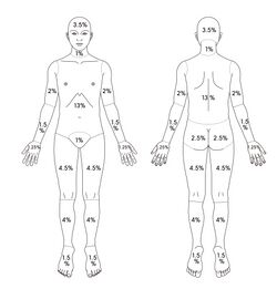 Rule of Nines Chart.
 -
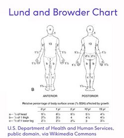 Lund and Browder Chart.


Important TBSA Considerations
Predicting TBSA is important for fluid resuscitation. The Parkland formula has historically been the most widely used protocol to determine appropriate fluid resuscitation over the first 24 hours.[10]
Parkland formula = 4 mL/kg/%TBSA (3 mL/kg/%TBSA in children) = total amount of crystalloid fluid during first 24 hours
There are, however, concerns associated with the Parkland formula, including the risk of over- or under-resuscitation,[11][12] [13] and other formulae have been proposed, including the American Burn Association (ABA) Clinical Practice Guidelines. The ABA formula recommends 2 mL/kg/%TBSA for adults.[12][14]
Please see Burn Wound Assessment for more information on the TBSA Assessment. Please see Systemic Response to Burns for more information on the whole-body response to burn injuries.
Burn Type and Depth
It is important to regularly re-examine the extent of tissue destruction as it can change for at least 48 hours post-burn. Burn injuries rarely present uniformly with a single depth throughout the affected area.[15] Early interventions and other patient factors (e.g. age and health) can influence the type and depth of a burn.[4]
Burn Site and Impact
The location of a burn injury can significantly impact functional outcomes and the level of trauma experienced by a patient. Burns to certain areas of the body require specialised treatment because of their functional importance and the risk for potential complications. These critical areas include the hands, face, perineum and joints.[1]
Subjective Assessment for a Patient with a Burn Injury
When taking a subjective history, it is essential to consider emotional trauma that may be associated with a burn injury. Consider if it is appropriate to involve family members or witnesses to fill in gaps in the history or to provide additional context.[1]
History of Presenting Complaint
History of the incident: Pay close attention to the events leading up to the injury and the mechanism of injury.[1]
First aid: Document any first aid administered. If the initial first aid seems inadequate, consider the possibility of a deeper burn injury. Include details of medications administered on-site, specifying amounts and times given.[1]
Falls: It is important to find out if there is any indication that the patient fell. If they did fall, find out what height they fell from and consider the potential for head injury, fractures, sprains, etc.[1]
Electrical injury: In cases of electrical injuries, find out what voltage was involved and which parts of the body were in contact with the ground or earth. Where there has been a high-voltage current, suspect nerve or deep muscle injury.[1]
Explosions: Often associated with falls and high-velocity injuries. They can also cause tympanic membrane injury, which can lead to hearing loss and affect communication.[1]
Passage to hospital: Document the mode of transportation and time to admission.[1]
Current Medical and Surgical History
Find out about any medical or surgical management:
- What pain medication has been given?
- What procedures so far? Debridement, escharotomy, flaps/grafts, etc
- What instructions are there from the multidisciplinary team?[16]
Past Medical History
Find out about the patient's general medical history, previous surgical interventions and medication, including the amount, duration and if their conditions are controlled or uncontrolled by medication.
Social History
In the social history, find out about a patient's pre-injury level of function, including their social supports and home situation, occupation (particularly relevant for patients with burns to their hands) and activities of daily living. Consider their basic activities of daily living (e.g. dressing, bathing, eating, shopping, driving, home maintenance), pre-injury physical function (mobility, including stair mobility and lifting ability) and pre-injury physical fitness (strength, flexibility, endurance and balance).[1]
Psychosocial Factors / Yellow Flags
For individuals with burn injuries, it is important to consider self-image, coping style, mental health and emotions.[1]
Key Aspects of the Objective Assessment of a Patient with a Burn Injury
The following sections discuss the key components of the objective assessment for a patient with a burn injury.
Pain Intensity Assessment
When assessing pain intensity in individuals with burn injuries, various pain assessment scales are used, depending on the patient's age.
Visual Analogue Scale (VAS): This scale is suitable for measuring pain in individuals aged 12 and older. Patients are asked to mark their level of pain on a horizontal line, where one end represents no pain, and the other end represents the worst pain imaginable.[1]
Wong-Baker FACES Pain Scale: This scale can be used in children aged three years and older. It is a self-report measure used to assess pain intensity. It consists of six pain assessment cards that show faces with different emotions, from smiling to crying.[1]
-
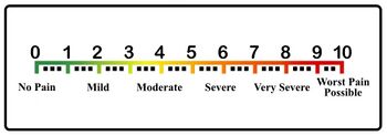 Visual Analogue Scale.
 -
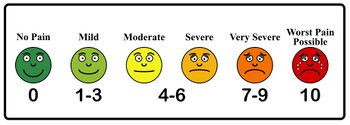 Wong-Baker FACES Scale.

.jpg)
Face, Legs, Activity, Cry, and Consolability (FLACC) scale: This observational pain scale is widely used in the paediatric population to assess pain in infants and children who are unable to express their pain verbally.[17] It scores pain intensity by rating five behaviours (face, legs, activity, cry, consolability) on a 0 to 2 scale: 0 = relaxed and comfortable; 1-3 = mild discomfort/pain; 4-6 = moderate pain; and 7-10 = severe discomfort/pain.
COMFORT Behavior Scale (CBS): The CBS is used to assess pain and distress in intubated and self-ventilating children in paediatric intensive care units.[18][19] It includes six categories, which are scored on a 1 to 5 scale, with a maximum score of 30. The categories are: alertness, calmness/agitation, respiratory response (in ventilated children) OR crying (in spontaneously breathing children), physical movement, muscle tone and facial tension. Higher scores indicate greater pain or distress.
Pain Observation Scale for Young Children (POCIS): The POCIS is also used to assess pain in children. It focuses on observing specific behaviours that indicate pain in children, particularly those aged between 1 and 4 years old. It typically has seven items, which represent observable behaviours associated with pain in young children (e.g. facial expressions, movement, crying, etc). A pain severity score is given, ranging from 0 to 7, with higher scores indicating greater pain.[20]
Behavioral Pain Scale (BPS) and Critical-Care Pain Observation Tool (CPOT): The BPS and CPOT are considered valid, reliable and useful measures for assessing acute pain in patients in intensive care units with burn injuries, including facial burns.[21]
For more information on Pain Assessment Tools, please see:
- British Pain Society: Outcome Measures
- Physiopedia page: Outcome Measures
Burn Outcome Measures and Scar Evaluation
When managing patients with burn injuries within a multidisciplinary team, it is beneficial to use outcome measures that can be retested as the condition progresses.[22][23] There are a range of measures available, but the following can be useful.
- The Burn Specific Health Scale-Brief (BSHS-B) evaluates the physical and psychosocial functioning of patients with burns and their quality of life.[24]
- The Burns Scar Index (Vancouver Scar Scale) was among the earliest tools developed to assess burn scars. It focuses on four indicators (scar height and thickness, pliability, vascularity, and pigmentation).[25] However, this index has low inter-rater reliability, inconsistent validity, and has had multiple modifications.[26]
- The Patient and Observer Scar Assessment Scale (POSAS) considers the patient's assessment of their scar alongside the clinical assessment.[27]
- The Burns Specific Pain Anxiety Scale is useful for evaluating pain-related anxiety in patients with burn injuries.[28]
Inhalation Assessment
"No consensus exists regarding the diagnosis, grading, and prognosis of inhalation injury [...] Full manifestation occurs up to 48 hours after the inhalation insult once the inflammation reaches its peak. Further, the clinical presentation (degree of respiratory failure) may not correspond with the intensity of the exposure."[29]
Most patients with inhalation injuries who present early to an emergency department will be conscious with patent airways. Their initial chest radiograph and arterial blood gases may "appear at most only slightly abnormal."[29] Because of delayed presentations or non-specific presentations, a number of diagnostic adjuncts are used to diagnose inhalation burn injuries.[29]
Physical findings: It is important to note that physical findings, while valuable, can sometimes be misleading and must be considered alongside other diagnostic tools. Key physical findings include sooty sputum, stridor (noisy breathing due to an obstructed airway), wheezing, facial burns, singed nasal/facial hairs, [Anxiety Disorder|anxiety], cough, stupor, [[1]], hoarse voice, oedema, erythema (superficial reddening of the skin, usually in patches), inspiratory and end expiratory crackles on auscultation, chest x-ray changes, and signs of hypoxia (headache, shortness of breath, fast heartbeat, coughing, wheezing, confusion, and a bluish colour in skin, fingernails, and lips).[29]
Bronchoscopy assessment: fiberoptic (flexible) bronchoscopy (FOB) is the gold standard for diagnosis of an inhalation injury, providing an immediate view of the airway.[29] While not universally available, it is widely used.[30] Signs of inhalation injury on bronchoscopy may include erythema, oedema (which may be seen as a blunting of the carina), mucosal blisters, erosions, haemorrhages, bronchial secretions, and soot deposits. Indirect laryngoscopy can serve as an alternative when bronchoscopy is unavailable, offering visualisation as far as the vocal cords.[29] Key determinants of inhalation injury severity include the duration of exposure to smoke, the temperature of the inhaled smoke, and smoke composition.[29]
For more information on inhalation injuries, please see:
- A Critical Update of the Assessment and Acute Management of Patients with Severe Burns[31]
- Inhalation Injury in the Burned Patient[29]
Oedema Assessment
Burn-induced inflammation causes rapid, massive swelling that can severely restrict joint movement and lead to long-term stiffness. It is important to note when the swelling began and whether it changes with position. To monitor swelling, use dry, non-invasive measurements, such as a flexible tape measure to take standard girth measurements or figure-of-eight measurements around the hand or foot.
Physical Assessment
When conducting a physical assessment, it can be helpful to divide the evaluation into two parts: 1) assessing the upper and lower limbs and trunk and 2) assessing general functional mobility. Clinicians should consider factors that may impact healing / recovery, such as prolonged bed rest, high levels of pain, and pre-existing comorbidities.[1][16][4]
Limbs and Trunk Assessment
Assess joint range of motion, muscle strength and muscle length. Specific limiting factors include pain, muscle length, trans-articular burns, scar contracture and the unique burn features.
General Functional Mobility Assessment
The mobility assessment should only be completed once the patient is medically stable. This assessment should focus on preventing complications associated with prolonged bed rest and restoring functional independence.
Assess the patient's functional transfers, gait, endurance and balance. Key factors to consider during the mobility assessment include posture, cardiovascular response to mobilisation, the patient's neurological status, concomitant injuries and their weight-bearing status.
References
- ↑ 1.00 1.01 1.02 1.03 1.04 1.05 1.06 1.07 1.08 1.09 1.10 1.11 1.12 1.13 1.14 1.15 1.16 1.17 1.18 1.19 Hale A, O’Donovan R, Diskin S, McEvoy S, Keohane C, Gormley G. Impairment and Disability Short Course. Physiotherapy in Burns, Plastics and Reconstructive Surgery, 2013.
- ↑ Zhou D, He L, Shi W, Ma P. Lessons from the similarities and differences in fluid resuscitation between burns and sepsis: a bibliometric analysis. Front Med (Lausanne). 2025 Mar 4;12:1561619.
- ↑ Folwell JS, Basel AP, Britton GW, Mitchell TA, Rowland MR, Cindass R, et al. Mechanical ventilation strategies in the critically ill burn patient: a practical review for clinicians. European Burn Journal. 2021; 2(3):140-151.
- ↑ 4.0 4.1 4.2 Siemionow MZ, Eisenmann-Klein M, editors. Plastic and reconstructive surgery. Springer Science & Business Media; 2010 Jan 13.
- ↑ Charles WN, Collins D, Mandalia S, Matwala K, Dutt A, Tatlock J, Singh S. Impact of inhalation injury on outcomes in critically ill burns patients: 12-year experience at a regional burns centre. Burns. 2022 Sep;48(6):1386-95.
- ↑ Wise B, Levine Z. Inhalation injury. Can Fam Physician. 2015 Jan;61(1):47-9.
- ↑ 7.0 7.1 Rangaiah PKB, Kumar BPP, Huss F, Augustine R. Precision diagnosis of burn injuries using imaging and predictive modeling for clinical applications. Sci Rep. 2025 Mar 4;15(1):7604.
- ↑ Woods A, Tan P, Mertz T, Lewis CJ. Advancements in burn size assessment: A systematic review of emerging technologies. Burns. 2026 Feb;52(1):107782.
- ↑ Giretzlehner M, Ganitzer I, Haller H. Technical and medical aspects of burn size assessment and documentation. Medicina. 2021; 57(3):242.
- ↑ Daniels M, Fuchs PC, Lefering R, Grigutsch D, Seyhan H, Limper U, et al. Is the Parkland formula still the best method for determining the fluid resuscitation volume in adults for the first 24 hours after injury? - A retrospective analysis of burn patients in Germany. Burns. 2021 Jun;47(4):914-21.
- ↑ Marín-Sánchez J, Cárdenas-Bolívar Y, Giraldo A, Fernandez Castro V, Carreño Hernandez F, Alvarado J. A56-19 impact of fluid resuscitation deviations from the Parkland formula on clinical outcomes in burn patients: a systematic review and meta-analysis. American Journal of Respiratory and Critical Care Medicine. 2026;212(1).
- ↑ 12.0 12.1 Wietlisbach L, Goff GK, O'Connor A, Gottlieb L, Vrouwe SQ. 850. Before-and-after adoption of a new fluid resuscitation protocol - Parkland formula versus ABA consensus formula. J Burn Care Res. 2026 Apr 6;47(Suppl 1):S278.
- ↑ Aigner A, Schiefer JL, Reinshagen K, Najem S; German Burn Registry; Vasileiadis V, Königs I. Too much or too little? Fluid resuscitation in the first 24 h after severe burns: Evaluating the Parkland formula - A retrospective analysis of adult burn patients in Austria, Germany, and Switzerland 2015-2022. Burns. 2025 May;51(4):107397.
- ↑ Cartotto R, Johnson LS, Savetamal A, Greenhalgh D, Kubasiak JC, Pham TN, Rizzo JA, Sen S, Main E. American Burn Association clinical practice guidelines on burn shock resuscitation. J Burn Care Res. 2024 May 6;45(3):565-589.
- ↑ Martin H. Immediate management of burn injury. 2007.
- ↑ 16.0 16.1 Hettiaratchy S, Papini R. Initial management of a major burn: II--assessment and resuscitation. BMJ. 2004;329(7457):101-103.
- ↑ Crellin DJ, Harrison D, Santamaria N, Babl FE. Systematic review of the Face, Legs, Activity, Cry and Consolability scale for assessing pain in infants and children. PAIN. 2015 Nov;156(11):2132–51.
- ↑ Boerlage AA, Ista E, Duivenvoorden HJ, de Wildt SN, Tibboel D, van Dijk M. The COMFORT behaviour scale detects clinically meaningful effects of analgesic and sedative treatment. Eur J Pain. 2015 Apr;19(4):473-9.
- ↑ Suprawoto DN, Nurhaeni N, Waluyanti FT. COMFORT Behavior Scale instrument: validity and reliability test for critically ill pediatric patients in Indonesia. Pediatr Rep. 2020 Jun 25;12(Suppl 1):8690.
- ↑ Voelker R. Diagnosing Pediatric Pain. JAMA. 1999;282(18):1713.
- ↑ de Jong AEE, Tuinebreijer WE, Hofland HWC, Van Loey NEE. Person-centred pain measurement in the ICU: a multicentre clinimetric comparison study of pain behaviour observation scales in critically ill adult patients with burns. Eur Burn J. 2024 Jun 17;5(2):187-197.
- ↑ Taal LA, Faber AW, Van Loey NE, Reynders CL, Hofland HW. The abbreviated burn specific pain anxiety scale: a multicenter study. Burns. 1999 Sep 1;25(6):493-7.
- ↑ Taal LA, Faber AW. The burn specific pain anxiety scale: introduction of a reliable and valid measure. Burns. 1997 Mar 1;23(2):147-50.
- ↑ Tyack Z, Simons M, Spinks A, Wasiak J. A systematic review of the quality of burn scar rating scales for clinical and research use. Burns. 2012 Feb 1;38(1):6-18.
- ↑ Nguyen TA, Feldstein SI, Shumaker PR, Krakowski AC. A review of scar assessment scales. Semin Cutan Med Surg. 2015 Mar;34(1):28-36.
- ↑ Knight D, Parks J. 108 An introductory systematic review of the Vancouver Scar Scale: versions, validations and utilizations. J Burn Care Res. 2025 Apr 1;46(Suppl 1):S86.
- ↑ Ross AR, McLawhorn MM, Bellon MA, Beynam M, Golding AB, Delatore CM, et al. 664. Patient and Observer Scar Assessment Scales 2.0 and 3.0 are correlated during burn scar evaluation. J Burn Care Res. 2026 Apr 6;47(Suppl 1):S460–1.
- ↑ Taal LA, Faber AW. The burn specific pain anxiety scale: introduction of a reliable and valid measure. Burns. 1997 Mar;23(2):147-50.
- ↑ 29.0 29.1 29.2 29.3 29.4 29.5 29.6 29.7 Foncerrada G, Culnan DM, Capek KD, González-Trejo S, Cambiaso-Daniel J, Woodson LC, Herndon DN, Finnerty CC, Lee JO. Inhalation injury in the burned patient. Annals of plastic surgery. 2018 Mar;80(3 Suppl 2):S98.
- ↑ Long B, Graybill JC, Rosenberg H. Just the facts: evaluation and management of thermal burns. Canadian Journal of Emergency Medicine. 2021 Nov 2:1-3.
- ↑ Lang TC, Zhao R, Kim A, Wijewardena A, Vandervord J, Xue M, Jackson CJ. A critical update of the assessment and acute management of patients with severe burns. Advances in wound care. 2019 Dec 1;8(12):607-33.