When refering to evidence in academic writing, you should always try to reference the primary (original) source. That is usually the journal article where the information was first stated. In most cases Physiopedia articles are a secondary source and so should not be used as references. Physiopedia articles are best used to find the original sources of information (see the references list at the bottom of the article).
If you believe that this Physiopedia article is the primary source for the information you are refering to, you can use the button below to access a related citation statement.
The shoulder is a complex anatomical structure consisting of four joints.[1] Movements at the shoulder enable us to move and position our arm and hand in space. The muscles of the shoulder provide a stable base for these upper extremity movements. The dynamic relationship between the shoulder muscles, ligaments and bony articulations allows for significant movement, but this is at the expense of stability. The shoulder is, therefore, susceptible to dysfunction and instability.
Common shoulder injuries include sprains, strains, dislocation or subluxation and other degenerative conditions. [2] It is essential to understand the anatomical features of the shoulder when assessing and treating these conditions. This article discusses the key anatomical structures of the shoulder complex, including the bony structures, articulations, ligaments, muscles, nerves and the vascular supply.
Key Terms
Axes: lines around which an object rotates. The rotation axis is a line that passes through the centre of mass. There are three axes of rotation: sagittal passing from posterior to anterior, frontal passing from left to right and vertical passing from inferior to superior. The rotation axes of the foot joints are perpendicular to the cardinal planes. Therefore, motion at these joints results in rotations within three planes. Example: supination involves inversion, internal rotation, and plantarflexion.
Bursae: reduces friction between the moving parts of the body joints. It is a fluid-filled sac. There are four types of bursae: adventitious, subcutaneous, synovial, and sub-muscular.
Capsule: one of the characteristics of the synovial joints. It is a fibrous connective tissue which forms a band that seals the joint space, provides passive and active stability and may even form articular surfaces for the joint. The capsular pattern is "the proportional motion restriction in range of motion during passive exercises due to tightness of the joint capsule."
Closed pack position: the position with the most congruency of the joint surfaces. In this position, joint stability increases. The closed pack position for interphalangeal joints is full extension.
Degrees of freedom: the direction of joint movement or rotation; there is a maximum of six degrees of freedom, including three translations and three rotations.
Ligament: fibrous connective tissue that holds the bones together.
Open (loose) pack position: position with the least joint congruency where joint stability is reduced.
Planes of movement: describe how the body moves. Up and down movements (flexion/extension) occur in the sagittal plane. Sideway movements (abduction/adduction) occur in the frontal plane. The transverse plane movements are rotational (internal and external rotation).
Shoulder Structure
The shoulder complex has four bones: the clavicle, sternum, scapula and proximal end of the humerus.[3]
Shoulder complex
The clavicle is situated anteriorly. It connects to the acromion, part of the scapula, and the sternum (which is part of the thoracic spine complex). The clavicle is a sigmoid-shaped long bone which lies horizontally. It provides a connection point between the axial skeleton and the shoulder girdle and, therefore, is able to transfer weight from the upper limbs to the axial skeleton.[4]
The sternum completes the anterior chest wall. It is a flat, cancellous bone with a thin layer of cortical bone. The sternum is slightly convex anteriorly, and it has multiple indentations along its lateral borders called costal notches.[5] It consists of three parts:
Scapular
Manubrium:
flat
has four-sides
widens superiorly
Body:
flat, rectangular bone
has facets on its lateral border for rib articulations
Xiphoid process:
a thin bone
projects inferiorly
The scapula is a thin, flat, triangular bone that sits on the postero-lateral aspect of the thoracic cage.
It has two surfaces:
Anterior surface (or the costal surface): the shallow subscapular fossa is located on the anterior surface of the scapula
Posterior surface: divided into the supraspinous fossa and infraspinous fossa by the spine of the scapula
It has three borders:
superior: extends from the superior angle of the scapula to the coracoid process[6]
medial: runs parallel to the spine
lateral: runs superolaterally towards the apex of the axilla, with the glenoid fossa located superiorly
The head of the humerus is at the proximal end of the humerus:[7]
Humerus
The head of the humerus is an irregular hemisphere
It articulates with the glenoid fossa of the scapula
The anatomical neck of the humerus is the point between the head and the tuberosities
The surgical neck is between the tuberosities and the shaft
The greater tuberosity is lateral to the head at the proximal end
The lesser tuberosity is located inferior to the head, on the anterior part of the humerus - the lesser tuberosity is very prominent and easily palpated
The intertubercular groove (bicipital groove or sulcus) is located between the two tuberosities
Three muscles attach in the intertubercular grove. From medial to lateral, they are:
teres major
latissimus dorsi
pectoralis major
The long head of biceps tendon is also contained in the intertubercular groove
Bones, Articulations and Kinematics of the Shoulder
The shoulder complex has many soft tissues, including joint capsules, the labrum, ligaments, bursae, tendons, and muscles. Because of its mobility, shoulder stability depends on a coordinated effort between the static tissues (i.e. non-contractile tissues, such as ligaments) and dynamic tissues (i.e. contractile tissues, such as tendons and muscles).
Bones and Articulations
Bones
Articulations
Characteristics
Key palpation points
Scapula,
Ribs
Scapulothoracic joint
The scapulothoracic joint is not a true anatomic joint. Instead, it is the articulation between the scapula and the thorax. One of its roles is to maintain the centralised position of the humeral head in the glenoid fossa during arm elevation.
The resting position of the scapula is as follows: approximately in the middle of upward-downward rotation, 35° internal rotation, and 10° anterior tilt.[8]
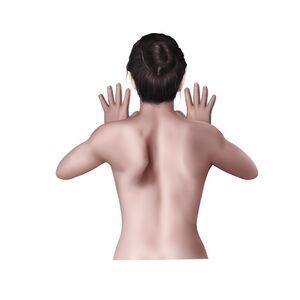
To palpate the scapula, position the patient in prone. If the patient can tolerate it, position the arm on the tested side at the small of the patient's back (i.e. in internal rotation). This will help you to visualise the borders of the scapula. With one hand, cup the shoulder anteriorly to lift the humerus. With this passive retraction manoeuvre, you can clearly identify the borders of the scapula. Please note that the lateral border is difficult to palpate, as it is covered with muscles.
To palpate the ribs posteriorly, position the patient in sitting or standing with their back towards you, trunk flexed, and scapula protracted. In order to palpate the first rib, locate the long spinous process of the C7 vertebra. The first rib is about two centimetres lateral and slightly inferior to the C7 spinous process. It can be difficult to palpate the first rib as it is covered by trapezius and levator scapulae. Use the same method to palpate the second rib located between T1 and T2.T2 can be found at the level of superior angle of scapula. Ribs three and four are located under the medial end of the spine of the scapula. Rib seven is located under the inferior angle of the scapula. The costal angle of rib nine is at the level of the spinous process of L1.
Sternum,
Clavicle
Sternoclavicular joint (SC)
The SC joint is the articulation between the sternal end of the clavicle, the manubrium of the sternum, and the first costal cartilage. It contains an articular disc, which separates the joint into two compartments.
The sternum is located on the anterior wall of the thorax. It is in the middle of the rib cage. The broadest part of the manubrium articulates with the costal cartilage of the first rib. The xiphoid process is located at the inferior end of the sternum, which is at the level of the seventh costal cartilage. You can palpate the xiphoid process by finding the small, pointed projection at the end of the sternum. The xiphoid process articulates directly with the costal cartilage of ribs seven and indirectly with the cartilage of ribs eight, nine, and ten.
When palpating the clavicle, start at the centre of the clavicle, as it is easy to find. Gently hold the posterior and anterior aspects of the clavicle, and move your fingers towards the medial end of the clavicle where the clavicle meets the manubrium of the sternum. This is where the sternoclavicular joint is located. You can confirm you are on the joint by passively moving the clavicle up and down, which creates a small motion at the joint.
Acromion of the scapula,
Clavicle
Acromioclavicular joint (AC)
The AC joint is the articulation between the lateral end of the clavicle and the acromion of the scapula. It moves with the glenohumeral joint, enabling functional movement at the shoulder girdle.[9]
To palpate the acromion of the scapula, position the patient in sitting. First, locate the spine of the scapula. Place your finger on the upper and the lower portion of the spine of the scapula and move it laterally. Pass the thickening portion of the spine called the tubercle and continue laterally to find a sharp edge. This is the acromial angle and the beginning of the acromion process, which turns upwards towards the front of the shoulder. From there, you can palpate the posterior, lateral, and anterior parts of the acromion process. The acromioclavicular joint (AC) is palpated at the lateral end of the clavicle. The joint line may be difficult to palpate as it is covered by trapezius. Place your finger on the AC joint to feel the joint line and passively move the clavicle up and down.
Humerus,
Glenoid fossa of the scapula
Glenohumeral joint
The glenohumeral joint is made up of the glenoid fossa of the scapula and the head of the humerus. It has the largest range of motion of all the joints in the body, relying on its surrounding muscles and connective tissue structures for stability.[10]
To palpate the glenohumeral joint, position the patient in supine. First, locate the coracoid process. To do this, slightly abduct the patient's arm. Ask the patient to flex their arm to activate the anterior deltoid. You will then be able to palpate the medial border of the anterior deltoid. Move your finger laterally and feel for a prominence, shaped like a knuckle, under the skin. It is the coracoid process of the scapula. The glenohumeral joint is palpated approximately 1 cm inferior to the coracoid process.
Shoulder Kinematics
Shoulder range of motion and strength can be affected by various factors.
A study by Pike et al.[11] found that shoulder range of motion (ROM) and strength significantly decrease as age increases, especially in the over-65 age group.
Increased body mass index (BMI) can also have a negative effect on shoulder flexibility.
The position of the thoracic spine must be considered when assessing shoulder range of motion as it significantly affects scapular kinematics during scapular plane abduction. A slouched posture is associated with decreased shoulder range of motion and muscle force.
Other factors influencing shoulder range of motion include biological sex, comorbidities, and hand dominance:[12][13]
active shoulder flexion and abduction are greater in males
active external rotation is greater in females
mean range of motion in all planes is lower among people with diabetes
mean range for all assessed movements except external rotation of the right shoulder is lower for those who are right-hand dominant compared to those who are left-hand dominant
The table below indicates values of the active range of motion in the shoulder joints:
Joint
Type of joint
Plane of movement
Motion
Kinematics
Closed pack position
Open pack position
Scapulothoracic joint
Not a true joint
Frontal
Transverse
Sagittal
Joint kinematics at the scapulothoracic joint include translation and rotation of the scapula on the ribcage.
Scapular movements include elevation, depression, adduction, and abduction. The scapula also rotates through upward and downward rotation, internal and external rotation, and anterior and posterior tilt.[10]
Scapular elevation: 40 degrees.
Scapular depression: 10 degrees.
Scapular protraction: 20 degrees.
Scapular retraction: 15 degrees.
Scapula rotation:
Upwards rotation : 60 degrees.
Downwards rotation : 30 degrees.
Internal rotation: 30-45 degrees.
External rotation: 10-20 degrees.
Anterior tilt: approximately 10 degrees.
Posterior tilt: approximately 30 degrees.
Because it is not a true joint, the scapulothoracic joint does not have a close-packed position.
Arms resting by the side of the body.
Sternoclavicular joint
Synovial- saddle-type
Sagittal
Frontal
Transverse
Anterior/posterior axial rotation,
Elevation/depression,
Protraction /retraction
Anterior rotation: 40-50 degrees.
Posterior rotation: 40-50 degrees.
Elevation: 45 degrees.
Depression: 10 degrees.
Combined protraction and retraction: 35 degrees.
Clavicle in maximal rotation, which is produced by maximal arm elevation and full scapular rotation.
Arms resting by the side of the body.
Acromioclavicular joint
Synovial-plane type
Acromioclavicular joint range of motion is difficult to measure as it combines the clavicle and glenohumeral joint.
Maximum rotation range of motion is achieved when the upper limb is resting in a horizontal position.[9]
Axial rotation: 25 degrees
90 degrees of shoulder abduction.
Arms resting by the side of the body.
Glenohumeral joint
Ball and socket
Sagittal
Frontal
Transverse
Shoulder flexion/extension,
Shoulder abduction/adduction,
Shoulder internal rotation/external rotation,
Shoulder circumduction: a combination of flexion, abduction, extension, and adduction.
Flexion/extension: 150-180 degrees/45 degrees.
Abduction/adduction: 160/30 degrees.
External/internal rotation: 85/85 degrees.
Abduction and external rotation.
40-50 degrees of abduction and 30 degress horizontal adduction.
Shoulder Passive Range of Motion Assessment
There are no set guidelines on selecting a testing position (e.g. sitting, supine, or standing) when assessing passive range of motion at the shoulder. The therapist must, therefore, consider various factors when choosing a testing position for the passive range of motion assessment. For example:[14]
if the therapist is shorter than the patient, it may be difficult to assess the patient in standing
if a patient has multiple disabilities, they may not be able to assume certain positions
It is recommended that the clinician document the testing position and use the same position when re-assessing passive range of motion.[14]
Shoulder flexion
The patient will ideally be in a sitting position to avoid compression on the scapula when supine
The patient's arm is positioned with the palm facing inwards, and the forearm in the mid-position
The therapist grasps the distal humerus, close to the elbow
The therapist moves the patient's humerus in an anterior and upward direction to the limit of shoulder flexion
If possible, the elbow should remain in extension to minimise the risk of passive insufficiency of the 2-joint triceps muscle affecting results
In the absence of pathology, the end feel will be firm
Normal range: pure glenohumeral flexion: 80-90 degrees, but with the clavicle and scapula moving: 180 degrees
Shoulder extension
The patient can be in sitting, standing or side-lying
The patient's arm is positioned with the palm facing inwards, and the forearm in the mid-position
The therapist grasps the patient’s humerus as distally as possible
The therapist moves the patient's arm into extension
In the absence of pathology, the end feel will be firm
Normal range: 60 degrees
Shoulder abduction
The patient can be in sitting, standing or supine
The patient's arm is positioned with the palm facing inwards, and the forearm in the mid-position
The therapist grasps the patient’s humerus as distally as possible
The therapist moves the patient's arm into abduction
In the absence of pathology, the end feel will be firm
Normal range: 90 degrees of pure glenohumeral abduction. Further motion is expected if the clavicle and scapula are not fixated
Shoulder adduction
The patient can be in sitting, standing or supine
The patient's arm is positioned with the palm facing inwards, and the forearm in the mid-position
The therapist grasps the patient’s humerus as distally as possible
The therapist moves the patient's arm into adduction
In the absence of pathology, the end feel will be firm
The patient can be sitting, standing, supine or side-lying
The therapist stabilises the patient's scapula with one hand and brings the patient's arm into 45 degrees of glenohumeral abduction. The therapist's other hand holds the patient's arm at the elbow, keeping it at 0 degrees of rotation
The therapist externally rotates the patient's arm while observing any scapular retraction
The movement is stopped when a firm end feel is reached or when the scapula starts to displace
Normal range: approximately 60 degrees
Shoulder internalrotation
The patient can be sitting, standing, side-lying
The therapist stabilises the patient's scapula with one hand and brings the patient's arm into 45 degrees of glenohumeral abduction. The therapist's other hand holds the patient's arm at the elbow, keeping it at 0 degrees of rotation
Next, the therapist brings the patient's hand behind the patient's back and lifts it off the thorax
In the absence of pathology, the end feel will be firm
Normal range: around 100 degrees from the anatomic position
An alternative testing position for internal and external glenohumeral joint rotation is prone with the shoulder at 90 degrees of abduction and the elbow at 90 degrees of flexion. The forearm hangs over the edge of the table. The therapist stabilises the distal part of the shoulder with one hand and moves the patient's forearm towards their head for external rotation or towards their hip for internal rotation.[16]
Shoulder Bursae
The bursae of the shoulder allow for functional movement in the shoulder joint. However, they are also a leading cause of shoulder pathology and dysfunction.
Shoulder Bursae
The shoulder has the following bursae:
Subscapular bursa or the scapulothoracic bursa: between the tendon of the subscapularis muscle and the shoulder joint capsule.
Subdeltoid bursa: between the deltoid muscle and the shoulder joint cavity.
Subacromial bursa: below the acromion process and above the greater tubercle of the humerus. This bursa is considered a primary source of shoulder pain,[17] and it is a well-vascularised structure.[18]
Subcoracoid bursa: between the coracoid process of the scapula and the shoulder joint capsule.
Infraspinatus bursa: between the infraspinatus tendon and the joint capsule.
Subcutaneous acromial bursa: above the acromion, beneath the skin.
The subacromial and the subdeltoid bursae are often considered a single bursa, referred to as the subacromial deltoid bursa.
Glenoid Labrum
The glenoid labrum is a fibrocartilaginous, ridge-like connective tissue that increases the articular surface area for the humeral head by deepening the glenoid fossa. It is also the primary attachment for the glenohumeral ligaments.
Labrum
Shoulder Joint Capsule
The glenohumeral joint capsule is an important passive stabiliser of the shoulder joint.[19] The glenohumeral joint capsule is thickened anteriorly, and it is twice the size of the humeral head. It provides most of the shoulder's extensibility anteriorly and inferiorly, and it “winds up” during abduction and external rotation. The joint capsule and glenohumeral ligaments function as stabilisers at the extremes of motion.
Ligaments of the Shoulder
Scapulothoracic Joint Ligaments
Shoulder Ligaments
The scapulothoracic joint has no ligaments as it is not a true joint. The attaching muscles and structures of the sternoclavicular and acromioclavicular joints support it.[10]
Sternoclavicular Joint Ligaments
Key ligaments
Origin
Insertion
Action/role
Key palpation points
Anterior sternoclavicular ligament
Anterosuperior surface of the sternal end of the clavicle
Anterosuperior surface of the manubrium and the adjacent part of the first costal cartilage
Reinforces the anterior aspect of the joint
Position the patient in supine. Find the centre of the clavicle. Palpate the posterior and anterior aspects of the clavicle and move your fingers towards the medial end of the clavicle, where the clavicle meets the manubrium of the sternum.
Posterior sternoclavicular ligament
Posterior aspect of the sternal end of the clavicle
Posterosuperior manubrium
Posterior reinforcement of the joint capsule
Interclavicular ligament
Sternal end of the left clavicle
Sternal end of the right clavicle
Superior reinforcement of the joint capsule.
Resists excessive depression or downward glide of the clavicle
Costoclavicular ligament
First rib and costal cartilage
Inferior surface of the clavicle
Limits elevation of the pectoral girdle.
Acts as a fulcrum for elevation-depression and protraction-retraction.
Limits the clavicular elevation and superior glide of the clavicle.
Position the patient in supine with their scapula depressed. To palpate the first rib, find the clavicle. Place your finger on the lateral end of the clavicle and roll behind it. Next, with the finger, push the belly of the trapezius muscle back and apply downward pressure to feel a bony prominence. This is the first rib. To confirm you are on the first rib, ask the patient to take a deep breath. The first rib should elevate during inhalation.
Acromioclavicular Joint Ligaments
Key ligaments
Origin
Insertion
Action/role
Key palpation points
Coracoclavicular ligament:
Conoid ligament (CL)
Trapezoid ligament (TL)
CL: coracoid process of the scapula
TL: coracoid process of the scapula
CL: conoid tubercle of the clavicle
TL: trapezoid line of the clavicle
Prevents superior dislocation of the scapula.
Resists scapular rotation.
Supports the weight of the upper limb suspended from the clavicle.
To palpate the coracoid process of the scapula, position the patient in a supine and slightly abduct the patient's arm. Ask the patient to flex their arm to activate the anterior deltoid. Palpate the medial border of the anterior deltoid. Move your finger laterally and feel for a bony prominence, shaped like a knuckle, under the skin. This is the coracoid process of the scapula. The conoid tubercle and the trapezoid line cannot be palpated as they are located on the underside of the clavicle.
Acromioclavicular ligament
Superior surface of the acromion
Acromial end of the clavicle
Resists posterior translation at the joint.
Resists posterior axial rotation of the joint.
Find the coracoid process of the scapula. The acromioclavicular joint is more lateral and superior to the coracoid process.
Glenohumeral Joint Ligaments
Key ligaments
Origin
Insertion
Action/role
Key palpation points
Coracoacromial ligament
Summit of the acromion of the scapula
Lateral border of the coracoid process of the scapula
Stabilises the head of the humerus.
Limits inferior translation and excessive external rotation of the humerus.
Transverse humeral ligament
Lesser tubercle of the humerus
Greater tubercle of the humerus
Holds the tendon of the long head of the biceps muscle in place.
To palpate the lesser tubercle of the humerus, start by finding the anterolateral portion of the acromion. Just below, you will find the greater tubercle of the head of the humerus. Next, place your finger on the anterior surface of the head of the humerus and passively move the patient’s arm into lateral rotation. You will feel the bicipital groove under your palpating finger as the patient's arm moves into lateral rotation. As you move the patient's arm further into lateral rotation, you will be able to palpate the lesser tubercle.
Superior glenohumeral ligament
Glenoid labrum
Lesser tubercle
Prevents posterior and inferior translation of the humeral head.
Middle glenohumeral ligament
Inferior to the superior ligament along the anterior margin of the glenoid
Anterior surface of the lesser tubercle
Provides anterior stability when the shoulder is abducted to between 45 and 60 degrees.
Inferior glenohumeral ligament
Glenoid labrum
Beyond the lesser tubercle
Prevents extremes of motion.
Stabilises against anteroinferior dislocation.
Muscles of the Shoulder
The dynamic stabilisers of the shoulder complex include the rotator cuff muscles, the deltoid, and the scapular muscles, which control scapulohumeral rhythm.[20] For optimal shoulder stabilisation, the dynamic stabilisers must function efficiently and synergistically. Dynamic stabilisation enables significant mobility at the shoulder and provides adequate stability when the shoulder complex is functioning normally. The rotator cuff muscles provide stability at the glenohumeral joint, primarily through the compression of the head of the humerus against the glenoid during movement. The rotator cuff muscles are supraspinatus, infraspinatus, teres minor, and subscapularis.[21]
Rotator cuff muscles
The following tables include muscles that act on the scapula and the humerus. Other functions of these muscles not related to the scapula and shoulder motion are not included in these tables (e.g. when the scapula is fixed, levator scapulae assists with the lateral flexion of the cervical spine).
Scapular Elevation
Muscle
Origin
Insertion
Innervation
Action
Levator scapulae
Transverse processes of C1 to C4
Medial border of the scapula
Dorsal scapular nerve C5
Cervical nerves C3-C4
Elevates the scapula.
Tilts the glenoid cavity inferiorly by rotating the scapula downward.
Upper trapezius
Occipital bone and the nuchal ligament
Lateral third of the clavicle
Accessory nerve (CN XI)
Spinal nerves C3-C4
Produces scapular elevation.
Scapular Depression
Muscle
Origin
Insertion
Innervation
Action
Pectoralis minor
Ribs three to five
Medial border and coracoid process of the scapula
Medial pectoral nerve C8-T1
The primary actions of pectoralis minor include stabilisation, depression, abduction or protraction, internal rotation and downward rotation of the scapula.
Lower trapezius
The spinous processes of T4 to T12
Lateral third of the clavicle, acromion, and the spine of the scapula
Accessory nerve (CN XI)
Spinal nerves C3-C4
Shoulder depression and scapula upward rotation.
Scapular Protraction
Muscle
Origin
Insertion
Innervation
Action
Serratus anterior
Ribs one to nine
Costal surface of the medial scapula
Long thoracic nerve C5-C6-C7
The main actions of serratus anterior are protraction and upward rotation of the scapulothoracic joint.
It is also a key scapular stabiliser, keeping the scapula against the ribcage at rest and during movement.
Known as the "Boxer's muscle", as it protracts the scapula during a punch.
Pectoralis minor
Ribs three to five
Medial border and coracoid process of the scapula
Medial pectoral nerve C8-T1
The primary actions of pectoralis minor include stabilisation, depression, abduction or protraction, internal rotation and downward rotation of the scapula.
Scapular Retraction
Muscle
Origin
Insertion
Innervation
Action
Rhomboids:
Rhomboid Major
Rhomboid Minor
Rhomboid Major: the spinous processes of T2 to T5
Rhomboid Minor: the spinous processes of C7 to T1
Rhomboid Major and Minor: Medial border of the scapula
Dorsal scapular nerve C4-C5
Retract, elevate and rotate the scapula.
Protract the medial border of the scapula, keeping it in position at the posterior thoracic wall.
Middle Trapezius
The spinous processes of T1-T4 (other sources indicate C7-T3)
Lateral third of the clavicle, as well as the acromion and the spine of the scapula.
Accessory nerve (CN XI),
Spinal nerves C3-C4
Scapula retraction.
Scapular Upward Rotation (Lateral Rotation)
Muscle
Origin
Insertion
Innervation
Action
Serratus anterior
Ribs one to nine
Costal surface of the medial scapula
Long thoracic nerve C5-C6-C7
The main actions of serratus anterior are protraction and upward rotation of the scapulothoracic joint.
It is also a key scapular stabiliser, keeping the scapula against the ribcage at rest and during movement.
Known as the "Boxer's muscle", as it protracts the scapula during a punch.
Upper trapezius
Occipital bone and the nuchal ligament
Lateral third of the clavicle
Accessory nerve (CN XI)
Spinal nerves C3-C4
The upper and lower fibres of trapezius work concurrently to produce rotation of the scapula.[10]
Lower trapezius
The spinous processes of the T4 to T12.
Lateral third of the clavicle, acromion, and the spine of the scapula
Accessory nerve (CN XI)
Spinal nerves C3-C4
Shoulder depression and scapula upward rotation
Scapular Downward Rotation (Middle Rotation)
Muscle
Origin
Insertion
Innervation
Action
Rhomboids:
Rhomboid Major
Rhomboid Minor
Rhomboid Major: the spinous processes of T2 to T5
Rhomboid Minor: the spinous processes of C7 to T1
Rhomboid Major and Minor: Medial border of the scapula.
Dorsal scapular nerve C4-C5
Retract, elevate and rotate the scapula.
Protract the medial border of the scapula, keeping it in position at the posterior thoracic wall.
Levator scapulae
Transverse processes of C1 to C4
Medial border of the scapula
Dorsal scapular nerve C5,
Cervical nerves C3-C4
Elevates the scapula
Tilts the glenoid cavity inferiorly by rotating the scapula downward.
Pectoralis minor
Ribs three to five
Medial border and coracoid process of the scapula
Medial pectoral nerve C8-T1
The primary actions of pectoralis minor include stabilisation, depression, abduction or protraction, internal rotation and downward rotation of the scapula.
Shoulder Flexion
Muscle
Origin
Insertion
Innervation
Action
Coracobrachialis
Coracoid process of the scapula
Medial surface of the humeral shaft
Musculocutaneous nerve
Assists with shoulder flexion and adduction.
Biceps brachii:
Short head (SH)
Long head (LH)
SH: coracoid process of the scapula
LH: supraglenoid tubercle of the scapula
SH/LH: radial tuberosity
Flexes the shoulder.
Helps to stabilise the head of the humerus in the glenoid cavity.
Deltoid:
Anterior deltoid (AD)
Lateral deltoid (LD)
Posterior deltoid (PD)
AD: lateral third of the clavicleLD: acromion of the scapula
PD: spine of the scapula
AD/LD/PD: deltoid tuberosity on the humerus
Axillary nerve
Only the anterior deltoid contributes to shoulder flexion.
Pectoralis major:
Clavicular head (CH)
Sternocostal head (SCH)
CH: anterior surface of the medial half of the clavicle
SCH: anterior surface of the sternum, superior six costal cartilages, and aponeurosis of external oblique muscle
CH/SCH: Lateral lip of intertubercular groove of the humerus
Lateral and medial pectoral nerves
When acting alone, the clavicular head flexes the shoulder.
Shoulder Extension
Muscle
Origin
Insertion
Innervation
Action
Latissimus dorsi
Spinous processes of T7 to T12
Iliac crest
Ribs nine to twelve
Intertubercular groove in the front of the humerus
Thoracodorsal nerve
Shoulder extension, adduction, and internal rotation
Teres major
Dorsal surface of the inferior angle of the scapula
Medial lip of intertubercular groove of the humerus
Lower subscapular nerve
Assists with shoulder extension.
Internally rotates the shoulder.
Contributes to the stabilisation of the shoulder joint.
Triceps brachii:
Long head (LH)
Medial head (MH)
Lateral head (LLH)
LH: infraglenoid tubercle of the scapula
MH: posterior surface of the humerus, inferior to the radial groove
LLH: posterior surface of the humerus
LH/MH/LLH: olecranon of the ulna
Radial nerve
Only the long head is involved in the shoulder extension.
Stabilises the humeral head during shoulder abduction.
Extends the elbow.
Deltoid:
Anterior deltoid (AD)
Lateral deltoid (LD)
Posterior deltoid (PD)
AD: lateral third of the clavicle
LD: acromion of the scapula
PD: spine of the scapula
AD/LD/PD: deltoid tuberosity on the humerus
Axillary nerve
The posterior deltoid helps with shoulder extension.
Shoulder Abduction
Muscle
Origin
Insertion
Innervation
Action
Supraspinatus
Supraspinous fossa in the scapula
Greater tubercle of the humerus
Suprascapular nerve
Initiate abduction up to 15 degrees, after which the lateral deltoid muscle takes over.
Deltoid:
Anterior deltoid (AD)
Lateral deltoid (LD)
Posterior deltoid (PD)
AD: lateral third of the clavicle
LD: acromion of the scapula
PD: spine of the scapula
AD/LD/PD: deltoid tuberosity on the humerus
Axillary nerve
The lateral deltoid abducts the shoulder.
Shoulder Adduction
Muscle
Origin
Insertion
Innervation
Action
Subscapularis
Subscapular fossa of the scapula
Lesser tuberosity of humerus
Upper and lower subscapular nerves
Internal rotation and adduction.
Assists in holding the humeral head in the glenoid cavity of the scapula.
Teres minor
Superior part of the lateral border of the scapula
Inferior facet on the greater tuberosity of humerus
Axillary nerve
Shoulder internal rotation.
Assists in holding the humeral head in the glenoid cavity of the scapula.
Teres major
Dorsal surface of the inferior angle of the scapula
Medial lip of intertubercular groove of the humerus
Lower subscapular nerve
Assists with shoulder extension.
Acts as a functional unit with latissimus dorsi to internally rotate, adduct and extend the shoulder.
Contributes to the stabilisation of the shoulder joint.
Latissimus dorsi
Spinous processes of T7 to T12
Iliac crest
Ribs nine to twelve
Intertubercular groove in the front of the humerus
Thoracodorsal nerve
Shoulder extension, adduction, and internal rotation.
Pectoralis major:
Clavicular head (CH)
Sternocostal head(SCH)
CH: anterior surface of the medial half of clavicle
SCH: anterior surface of sternum, superior six costal cartilages, and aponeurosis of the external oblique muscle
CH/SCH: Lateral lip of intertubercular groove of the humerus
Lateral and medial pectoral nerves
Shoulder adduction and internal rotation.
Draws the scapula anteriorly and inferiorly.
External Rotation
Muscle
Origin
Insertion
Innervation
Action
Infraspinatus
Infraspinous fossa on the scapula
Middle facet on the greater tuberosity of humerus
Suprascapular nerve
Shoulder external rotation.
Assists in holding the humeral head in the glenoid cavity of the scapula.
Internal Rotation
Muscle
Origin
Insertion
Innervation
Action
Subscapularis
Subscapular fossa of scapula
Lesser tuberosity of humerus
Upper and lower subscapular nerves
Internal rotation and adduction.
Assists in holding the humeral head in the glenoid cavity of the scapula.
Teres major
Dorsal surface of the inferior angle of the scapula
Medial lip of intertubercular groove of the humerus
Lower subscapular nerve
Assists with shoulder extension.
Acts as a functional unit with the latissimus dorsi to internally rotate, adduct and extend the shoulder.
Contributes to the stabilisation of the shoulder joint.
Latissimus dorsi
Spinous processes of T7 to T12
Iliac crest
Ribs nine to twelve
Intertubercular groove in the front of the humerus
Thoracodorsal nerve
Shoulder extension, adduction, and internal rotation.
Pectoralis major:
Clavicular head (CH)
Sternocostal head(SCH)
CH: anterior surface of the medial half of clavicle
SCH: anterior surface of sternum, superior six costal cartilages, and aponeurosis of external oblique muscle
CH/SCH: Lateral lip of intertubercular groove of the humerus
Lateral and medial pectoral nerves
Shoulder adduction and internal rotation.
Draws the scapula anteriorly and inferiorly.
Deltoid:
Anterior deltoid (AD)
Lateral deltoid (LD)
Posterior deltoid (PD)
AD: the lateral third of the clavicle
LD: acromion of the scapula
PD: spine of the scapula
AD/LD/PD: deltoid tuberosity on the humerus
Axillary nerve
The anterior deltoid contributes to shoulder internal rotation.
Innervation of the Shoulder
The majority of the muscles in the shoulder are innervated by nerves originating from the brachial plexus. The brachial plexus is a network of nerves formed from the ventral rami of nerve roots C5 to T1.[10] From proximal to distal, the brachial plexus is organised by roots, trunks, divisions, and cords.
Nerve
Origin
Branches
Motor fibres
Sensory fibres
Accessory nerve (cranial nerve XI)
Upper spinal cord (C1-C5/C6).
Lateral aspect of the medulla oblongata.
Internal branch
External branch
Purely somatic motor function, innervating trapezius
None
Dorsal scapular nerve
Anterior ramus of spinal nerve C5
None
Rhomboid major, rhomboid minor and levator scapulae
None
Long thoracic nerve
Spinal nerves C5, C6, and C7
Serratus anterior
None
Subclavian nerve
Anterior ramus of spinal nerves C5-C6
Subclavius
None
Suprascapular nerve
Superior trunk of brachial plexus
Motor muscular branches
Sensory articular branches
Supraspinatus and
infraspinatus
Acromioclavicular joint
Glenohumeral joint
Medial pectoral nerve
Medial cord of brachial plexus (C8, T1)
Muscular branches
Pectoralis major and minor
None
Lateral pectoral nerve
Lateral cord of brachial plexus (C5, C6, C7)
Pectoralis major and minor
None
Musculocutaneous nerve
Lateral cord of brachial plexus (C5-C7)
Coracobrachialis, biceps brachii
Anterolateral skin of the forearm
Upper subscapular nerve
Posterior cord of the brachial plexus (C5-C6)
Superior portion of the subscapularis
None
Lower subscapular nerve
Posterior cord of the brachial plexus (C5-C6)
Inferior portion of the subscapularis
Teres major
Thoracodorsal nerve
Posterior cord of the brachial plexus (C6-C8)
Small terminal muscular branches
Latissimus dorsi
None
Axillary nerve
Posterior cord of brachial plexus (C5-C6)
Anterior, posterior, and articular branches
Deltoid, teres minor, lateral head of triceps brachii
Glenohumeral joint,
and the skin of the deltoid region/upper arm
Radial nerve
Posterior cord of brachial plexus (C5-T1)
Posterior brachial cutaneous nerve, inferior lateral brachial cutaneous nerve, posterior antebrachial cutaneous nerve, muscular branches, deep branch of radial nerve, superficial branch of radial nerve
Triceps brachii
Lower outer aspect and posterior surface of the arm
Vascular Supply of the Shoulder
The blood supply for the shoulder is provided primarily by the subclavian artery. As it enters the shoulder region and axilla, the subclavian artery becomes the axillary artery.[10]
Latissimus dorsi, deltoid, long head of triceps brachii, subscapularis, supraspinatus, infraspιnatus, and serratus anterior and adjacent skin
Anterior circumflex humeral artery (AC)
Posterior circumflex humeral artery (PC)
AC: ascending branch
PC: descending branch
AC: Glenohumeral joint, teres major and minor, deltoid, and the head of the humerus.
PC: Glenohumeral joint, deltoid, teres major, teres minor, and long and lateral heads of triceps brachii
Clinical Relevance
Forward head posture increases tension on the levator scapulae muscles. Increased tightness in the levator scapulae due to increased activity from a forward head posture can lead to cervicogenic headaches.
Upper trapezius is frequently affected in neck injuries, including high-velocity accidents, such as motor vehicle accidents.
Winged ScapularScapular winging occurs when the muscles of the scapula are weak or paralysed, resulting in reduced ability to stabilise the scapula. Damage to the long thoracic nerve, which provides sole innervation to the serratus anterior muscle, can lead to scapula winging.
When the position and motion of the patient's scapula are altered, they can experience a condition called scapular dyskinesia or scapular dysrhythmia. It is characterised by inferior angle prominence, medial border prominence, excessive superior border elevation, or symmetric scapular motion during arm movement. Learn more about scapular dyskinesia from here.
Shoulder instability is defined as loss of shoulder comfort and function due to undesirable translation of the humeral head on the glenoid fossa.
Rotator cuff tears are the leading cause of shoulder pain and shoulder-related disability. Degenerative changes, repetitive micro-traumas, severe traumatic injuries, atraumatic injuries and secondary dysfunctions can result in rotator cuff tears. You can learn more about shoulder conditions in the shoulder programme.
Erb’s palsy occurs when there is an injury to the brachial plexus, specifically the upper brachial plexus, at birth. Backpackers, or people carrying heavy weights while hiking, can also experience compression of the upper trunks of the brachial plexus.
↑Levangie PK, Norkin CC. Joint Structure and Function: A Comprehensive Analysis. 4th ed. Philadelphia, PA: F.A. Davis Co., 2005.
↑Hyland S, Charlick M, Varacallo M. Anatomy, Shoulder and Upper Limb, Clavicle. [Updated 2023 Jul 24]. In: StatPearls [Internet]. Treasure Island (FL): StatPearls Publishing; 2023 Jan-. Available from: https://www.ncbi.nlm.nih.gov/books/NBK525990/
↑Graeber GM, Nazim M. The anatomy of the ribs and the sternum and their relationship to chest wall structure and function. Thoracic surgery clinics. 2007 Nov 1;17(4):473-89.
↑ 6.06.16.2Miniato MA, Mudreac A, Borger J. Anatomy, Thorax, Scapula. [Updated 2023 Jul 24]. In: StatPearls [Internet]. Treasure Island (FL): StatPearls Publishing; 2023 Jan-. Available from: https://www.ncbi.nlm.nih.gov/books/NBK538319/
↑Mostafa E, Imonugo O, Varacallo M. Anatomy, Shoulder and Upper Limb, Humerus. [Updated 2023 Aug 7]. In: StatPearls [Internet]. Treasure Island (FL): StatPearls Publishing; 2023 Jan-. Available from: https://www.ncbi.nlm.nih.gov/books/NBK534821/
↑Kennedy MS, Nicholson HD, Woodley SJ. The morphology of the subacromial and related shoulder bursae. An anatomical and histological study. J Anat. 2022 May;240(5):941-958.
↑Lee SB, Kim KJ, O'Driscoll SW, Morrey BF, An KN. Dynamic glenohumeral stability provided by the rotator cuff muscles in the mid-range and end-range of motion. A study in cadavera. J Bone Joint Surg Am. 2000 Jun;82(6):849-57.