
Freiberg's test
Top Contributors - Uchechukwu Chukwuemeka, Marlies Verbruggen, Alexandra Stead, George Prudden, Kim Jackson, Vidya Acharya and Wanda van Niekerk
Definition / Description
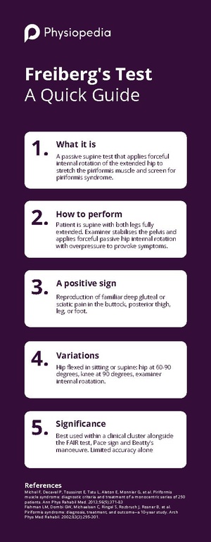
The Freiberg's test, commonly known as the Freiberg sign or the Freiberg manoeuvre, is a passive provocation test used to evaluate the hip and gluteal area clinically. It is mostly used to check for piriformis syndrome, a painful neuromuscular disorder when the sciatic nerve is irritated or compressed by the piriformis muscle.[1][2][3]
While the patient is in the supine position, the test applies forceful internal rotation of the extended hip passively to induce pain. Stretching the inflamed piriformis muscle and causing compression of the sciatic nerve at the sacrospinous ligament level are the two goals of this manoeuvre.[2] When combined with other provocation tests, the Freiberg test is a reasonably sensitive clinical tool, since a positive result is reported in roughly 56.2% of patients with verified piriformis syndrome.[4]
Clinical Relevant Anatomy
The flat, triangular piriformis muscle is situated deep within the gluteal area. It comes from the superior margin of the larger sciatic notch, the pelvic (anterior) surface of the sacral segments S2 to S4, and the areas lateral to and between the anterior sacral foramina. It attaches onto the superior aspect of the greater trochanter of the femur after leaving the pelvis through the greater sciatic foramen.[5][6]
The piriformis muscle is responsible for the hip joint's partial extension, abduction, and external rotation. It is located nearly parallel to the posterior border of the gluteus medius and is the most superficial of the deep gluteal muscles.[7]
Although there are several anatomical variances, the sciatic nerve (L4 to S3) normally exits the pelvis inferior to the piriformis muscle's belly. A six-category classification scheme described by Beaton and Anson shows the connection between the sciatic nerve and the piriformis muscle:[5]
- Type A (Normal): The piriformis muscle is superior to the sciatic nerve.
- Type B: The nerve is divided, with the tibial division passing beneath the piriformis muscle and the peroneal division passing through it.
- Type C: The undivided muscle is passed over by the peroneal division.
- Type D: The piriformis muscle belly is where the entire sciatic nerve travels.
- Type E: The piriformis muscle is inferior to the entire sciatic nerve.
- Type F: The nerve splits proximally and travels through a congenitally bifid muscle or tendons.
The sciatic nerve may be directly compressed when the piriformis muscle is tight or in spasm, hypertrophied, resulting in the distinctive pain pattern linked to piriformis syndrome. In Freiberg's test, forced internal rotation of the hip puts the piriformis in a passive stretch and may replicate this compression.[1][6]
Purpose
Freiberg's test is primarily intended to:
- Use passive piriformis muscle stretching to replicate the patient's familiar gluteal or sciatic pain to screen for piriformis syndrome.
- Analyse sciatic nerve irritation brought on by the piriformis muscle's mechanical compression at the sacrospinous ligament.
- Participate in a clinical cluster of tests to improve the diagnostic precision for piriformis syndrome, including the FAIR test, Pace sign, Beatty's manoeuvre, and Lasegue sign.[4][8]
Test instructions
The following is a common way to perform the Freiberg's test:
- The patient is positioned on the examination table in a supine position, with both lower limbs in full extension
- The examiner stands on the side being evaluated and places one hand on the pelvis to stabilise and the other hand is controlling the ankle of the tested side.
- The tested leg is maintained in a straight position.
- The tested leg is brought into medial rotation by a passive, forceful internal rotation of the hip joint by the examiner.
- To replicate symptoms, the examiner gradually applies overpressure in the end range.
If the patient experiences normal pain or discomfort in the buttock, sciatic notch area, or down the sciatic nerve distribution (posterior thigh, posterior leg, or foot), the test is deemed positive, while no reproduction of gluteal or sciatic symptoms depicts the test is negative.[9]
It should be noted that this manoeuvre stretches the piriformis muscle and the other lateral rotators, including the gemelli, obturator internus, and quadratus femoris. It also produces pressure on the sciatic nerve at the sacrospinous ligament.[1] [4] Additionally, the test may also be performed with the hip in flexion (60 to 90 degrees) with the knee flexed to 90 degrees, with the examiner pushing the heel laterally to induce internal rotation of the thigh. This is particularly noted in prone variants described by some authors.[8] Moreover, a seated variation has also been described, where the examiner prevents the knees from adducting while the patient attempts to bring them together.[9]
Evidence
About 56.2% of patients with piriformis syndrome have a positive Freiberg's test.[4] The intricacy of the illness and the fact that piriformis syndrome is an excluding diagnosis, are reflected in this moderate sensitivity. In addition, Freiberg's test has a limited diagnostic accuracy when used alone, as is the case with the majority of individual clinical tests for piriformis syndrome. Diagnostic reliability is increased by combining provocative tests, such as Beatty's manoeuvre, Pace sign, and the FAIR test. In contrast, the FAIR test has revealed a positive likelihood ratio of 5.2, a negative likelihood ratio of 0.14, a sensitivity of 0.88, and a specificity of 0.83.[7]
The diagnostic accuracy results in the literature are often generated from studies using a multi-test cluster strategy for piriformis syndrome; no large-scale research has independently revealed sensitivity and specificity values specific to Freiberg's test alone.[4][11]
Clinical Bottom Line
Freiberg's test is an easy, cost-effective passive provocation test intended to examine the hip and gluteal area clinically.It targets the piriformis muscle and the sciatic nerve by forcing passive internal rotation of the extended hip while the patient is supine. Although the test is positive in only around 56% of confirmed cases, a positive result, or the recreation of a characteristic deep gluteal or sciatic pain, is suggestive of piriformis syndrome.
In addition to the FAIR test, the Pace sign, Beatty's manoeuvre, and palpation findings, clinicians are recommended to perform Freiberg's test as part of a clinical cluster. To rule out alternative causes of sciatic and gluteal pain, such as lumbar radiculopathy or FAI, a comprehensive neurological and musculoskeletal assessment is necessary. Imaging, diagnostic injections, or electrophysiological tests should be considered in cases when the diagnosis is still unclear.[4][5] [6]
Resources
References
- ↑ 1.0 1.1 1.2 Shah S,Wang TW. Piriformis syndrome. eMedicine specialities :Sports medicine: hip 2009. Available from:http://emedicine.medscape.com/article/87545-overview (accessed 27 February 2026)
- ↑ 2.0 2.1 Kirschner JS, Foye PM, Cole JL. Piriformis syndrome, diagnosis and treatment. Muscle Nerve. 2009; 40(1):10-8. doi: 10.1002/mus.21318.
- ↑ Boyajian-O'Neill LA, McClain RL, Coleman MK, Thomas PP. Diagnosis and management of piriformis syndrome: an osteopathic approach. J Am Osteopath Assoc. 2008 Nov;108(11):657-64. doi: 10.7556/jaoa.2008.108.11.657.
- ↑ 4.0 4.1 4.2 4.3 4.4 4.5 Michel F, Decavel P, Toussirot E, Tatu L, Aleton E, Monnier G, Garbuio P, Parratte B. Piriformis muscle syndrome: diagnostic criteria and treatment of a monocentric series of 250 patients. Ann Phys Rehabil Med. 2013 Jul;56(5):371-83. doi: 10.1016/j.rehab.2013.04.003.
- ↑ 5.0 5.1 5.2 Hopayian K, Song F, Riera R, Sambandan S. The clinical features of the piriformis syndrome: a systematic review. Eur Spine J. 2010;19(12):2095-109. doi: 10.1007/s00586-010-1504-9.
- ↑ 6.0 6.1 6.2 Tonley JC, Yun SM, Kochevar RJ, Dye JA, Farrokhi S, Powers CM. Treatment of an individual with piriformis syndrome focusing on hip muscle strengthening and movement reeducation: a case report. J Orthop Sports Phys Ther. 2010 Feb;40(2):103-11. doi: 10.2519/jospt.2010.3108.
- ↑ 7.0 7.1 Fishman LM, Dombi GW, Michaelsen C, Ringel S, Rozbruch J, Rosner B, et al. Piriformis syndrome: diagnosis, treatment, and outcome--a 10-year study. Arch Phys Med Rehabil. 2002 Mar;83(3):295-301. doi: 10.1053/apmr.2002.30622.
- ↑ 8.0 8.1 Bârzu M. Diagnostic Methods in Piriformis Syndrome. Timisoara Physical Education and Rehabilitation Journal. West University of Timisoara, 2013;6(11): 22-28.
- ↑ 9.0 9.1 Levin SM. Piriformis syndrome. Orthopedics. 2000 Mar;23(3):183-4. doi: 10.3928/0147-7447-20000301-05.
- ↑ Ortho Eval Pal with Paul Marquis PT. Freiberg Sign for Piriformis Syndrome. Available from: http://www.youtube.com/watch?v=oV1rJn1xdGc[last accessed 27/2/2026]
- ↑ Hopayian K, Danielyan A. Four symptoms define the piriformis syndrome: an updated systematic review of its clinical features. Eur J Orthop Surg Traumatol. 2018 Feb;28(2):155-164. doi: 10.1007/s00590-017-2031-8.