Knee: Difference between revisions
Elvira Muhic (talk | contribs) No edit summary |
Elvira Muhic (talk | contribs) No edit summary |
||
| Line 8: | Line 8: | ||
= Anatomy of the Knee = | = Anatomy of the Knee = | ||
== Bones | == Bones<br> == | ||
The knee joint is one of the largest and most complex joints in the body. It is constructed by 4 bones and an extensive network of ligaments and muscles. | [[Image:Knee-patella.jpg|right|450x400px]] | ||
The knee joint is one of the largest and most complex joints in the body. It is constructed by 4 bones and an extensive network of ligaments and muscles. | |||
The thigh bone ([http://www.physio-pedia.com/Femur Femur]), the shin bone ([http://www.physio-pedia.com/Tibia Tibia]) and the kneecap ([http://www.physio-pedia.com/Patella Patella]) articulate through tibiofemoral and patellofemoral joints. These three bones are covered in articular cartilage which is an extremely hard, smooth substance designed to decrease the friction forces. The patella lies in an indentation of the femur known as the intercondylar groove. | The thigh bone ([http://www.physio-pedia.com/Femur Femur]), the shin bone ([http://www.physio-pedia.com/Tibia Tibia]) and the kneecap ([http://www.physio-pedia.com/Patella Patella]) articulate through tibiofemoral and patellofemoral joints. These three bones are covered in articular cartilage which is an extremely hard, smooth substance designed to decrease the friction forces. The patella lies in an indentation of the femur known as the intercondylar groove. | ||
The smaller shin bone that runs alonside the tibia (Fibula) is not directly involved in the knee joint<ref>Keith L. Moore. Clinically Oriented Anatomy 6e edition. P 634</ref>, but provides a surface for important muscles and ligaments to attach to. <br> | The smaller shin bone that runs alonside the tibia (Fibula) is not directly involved in the knee joint<ref>Keith L. Moore. Clinically Oriented Anatomy 6e edition. P 634</ref>, but provides a surface for important muscles and ligaments to attach to. <br> | ||
== Menisci == | == Menisci == | ||
| Line 20: | Line 32: | ||
There are two menisci in the space between the femoral and tybial condyles. They are crescent-shaped lamellae, each with anterior and posterior horn, and are triangular in cross section. The surface of each meniscus is concave superiorly, providing a congruous surface to the femoral condyles and is flat inferiorly to accompany the relatively flat tibial plateau. The horns of the medial meniscus are further appart and meniscus appears ‘C’ shaped, than those of the lateral one where meniscus appears more ‘O’ shaped. | There are two menisci in the space between the femoral and tybial condyles. They are crescent-shaped lamellae, each with anterior and posterior horn, and are triangular in cross section. The surface of each meniscus is concave superiorly, providing a congruous surface to the femoral condyles and is flat inferiorly to accompany the relatively flat tibial plateau. The horns of the medial meniscus are further appart and meniscus appears ‘C’ shaped, than those of the lateral one where meniscus appears more ‘O’ shaped. | ||
[[Image:Menisci top 95147470.jpg|right| | [[Image:Menisci top 95147470.jpg|right|450x300px]] | ||
<div> | <div> | ||
'''The menisci correct the lack of congruence between the articular surfaces of tibia and femur, increase the area of contact and improve weight distribtion and shock apsorption. They also help to guide and coordinate knee motion, making them very important stabilizers of the knee.''' | '''The menisci correct the lack of congruence between the articular surfaces of tibia and femur, increase the area of contact and improve weight distribtion and shock apsorption. They also help to guide and coordinate knee motion, making them very important stabilizers of the knee.''' | ||
Revision as of 21:57, 29 July 2016
Original Editor - Rachael Lowe
Top Contributors - Elvira Muhic, Rachael Lowe, Admin, Laura Ritchie, WikiSysop, Simisola Ajeyalemi, Michelle Lee, Kim Jackson, Andeela Hafeez, Leana Louw, Evan Thomas, Scott Buxton, Bert Lasat, William Jones, Eric Robertson, Joao Costa, Kristen Mason, Blessed Denzel Vhudzijena, George Prudden, 127.0.0.1, Tony Lowe, Vidya Acharya, Rucha Gadgil, Ernest Gamble, Jess Bell, Sai Kripa and Aminat Abolade
Anatomy of the Knee[edit | edit source]
Bones
[edit | edit source]
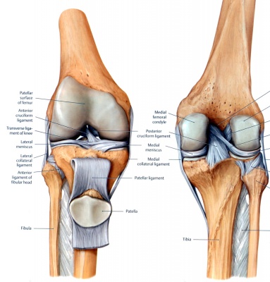
The knee joint is one of the largest and most complex joints in the body. It is constructed by 4 bones and an extensive network of ligaments and muscles.
The thigh bone (Femur), the shin bone (Tibia) and the kneecap (Patella) articulate through tibiofemoral and patellofemoral joints. These three bones are covered in articular cartilage which is an extremely hard, smooth substance designed to decrease the friction forces. The patella lies in an indentation of the femur known as the intercondylar groove.
The smaller shin bone that runs alonside the tibia (Fibula) is not directly involved in the knee joint[1], but provides a surface for important muscles and ligaments to attach to.
Menisci[edit | edit source]
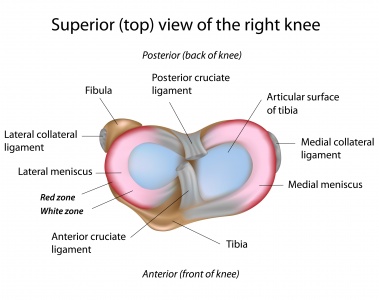
There are two menisci in the space between the femoral and tybial condyles. They are crescent-shaped lamellae, each with anterior and posterior horn, and are triangular in cross section. The surface of each meniscus is concave superiorly, providing a congruous surface to the femoral condyles and is flat inferiorly to accompany the relatively flat tibial plateau. The horns of the medial meniscus are further appart and meniscus appears ‘C’ shaped, than those of the lateral one where meniscus appears more ‘O’ shaped.
The menisci correct the lack of congruence between the articular surfaces of tibia and femur, increase the area of contact and improve weight distribtion and shock apsorption. They also help to guide and coordinate knee motion, making them very important stabilizers of the knee.
The menisci are connected with the tibia by coronary ligaments. The medial meniscus is much less mobile during joint motion than the lateral meniscus owing in large part to its firm attachment to the knee joint capsule and medial collateral ligament (MCL). On the lateral side, the meniscus is less firmly attached to the joint capsule and has no attachment to the lateral collateral ligament (LCL). In fact, the posterior horn of the lateral meniscus is separated entirely from the posterolateral aspect of the joint capsule by the tendon of the popliteus muscle as it descends from the lateral epicondyle of the femur.[2]
Menisci do not contain pain-sensitive structures and are consequently insensitive to trauma. Their outher third (red zone) has some blood supply and therefore a slight ability to heal. The inner non-vascularized part (white zone) receives nutrition through diffusion of synovial fluid.The Joint Capsule
[edit | edit source]
The joint capsule is a thick ligamentous structure that surrounds the entire knee. Inside this capsule is a specialized membrane known as the synovial membrane which provides nourishment to all the surrounding structures. The synovial membrane produces synovial fluid which lubricates the knee joint. Other structures include the infrapatellar fat pad and bursa which function as cushions to exterior forces on the knee. The capsule itself is strengthened by the surrounding ligaments.
Ligaments [edit | edit source]
The ligaments of the knee maintain the stability of the knee. Each ligament has a particular function in helping to maintain optimal knee stability.
- Medial Collateral Ligament (MCL) - This band runs from the medial epicondyle of the femur to the medial condyl and the superior part of the medial surface of the tibia. In the middle of the ligament the deep fibers are attached to the medial meniscus[3]. MCL resists forces acting from the outer surface of the knee, valgus forces.
- Lateral Collateral Ligament (LCL) – a cord like ligament that begins on the lateral epicoondyle of the femur to the lateral surface of the fibula head. It resists impacts from the inner surface of the knee, varus forces.
- Anterior Cruciate Ligament (ACL) - The ACL is one of the most important structures in the knee. The cruciate ligaments are so called because they form a cross in the middle of the knee joint. The ACL, runs from the anterior of the tibia to the posterior of the femur and prevents the tibia moving forward. It is most commonly injured in twisting movements[4].
- Posterior Cruciate Ligament (PCL) - This ligament runs from the posterior surface of the tibia to the anterior surface of the femur and so wraps around the ACL. The PCL limits anterior rolling of the femur on the tibial plateau during extension[5].
Muscles and Tendons[edit | edit source]
| Muscles | Function | Periferal nerve | Spinal inervation |
|---|---|---|---|
| M. Quadriceps femoris | - strong extensor of the knee | Femoral | L2, L3, L4 |
| M. Semitendinosus* |
- flexor and internal rotator of the knee |
Tibial | L5, S1, S2 |
| M. Semimembranosus |
- flexor and internal rotator of the knee |
Tibial | L5, S1, S2 |
| M. Gracilis* |
- flexor and internal rotator of the knee |
Obturator | L2, L3, L4 |
| M. Sartorius* |
- flexor and internal rotator of the knee |
Femoral | L2, L3 |
| M. Popliteus |
- flexor and internal rotator of the knee - prevents the femur from slipping forwards on the tibia during squatting |
Tibial | L4, L5, S1 |
| M. Tensor fasciae latae |
- weak extensor when knee is extended - weak flexor and external rotator of the knee in flexion greater than 30o |
Superior gluteal | L4, L5 |
| M. Gastrocnemius |
- weak flexor of the knee - weak internal and external rotator of the knee - strong plantiflexor and inventor of the heel |
Tbial | S1, S2 |
| M. Biceps femoris |
- strong flexor and external rotator of the knee |
Sciatic | L5, S1 |
* - These three muscles formes so-called pes anserinus.
Function:[edit | edit source]
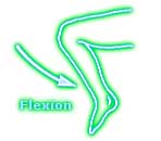
The ligaments and menisci provide static stability and the muscles and tendons dynamic stability. The main movement of the knee is flexion - extension. For that matter, knee act as a hinge joint, whereby the articular surfaces of the femur roll and glide over the tibial surface.During flexion and extension, tibia and patella act as one structure in relation to the femur.
The quadriceps muscle group is made up of four different individual muscles. They join together forming one single tendon which inserts into the anterior tibial tuberosity. Imbelded in the tendon is patella, a triangular sesamoid bone and its function is to increase the efficiency of the quadriceps contractions. Contraction of the quadriceps pulls the patella upwards and extends the knee. Range of motion: extension 0o.
The hamstring muscle group consist m.biceps femoris, m.semitendinosus and m.semimembranosus. They are situated at the back of the thigh and their function is flexing or bending the knee as well as providing stability on either side of the joint line. Range of motion: flexion 140o.
Secondary movement isinternal - external rotation of the tibia in relation to the femur, but it is possible only when the knee is flexed.
| [6] | [7] |
Closed Packed Position:[edit | edit source]
Near full extension. There is a medial rotation of the femoral condyles on the tibial plateau[8].
Open Packed Position:[edit | edit source]
Midrange flexion[9].
Clinical Examination[edit | edit source]
- Knee Examination
- Special Tests
- Outcome Measures
Conditions
[edit | edit source]
- Anterior Cruciate Ligament Injury
- Articular Cartilage Lesions
- Baker's Cyst
- Chondromalacia Patellae
- Femoral Fractures
- Hamstring Strain
- Iliotibial Band Syndrome
- Knee Osteoarthritis
- Knee Bursitis
- Lateral Collateral Ligament Injury
- Medial Collateral Ligament Injury
- Meniscal Lesions
- Ober's Test
- Osteochondritis Dissecans of the Knee
- Osgood-Schlatter's Disease
- Plica Syndrome
- Quadriceps Muscle Strain
- Quadricpes Muscle Contusion
- Patellofemoral Pain Syndrome
- Patellofemoral Instability
- Patellar Tendinitis
- Patellar Fractures
- Pes Anserinus Bursitis
- Plica Syndrome
- PrePatellar Bursitis
- Popliteus Tendinitis
- Posterior Cruciate Ligament Injury
- Sinding Larsen Johansson Syndrome
- Tibial Plateau Fractures
Procedures
[edit | edit source]
- Arthroscopic Meniscectomy
- Meniscal Repair
- Articular Cartilage Debridement & Microfracture
- ACL Reconstruction
- Patellar Tendon Autograph
- Hamstring Tendon Autograph
- Allograph
- Patellar Tendon Autograph
- PCL Reconstruction
- ORIF Post-Fracture
- Arthroscopic Debridement & Manipulation
- Lateral Release of the Patella
- Tibial Osteotomy
Interventions[edit | edit source]
Recent Related Research (from Pubmed)[edit | edit source]
Reference[edit | edit source]
- ↑ Keith L. Moore. Clinically Oriented Anatomy 6e edition. P 634
- ↑ Dr. Michael D. Chivers. Anatomy and physical examination of the knee menisci: a narrative review of the orthopedic literature. JCCA 2009. (level A1)
- ↑ Keith L. Moore. Clinically Oriented Anatomy 6e edition. P 636
- ↑ Lam MH et al. Knee rotational stability during pivoting movement is restored after anatomic double-bundle anterior cruciate ligament reconstruction. Am J Sports Med. 2011. (level b)
- ↑ Shingo Fukagawa. Posterior Displacement of the Tibia Increases in Deep Flexion of the Knee. 2009. (level C)
- ↑ Anatomy Zone. Knee Joint - Part 1 - 3D Anatomy Tutorial. Available from: http://www.youtube.com/watch?v=ve448qTT_-4 [last accessed 21/09/14]
- ↑ Anatomy Zone. Knee Joint - Part 2 - 3D Anatomy Tutorial. Available from: http://www.youtube.com/watch?v=58g4nWqbHAc [last accessed 21/09/14]
- ↑ Lentell G. The effect of knee position on torque output during inversion and eversion movements at the ankle. J Orthop Sports Phys Ther. 1988.
- ↑ Lentell G. The effect of knee position on torque output during inversion and eversion movements at the ankle. J Orthop Sports Phys Ther. 1988.
</div></div></div>
</div>


