Running Mechanics for Clinicians: Difference between revisions
No edit summary |
No edit summary |
||
| Line 92: | Line 92: | ||
Bramah et al found that similar mechanical patterns were associated with multiple injuries<ref name=":0">Bramah C, Preece SJ, Gill N, Herrington L. Is there a pathological gait associated with common soft tissue running injuries?. The American journal of sports medicine. 2018 Oct;46(12):3023-31.</ref>. | Bramah et al found that similar mechanical patterns were associated with multiple injuries<ref name=":0">Bramah C, Preece SJ, Gill N, Herrington L. Is there a pathological gait associated with common soft tissue running injuries?. The American journal of sports medicine. 2018 Oct;46(12):3023-31.</ref>. | ||
Looking from the saggital plan, we can identify the following patterns: | '''Looking from the saggital plan, we can identify the following patterns:''' | ||
1- Foot Inclination Angle at Initial Contact:by drawing lines to compare the angle between the sole of the shoes and the treadmill. A great angle indicates greater foot inclination. It can potentially be caused by rear foot strike if the runner's toes are too high compared to the heel or a forefoot strike when the inclincation is mainly due to a high angle at the heel. Neither strikes are considered to be superior to the other. Landing with high inclination will limit the ability to engage in dorsiflexion which serves as shock absorption. High inclined foot will take longer time to get the foot flat on floor to start the shock absorption mechanics resulting in high impact vertical loading<ref name=":1">Souza RB. An evidence-based videotaped running biomechanics analysis. Physical medicine and rehabilitation clinics. 2016 Feb 1;27(1):217-36.</ref>. [https://www.ncbi.nlm.nih.gov/pmc/articles/PMC4714754/figure/F3/ Fig 3 in the study by Souza RB] shows how to identify foot inclination. | 1- Foot Inclination Angle at Initial Contact:by drawing lines to compare the angle between the sole of the shoes and the treadmill. A great angle indicates greater foot inclination. It can potentially be caused by rear foot strike if the runner's toes are too high compared to the heel or a forefoot strike when the inclincation is mainly due to a high angle at the heel. Neither strikes are considered to be superior to the other. Landing with high inclination will limit the ability to engage in dorsiflexion which serves as shock absorption. High inclined foot will take longer time to get the foot flat on floor to start the shock absorption mechanics resulting in high impact vertical loading<ref name=":1">Souza RB. An evidence-based videotaped running biomechanics analysis. Physical medicine and rehabilitation clinics. 2016 Feb 1;27(1):217-36.</ref>. [https://www.ncbi.nlm.nih.gov/pmc/articles/PMC4714754/figure/F3/ Fig 3 in the study by Souza RB] shows how to identify foot inclination. | ||
| Line 102: | Line 102: | ||
2- Knee flexion angle at mid stance: compare a straight line drawn through femur to the floor to a line form the lateral condyle of femur to lateral malleoulus [https://www.ncbi.nlm.nih.gov/pmc/articles/PMC4714754/figure/F5/ (Figure).] A greater angle indicates more knee flexion. Injured runners tend to land with more knee extension at intial contact<ref name=":0" /> This influences tissue stress and the ability to absorb shocks. | 2- Knee flexion angle at mid stance: compare a straight line drawn through femur to the floor to a line form the lateral condyle of femur to lateral malleoulus [https://www.ncbi.nlm.nih.gov/pmc/articles/PMC4714754/figure/F5/ (Figure).] A greater angle indicates more knee flexion. Injured runners tend to land with more knee extension at intial contact<ref name=":0" /> This influences tissue stress and the ability to absorb shocks. | ||
During running, knee and ankle function as suspensions. Landing with knee in flexion and foot flat engages the suspension spring from the moment the foot touches the ground<ref name=":1" />. On the other hand, runners with extended knee and high inclined foot at intial contact are less likely to engage the shock absorption mechanism within knee and ankle resulting in higher shocks. This places greater eccentric demands on the quadriceps and is linked to the development of PFPS<ref>Dierks TA, Manal KT, Hamill J, Davis I. Lower extremity kinematics in runners with patellofemoral pain during a prolonged run. Medicine and science in sports and exercise. 2011 Apr;43(4):693-700.</ref>. The body can respond by compensating on different levels leading to further complications. From a managemenet prespective, this can e addressed by gait re-education to alter the mechanical pattern and/or eccentric training of quadriceps to meet the shock absorption demands. | During running, knee and ankle function as suspensions. Landing with knee in flexion and foot flat engages the suspension spring from the moment the foot touches the ground<ref name=":1" />. On the other hand, runners with extended knee and high inclined foot at intial contact are less likely to engage the shock absorption mechanism within knee and ankle resulting in higher shocks. This places greater eccentric demands on the quadriceps and is linked to the development of PFPS<ref>Dierks TA, Manal KT, Hamill J, Davis I. Lower extremity kinematics in runners with patellofemoral pain during a prolonged run. Medicine and science in sports and exercise. 2011 Apr;43(4):693-700.</ref>. The body can respond by compensating on different levels leading to further complications. From a managemenet prespective, this can e addressed by gait re-education to alter the mechanical pattern and/or eccentric training of quadriceps to meet the shock absorption demands. | ||
'''Frontal plane:''' | |||
1- Trunk side flexion : the angle between a vertical line drawn through the central line (starting mid-way between Posterior superior iliac spines PSISs upwards through the trunk) and a vertical line starting between PSIS to C7. A greater angle indicates greater side flexion. There is no evidence suggesting an associated pathology with increased trunk side flexion, however, it may indicates compensation from a dysfunction in a distal joint. As the trunk shifts greatly side to side it shifts the body's centre of mass COM. This could result in excessive pelvis drop opposite to the wight bearing leg. To address this patttern we need to think of possible causes for trunk shifting as a compensation to off-load hip muscles by shifting COM away. | |||
2- contralateral pelvic drop away from weigh-bearing leg (hip dip): an angle between a horizontal line between PSISs and another horizontal line across the body. [https://journals.sagepub.com/doi/full/10.1177/0363546518793657?url_ver=Z39.88-2003&rfr_id=ori%3Arid%3Acrossref.org&rfr_dat=cr_pub%3Dpubmed Refer to (Figure3) in this study.] Healthy runners show some degree of pelvic drop, ranging from 3-4 degrees but it's usually controlled. Injured runners demonestrates contralateral pelvic drop compared to healthy runners which refers to a link between this pattern and multiple injuries. Different compensations can be expected to keep the body's balance as the COM is shifting away such as increased hip adduction resulting in different presentations such as ITB, patellar maltracking and/or PFPS<ref name=":0" />. | |||
== References == | == References == | ||
<references /> | <references /> | ||
Revision as of 21:09, 6 October 2019
Original Editor - Mariam Hashem
Top Contributors - Mariam Hashem, Kim Jackson, Tarina van der Stockt, Jess Bell and Robin Tacchetti
This article or area is currently under construction and may only be partially complete. Please come back soon to see the finished work!
Overview of Running Injuries[edit | edit source]
Lower extremity running-related injuries range from 19.4 to 79.3 percent[1]. The most common injnuries are[2]:
- Patellofemoral pain
- Medial tibial stress syndrome (shin splints)
- Achilles tendinopathy
- Iliotibial band syndrome
- Plantar fasciitis
- Stress fractures of the metatarsals and tibia
Hamstrings and calf problems were reported by men marathon runners, while hip pain problems were common among women[3]. Most of these injuries have a high recurrence rates.
A 2015 systematic review[4] of 15 studies identified different risk factors for women and men:
| Women | Men contributing factors to tissue stress | |
|---|---|---|
| Age | History of previous injury | |
| History of previous sports activity | Running experience for 2 years | |
| running on a concrete surface | History of previous injury | |
| Participating in a marathon | Average weekly running distance (20–29 miles) | |
| weekly running distance (30–39 miles) | ||
| wearing running shoes for 4 to 6 months |
Stress Frequency Model[edit | edit source]
Running Injuries are caused by an inter-relation of multiple factors. A simple injury causation model using the stress frequency curve can help us to develop an idea on the contributing factors and how to address them in the management plan.
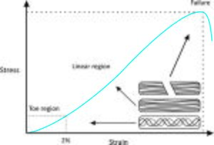
A tissue is influenced by the applied stress and the frequency of application. If the stress and it's frequency are below the injury threshold, the tissue will function normally within it's capacity. However, if either or both of these factors exceeded the injury threshold, the tissue is moer likely to be injured. This explains why some runners may not experience injury for long time and develop one as soon as they increase their frequency of training such as training for a marathon.
Considering stress and frequecny is important to understand individual tissue's capacity.
Running mechanics influence the stress applied to the body, magnitude, type (bending, shear or tension) and the speed of application on each foot contact. Assessing running mechanics leads us to think about the stressed structure and explains the presented symptoms. For example; landing on toes -forefoot strike- results in greater stresses on achillies tendons and the calf muscle forces.
Assessing frequency of running and training volume to understand the effect of the accumulated tissue stress . If falling below the tissue's threshold the likelihood of developing injury will be low and vice versa. Subjective examination can help us to understand the frequency, ask your client about them about their weekly training and what is your standard trainig like? How much running they do? what wa the frequency of applied stress before the injury occured?
the next thing is wehre is identifying the injury threashold which refers to the interaction between the tissue capacity to tolerate the stress an the tissue capacity to tolerate the frequncy of the applied stress. Tissue capacity refers to the functional capabilities of a specific tiussue to cope with stress type and frequency. A muscle capable of producing high peak force may be able to tolerate high level of stress on an individual foot contact. On the contrary , if the mscule's capability is low, applying stress with high frequency the muscle may not be able to cope well leading to injuries. To translate this into practical application, when assessing a runner we should think of adjusting the mechanical pattern or push up the tissue's endurance to tolerate the applied load. Lowering the applied stress by reducing the amoint of running can be a method of off-loading the injured tissue while building up the tissue resileince to cope with the functional aspiration.
Clinical Running Assessment Set Ups[edit | edit source]
Many of the common running-related biomechanical patterns can be identified by 2D analysis using inexpensive tools[1] such as a mobile phone or tablet camera. Satndaridizing the method of assessment is improtant for accuracy of identifying the patterns and to make sure your findings are not due to viewing angles.
Settings:
Tools/equipment: High speed camera or mobile phone/table camera and a tripod.
Distance: 1.5-2 meters from the treadmill
Height:0.8-1 meter-pelvic height
Views: side (saggital) and rear (frontal)
Timing: initial contact and mid-stance
Joints/regions: thorax, pelvis. hips, knees and ankles.
Follow a structured process of assessment by looking at one joint/region at a time. To end up with a structured problem list, slow down the speed of the camera to allow you to go backward and forward and take still pictures to draw lines and identify areas of stress.
Common Mechanical Patterns[edit | edit source]
Bramah et al found that similar mechanical patterns were associated with multiple injuries[6].
Looking from the saggital plan, we can identify the following patterns:
1- Foot Inclination Angle at Initial Contact:by drawing lines to compare the angle between the sole of the shoes and the treadmill. A great angle indicates greater foot inclination. It can potentially be caused by rear foot strike if the runner's toes are too high compared to the heel or a forefoot strike when the inclincation is mainly due to a high angle at the heel. Neither strikes are considered to be superior to the other. Landing with high inclination will limit the ability to engage in dorsiflexion which serves as shock absorption. High inclined foot will take longer time to get the foot flat on floor to start the shock absorption mechanics resulting in high impact vertical loading[7]. Fig 3 in the study by Souza RB shows how to identify foot inclination.
Conversely, landing with forefoot strike (on tip-toes) allows less time to engage in dorsiflexion utilizing the calf complex and possibly stressing the achillies tendon. Refer to this link to see the difference between different foot strike patterns.
In the management plan, a relatively low inclincation angle where foot is low to the ground regardless of the type of strike (heel or toe) minimzes the stress on achillies for forefoot runners or engage dorsiflexion for heelstrike runners.
2- Knee flexion angle at mid stance: compare a straight line drawn through femur to the floor to a line form the lateral condyle of femur to lateral malleoulus (Figure). A greater angle indicates more knee flexion. Injured runners tend to land with more knee extension at intial contact[6] This influences tissue stress and the ability to absorb shocks.
During running, knee and ankle function as suspensions. Landing with knee in flexion and foot flat engages the suspension spring from the moment the foot touches the ground[7]. On the other hand, runners with extended knee and high inclined foot at intial contact are less likely to engage the shock absorption mechanism within knee and ankle resulting in higher shocks. This places greater eccentric demands on the quadriceps and is linked to the development of PFPS[8]. The body can respond by compensating on different levels leading to further complications. From a managemenet prespective, this can e addressed by gait re-education to alter the mechanical pattern and/or eccentric training of quadriceps to meet the shock absorption demands.
Frontal plane:
1- Trunk side flexion : the angle between a vertical line drawn through the central line (starting mid-way between Posterior superior iliac spines PSISs upwards through the trunk) and a vertical line starting between PSIS to C7. A greater angle indicates greater side flexion. There is no evidence suggesting an associated pathology with increased trunk side flexion, however, it may indicates compensation from a dysfunction in a distal joint. As the trunk shifts greatly side to side it shifts the body's centre of mass COM. This could result in excessive pelvis drop opposite to the wight bearing leg. To address this patttern we need to think of possible causes for trunk shifting as a compensation to off-load hip muscles by shifting COM away.
2- contralateral pelvic drop away from weigh-bearing leg (hip dip): an angle between a horizontal line between PSISs and another horizontal line across the body. Refer to (Figure3) in this study. Healthy runners show some degree of pelvic drop, ranging from 3-4 degrees but it's usually controlled. Injured runners demonestrates contralateral pelvic drop compared to healthy runners which refers to a link between this pattern and multiple injuries. Different compensations can be expected to keep the body's balance as the COM is shifting away such as increased hip adduction resulting in different presentations such as ITB, patellar maltracking and/or PFPS[6].
References[edit | edit source]
- ↑ Van Gent RN, Siem D, van Middelkoop M, Van Os AG, Bierma-Zeinstra SM, Koes BW. Incidence and determinants of lower extremity running injuries in long distance runners: a systematic review. British journal of sports medicine. 2007 Aug 1;41(8):469-80.
- ↑ Callahan LR, Sheon RP. Overview of running injuries of the lower extremity. UpToDate, Grayzel J.(Accessed on July 06, 2017). 2002.
- ↑ Fredericson M, Misra AK. Epidemiology and aetiology of marathon running injuries. Sports Medicine. 2007 Apr 1;37(4-5):437-9.
- ↑ Van der Worp MP, Ten Haaf DS, van Cingel R, de Wijer A, Nijhuis-van der Sanden MW, Staal JB. Injuries in runners; a systematic review on risk factors and sex differences. PLoS One. 2015 Feb 23;10(2):e0114937.
- ↑ How to get your running gait analysed. Available from: https://www.youtube.com/watch?v=gmBz3QC5JAg. [last access:06/10/2019]
- ↑ 6.0 6.1 6.2 Bramah C, Preece SJ, Gill N, Herrington L. Is there a pathological gait associated with common soft tissue running injuries?. The American journal of sports medicine. 2018 Oct;46(12):3023-31.
- ↑ 7.0 7.1 Souza RB. An evidence-based videotaped running biomechanics analysis. Physical medicine and rehabilitation clinics. 2016 Feb 1;27(1):217-36.
- ↑ Dierks TA, Manal KT, Hamill J, Davis I. Lower extremity kinematics in runners with patellofemoral pain during a prolonged run. Medicine and science in sports and exercise. 2011 Apr;43(4):693-700.


