Medial Collateral Ligament of the Knee
Original Editor - Rebecca Wilson
Top Contributors - George Prudden, Kim Jackson, 127.0.0.1, WikiSysop, Vidya Acharya, Rucha Gadgil, Saimat Lachinova and Lucinda hampton
Description[edit | edit source]
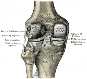
The MCL is one of four major ligaments that supports the knee. This ligament is a strong broad band[1] found on the inner aspect of the knee joint and is the largest structure situated on the medial side.[2] The MCL is understood as being the most common ligament injury of the knee.[3] This structure is divided into superficial and deep ligaments. The superficial ligament is also known as the tibiofemoral ligament[2] whereas the deep ligament is identified as the mid-third capsular ligament.[4]
Attachments[edit | edit source]
The superficial medial collateral ligament (sMCL) has one femoral and two tibial attachments.[2] The femoral attachment is situated on the medial epicondyle. The proximal attachment blends into the semimembranosus tendon and the insertion of the distal attachment is at the posteromedial crest of the tibia.[2]
The Deep medial ligament (dMCL) is divided into two, the meniscofemoral and meniscotibial ligaments.[5] The origin of the meniscofemoral comes from the femur just distal to the superficial medial collateral, inserting into the medial menisci.[5] The meniscotibial ligament is thicker and shorter with an attachment forming on the distal edge of the articular cartilage of the medial tibial plateau[2] coming from the medial meniscus.[5]
Function[edit | edit source]
The medial collateral ligament is recognised as being a primary static stabiliser of the knee[3][6] and assists in passively stabilising the joint. When stress is applied this ligament aids control in transferring the joint through a normal range of movement.[7] The MCL also prevents an anterior movement of the tibia and hyperextension.[1] The ligaments role also includes joint proprioception, when stretched beyond ability or are exposed to an excessive load, proprioceptive feedback generates a muscle contraction.[7] The sMCL resists valgus forces applied to the knee through all degrees of flexion with the deep medial collateral acting as a secondary resistance.[5] Specifically the primary valgus stabilizer is identified as the proximal division of the sMCL.[8] The dMCL assists the knee in rotational stability primarily in extension moving through into early flexion.[9]
Clinical Relevance[edit | edit source]
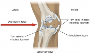
Through the predominance in injury to the MCL it is important to understand the functions of the structure so that the correct procedure or treatment can be applied. Injuries to the MCL can have detrimental effects to surrounding structures. It is recognised that either partial or complete ruptures in the ligament significantly increases the load on the ACL. Partial tears show that increases in ACL load were identified at 30 degree knee flexion and valgus load and internal torque.[10]
Assessment[edit | edit source]
Palpation[edit | edit source]
The anterior aspect of the ligament can be palpated moving vertically, roughly midway along the medial joint line.[1]
Special test[edit | edit source]
The valgus stress test allows the therapist to examine the medial aspect of the joint for any laxity. An increase in laxity and joint space usually distinguishes damage to the medical collateral ligament.[11] The patient should be positioned supine. By performing the test with the knee in approximately 30 degrees flexion rather than extension, this is due to the fact that flexion helps to relax surrounding structures including the posterior capsule. This ensures there is isolated testing of the MCL.[12] Therapists position one hand on the lateral aspect of the joint line of the knee with the other hand on the medial aspect of the ankle. A valgus force is then applied, a positive result of the knee in this position would be an increase in joint space medially.[12]
Resources[edit | edit source]
See also[edit | edit source]
- Knee
- Anterior cruciate ligament
- Medial meniscus
- MCL injuries
- Pellegrini-Stieda syndrome
- Diagnostic imaging of the knee
References
[edit | edit source]
- ↑ 1.0 1.1 1.2 Atkins, E., Kerr, J. & Goodland, E., 2015. A Practical Approach to Musculoskeletal Medicine: Assessment, Diagnosis, Treatment. 4th ed. China: Elsevier.
- ↑ 2.0 2.1 2.2 2.3 2.4 Laprade, R. F., et al., 2007. The Anatomy of the Medial Part of the Knee. Journal of Bone and Joint Surgery [online]. 89(9), pp. 2000-2010. [viewed 12 September 2016]. Available from: http://jbjs.org/content/89/9/2000
- ↑ 3.0 3.1 Chen, L. et al., 2008. Medial collateral ligament injuries of the knee: current treatment concepts. Current Reviews in Musculoskeletal Medicine [online]. 1(2), pp. 108-113. [viewed 12 September 2016]. Available from: http://www.ncbi.nlm.nih.gov/pmc/articles/PMC2684213/
- ↑ Phisitkul, P. et al., 2006. MCL Injuries of the Knee: Current Concepts Review. The IOWA Orthopaedic Journal [online]. 26, pp. 77-90. [viewed 12 September 2016]. Available from: http://www.ncbi.nlm.nih.gov/pmc/articles/PMC1888587/
- ↑ 5.0 5.1 5.2 5.3 Duffy, P. & Miyamoto, R. G. 2010. Management of Medial Collateral Ligament Injuries in the Knee: An Update and Review. The Physician and Sportsmedicine [online]. 38(2), pp. 39-54. [viewed 12 September 2016]. Available from: http://www.ncbi.nlm.nih.gov/pubmed/20631463
- ↑ Markatos, K. & Tzagk, G. 2016. The anatomy of the medial collateral ligament of the knee and its significance in joint stability. Journal of Anatomy and Embryology [online]. 121(2), pp. 198-204. [viewed 12 September 2016]. Available from: http://www.fupress.net/index.php/ijae/article/view/18495
- ↑ 7.0 7.1 Frank. C. B. 2004. Ligament structure, physiology and function. Journal of Musculoskeletal and Neuronal Interactions [online]. 4(2), pp. 199-201. [viewed 13 September 2016]. Available from: http://www.ncbi.nlm.nih.gov/pubmed/15615126
- ↑ Griffith, C. et al., 2009. Medial Knee Injury: Part 1, Static Function of the Individual Components of the Main Medial Knee Structures. The American Journal of Sports Medicine [online]. 37(9), pp. 1762-1770. [viewed 13 September 2016]. Available from: http://www.ncbi.nlm.nih.gov/pubmed/19609008
- ↑ Cavignac, E. et al., 2015. The Role of the Deep Medial Collateral Ligament in controlling rotational stability of the knee. Knee Surgery, Sports Traumatology, Arthoscopy [online]. 23(10), pp. 3101-3107. [viewed 20 September 2016]. Available from: http://www.ncbi.nlm.nih.gov/pubmed/24894123
- ↑ Battaglia, M. J. et al., 2009. Medial Collateral Ligament Injuries and Subsequent Load on the Anterior Cruciate Ligament: A Biomechnical Evaluation in a Cadaveric Model. The American Journal of Sports Medicine [online], 37(2), pp. 305-311. [viewed 13 September 2016]. Available from: http://www.ncbi.nlm.nih.gov/pubmed/19098154
- ↑ Luke, A., no date. Sports Medicine: Knee Physical Examination [online]. Department of Orthopedic Surgery. [viewed 14 September 2016]. Available from: http://orthosurg.ucsf.edu/patient-care/divisions/sports-medicine/conditions/physical-examination-info/knee-physical-examination/
- ↑ 12.0 12.1 Rossi, R. et al., 2011. Clinical examination of the knee: know your tools for diagnosis of knee injuries. BMC Sports Science, Medicine and Rehabilitation [online]. 3(25), pp. 1-10. [viewed 13 September 2016]. Available from: http://link.springer.com/article/10.1186/1758-2555-3-25


