Jefferson fracture: Difference between revisions
Bert Lasat (talk | contribs) No edit summary |
Kim Jackson (talk | contribs) No edit summary |
||
| (35 intermediate revisions by 10 users not shown) | |||
| Line 1: | Line 1: | ||
= | <div class="editorbox"> | ||
'''Original Editor '''- [[User:Bert Lasat|Bert Lasat]] | |||
'''Top Contributors''' - {{Special:Contributors/{{FULLPAGENAME}}}} | |||
</div> | |||
== Description == | |||
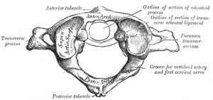
A Jefferson fracture is a bone [[fracture]] of the vertebra C1. The vertebra C1 is a bony ring, with two wedge-shaped lateral masses, connected by relatively thin anterior and posterior arches and a transverse ligament. The lateral mass on vertebra C1, who is taller, is directed laterally. Therefore, vertical forces compressing the lateral masses between the occipital condyles and the axis drive them apart, fracturing one or both of the anterior or posterior arches. The impact forces cause an outward spread of the lateral masses of C1. | |||
A Jefferson fracture doesn’t always result in a spinal cord injury, because the dimensions of the bony ring increase. [[Spinal Cord Injury|Spinal cord injury]] is more expected if the [[Transverse Ligament of the Atlas|transverse ligament]] has also been ruptured. | |||
= | == Clinically Relevant Anatomy == | ||
[[File:Altas-2.png|thumb]] | |||
The first vertebra of the Cervical spine is also called the [[atlas]]. The atlas has in contrast to the other vertebra no body. The upper surface of the atlas bears a superior articular facet on a thick lateral mass on each side. The lateral masses articulates with the occipital condyles<ref name="p2">Clinical Anatomy. 11e edition, Harold Ellis. p 325-326. 2006</ref>. | |||
== Epidemiology /Etiology == | |||
< | 10% of injuries to the cervical spine are fractures of the atlas and in 2% of all spinal injuries<ref name="p3" />. Everyone can have a cervical spine injury but it is rare in children, the incidence is only 1,9% to 9,5% of all cervical injuries<ref name="p3">Fracture of the atlas through a synchondrosis of the anterior arch complicated by atlantoaxial rotatory fixation in a four-yearold child. T. Hagino et al. 2006 British Editorial Society of Bone and Joint Surgery.</ref>. The injury in children due to falling at the playground. | ||
= | The Jefferson fracture occurs most likely because of a diving accident (striking the bottom of the pool) with hyperextension of the cervical spine or may result from an axial load on the posterior side of the head<ref name="p4">Clinically oriented anatomy. 6th edition. Keith L.Moore. 2010.</ref>. It may also result from an impact against the roof of a vehicle. | ||
The fracture can be complicated or can occur by an atlantoaxial rotator fixation or a strong rotation of the head; this is only reported in one adult case<ref>Fracture of the atlas through a synchondrosis of the anterior arch complicated by atlantoaxial rotatory fixation in a four-yearold child. T. Hagino et al. 2006 British Editorial Society of Bone and Joint Surgery.</ref>. | |||
== Clinical Presentation == | |||
There are different kinds of fractures of the Atlas. The study from Gebauer M. et al says that there are 4 types of fractures pending on the speed of axial pressure. They are different from each other because the place of the fracture isn’t the same. The 4 types are: | |||
#Type-I: anterior arch fracture. | |||
#Type-II: posterior arch fracture. | |||
#Type-III: anterior and posterior arch double fracture. | |||
#Type-IV: lateral mass fracture. | |||
The study says that the fractures result from an axial force on the vertebra at slow speed and at high speed. They used an axial force application at constant speeds of either 0,5mm/s or 300mm/s. The results show us that at slow speed 13 of 20 fractures where type-IV fractures. The results at high speed are Type-III fractures, burst fractures of 2 to 4 parts. Type-III fracture occurred in all 20 tested specimens. | |||
<br> | There conclusion is that the Type of atlas fracture depends on the speed of axial force impact<ref>Gebauer M et al. Biomechanical analysis of atlas fractures: a study on 40 human atlas specimens. Center for Biomechanics and Skeletal Biology UKE. 2008.</ref>.<br> | ||
= | {{#ev:youtube|xlv6Vo218Ps}} <ref>Radiology ChannelJefferson fracture - radiology video tutorial (x-ray, CT) Available from https://www.youtube.com/watch?time_continue=1&v=xlv6Vo218Ps&feature=emb_logo</ref> | ||
== Physical Therapy Management== | |||
Patients with a Jefferson fracture must be diagnosed for the injury with [[X-Rays|X-rays]] films. Patients may present with unstable or stable Jefferson fractures | |||
<br>In the study of Kesterson et al. four patients with an unstable fracture | Patients with an atlas fracture are most likely to undergo a surgical treatment. A case report found that the treatment of unstable burst fractures of the atlas (Jefferson fractures) is controversial. Unstable Jefferson fractures have been managed successfully with immobilization, typically halo traction or halo vest, or surgery<ref>Nonoperative Treatment of an Unstable Jefferson Fracture Using a Cervical Collar. Brian M et al. The Association of Bone and Joint Surgeons 2008</ref>. The study had a patient with an unstable fracture who was treated non-operatively with a cervical collar. The patient had frequent clinical examinations and flexion-extension radiographs and twelve months after treatment; the patient achieved a painless union of his fracture<ref>Nonoperative Treatment of an Unstable Jefferson Fracture Using a Cervical Collar. Brian M et al. The Association of Bone and Joint Surgeons 2008</ref>. <br>Patients with an unstable fracture needing surgical treatment can get the following surgeries: occiput-C2 wiring and fusion<ref>Evaluation and treatment of atlas burst fractures (Jefferson fractures). Lee Kesterson et al. Journal of Neurosurgery. August 1991.</ref>, C1-ring osteosynthesis<ref>A biomechanical rationale for C1-ring osteosynthesis as treatment for displaced Jefferson burst fractures with incompetency of the transverse atlantal ligament. Heiko Koller et al. Springer-Verlag 2010.</ref>, or anterior or posterior screw fixation<ref>Outcomes of C1 and C2 posterior screw fixation for upper cervical spine fusion. F. De Iure et al. Springer-Verlag 2009.</ref><ref>Ideal screw entry point and projection angles for posterior lateralfckLRmass fixation of the atlas: an anatomical study. Serkan Simsek et al. 31 July 2009.</ref>. <br>In the study of Kesterson et al. four patients with an unstable fracture were treated with the occiput-C2 wiring and fusion; patients with stable fracture were treated with Minerva jackets or [[Cervical Collar|rigid collar]] stabilization. The result of this study is that the patients had an excellent long-term result for spinal stability and the resolution of subjective complaint<ref>Evaluation and treatment of atlas burst fractures (Jefferson fractures). Lee Kesterson et al. Journal of Neurosurgery. August 1991.</ref>. | ||
== References == | |||
<references /> | |||
[[Category:Cervical Spine]] | |||
[[Category:Cervical Spine - Conditions]] | |||
[[Category:Fractures]] | |||
[[Category:Conditions]] | |||
Latest revision as of 17:46, 2 January 2021
Original Editor - Bert Lasat
Top Contributors - Bert Lasat, Kim Jackson, Priyanka Chugh, Rachael Lowe, Vidya Acharya, Admin, 127.0.0.1, Evan Thomas, Venugopal Pawar and WikiSysop
Description[edit | edit source]
A Jefferson fracture is a bone fracture of the vertebra C1. The vertebra C1 is a bony ring, with two wedge-shaped lateral masses, connected by relatively thin anterior and posterior arches and a transverse ligament. The lateral mass on vertebra C1, who is taller, is directed laterally. Therefore, vertical forces compressing the lateral masses between the occipital condyles and the axis drive them apart, fracturing one or both of the anterior or posterior arches. The impact forces cause an outward spread of the lateral masses of C1.
A Jefferson fracture doesn’t always result in a spinal cord injury, because the dimensions of the bony ring increase. Spinal cord injury is more expected if the transverse ligament has also been ruptured.
Clinically Relevant Anatomy[edit | edit source]
The first vertebra of the Cervical spine is also called the atlas. The atlas has in contrast to the other vertebra no body. The upper surface of the atlas bears a superior articular facet on a thick lateral mass on each side. The lateral masses articulates with the occipital condyles[1].
Epidemiology /Etiology[edit | edit source]
10% of injuries to the cervical spine are fractures of the atlas and in 2% of all spinal injuries[2]. Everyone can have a cervical spine injury but it is rare in children, the incidence is only 1,9% to 9,5% of all cervical injuries[2]. The injury in children due to falling at the playground.
The Jefferson fracture occurs most likely because of a diving accident (striking the bottom of the pool) with hyperextension of the cervical spine or may result from an axial load on the posterior side of the head[3]. It may also result from an impact against the roof of a vehicle.
The fracture can be complicated or can occur by an atlantoaxial rotator fixation or a strong rotation of the head; this is only reported in one adult case[4].
Clinical Presentation[edit | edit source]
There are different kinds of fractures of the Atlas. The study from Gebauer M. et al says that there are 4 types of fractures pending on the speed of axial pressure. They are different from each other because the place of the fracture isn’t the same. The 4 types are:
- Type-I: anterior arch fracture.
- Type-II: posterior arch fracture.
- Type-III: anterior and posterior arch double fracture.
- Type-IV: lateral mass fracture.
The study says that the fractures result from an axial force on the vertebra at slow speed and at high speed. They used an axial force application at constant speeds of either 0,5mm/s or 300mm/s. The results show us that at slow speed 13 of 20 fractures where type-IV fractures. The results at high speed are Type-III fractures, burst fractures of 2 to 4 parts. Type-III fracture occurred in all 20 tested specimens.
There conclusion is that the Type of atlas fracture depends on the speed of axial force impact[5].
Physical Therapy Management[edit | edit source]
Patients with a Jefferson fracture must be diagnosed for the injury with X-rays films. Patients may present with unstable or stable Jefferson fractures
Patients with an atlas fracture are most likely to undergo a surgical treatment. A case report found that the treatment of unstable burst fractures of the atlas (Jefferson fractures) is controversial. Unstable Jefferson fractures have been managed successfully with immobilization, typically halo traction or halo vest, or surgery[7]. The study had a patient with an unstable fracture who was treated non-operatively with a cervical collar. The patient had frequent clinical examinations and flexion-extension radiographs and twelve months after treatment; the patient achieved a painless union of his fracture[8].
Patients with an unstable fracture needing surgical treatment can get the following surgeries: occiput-C2 wiring and fusion[9], C1-ring osteosynthesis[10], or anterior or posterior screw fixation[11][12].
In the study of Kesterson et al. four patients with an unstable fracture were treated with the occiput-C2 wiring and fusion; patients with stable fracture were treated with Minerva jackets or rigid collar stabilization. The result of this study is that the patients had an excellent long-term result for spinal stability and the resolution of subjective complaint[13].
References[edit | edit source]
- ↑ Clinical Anatomy. 11e edition, Harold Ellis. p 325-326. 2006
- ↑ 2.0 2.1 Fracture of the atlas through a synchondrosis of the anterior arch complicated by atlantoaxial rotatory fixation in a four-yearold child. T. Hagino et al. 2006 British Editorial Society of Bone and Joint Surgery.
- ↑ Clinically oriented anatomy. 6th edition. Keith L.Moore. 2010.
- ↑ Fracture of the atlas through a synchondrosis of the anterior arch complicated by atlantoaxial rotatory fixation in a four-yearold child. T. Hagino et al. 2006 British Editorial Society of Bone and Joint Surgery.
- ↑ Gebauer M et al. Biomechanical analysis of atlas fractures: a study on 40 human atlas specimens. Center for Biomechanics and Skeletal Biology UKE. 2008.
- ↑ Radiology ChannelJefferson fracture - radiology video tutorial (x-ray, CT) Available from https://www.youtube.com/watch?time_continue=1&v=xlv6Vo218Ps&feature=emb_logo
- ↑ Nonoperative Treatment of an Unstable Jefferson Fracture Using a Cervical Collar. Brian M et al. The Association of Bone and Joint Surgeons 2008
- ↑ Nonoperative Treatment of an Unstable Jefferson Fracture Using a Cervical Collar. Brian M et al. The Association of Bone and Joint Surgeons 2008
- ↑ Evaluation and treatment of atlas burst fractures (Jefferson fractures). Lee Kesterson et al. Journal of Neurosurgery. August 1991.
- ↑ A biomechanical rationale for C1-ring osteosynthesis as treatment for displaced Jefferson burst fractures with incompetency of the transverse atlantal ligament. Heiko Koller et al. Springer-Verlag 2010.
- ↑ Outcomes of C1 and C2 posterior screw fixation for upper cervical spine fusion. F. De Iure et al. Springer-Verlag 2009.
- ↑ Ideal screw entry point and projection angles for posterior lateralfckLRmass fixation of the atlas: an anatomical study. Serkan Simsek et al. 31 July 2009.
- ↑ Evaluation and treatment of atlas burst fractures (Jefferson fractures). Lee Kesterson et al. Journal of Neurosurgery. August 1991.


