Functional Anatomy of the Hip - Neural and Vascular: Difference between revisions
Kim Jackson (talk | contribs) m (Kim Jackson moved page Hip Anatomy- Neural and Vascular to Functional Anatomy of the Hip - Neural and Vascular: More appropriate title) |
mNo edit summary |
||
| Line 8: | Line 8: | ||
== Nerve Supply == | == Nerve Supply == | ||
Hilton's law states “The same trunks of nerves, whose branches supply the groups of muscles moving a joint, furnish also a distribution of nerves to the skin over the insertions of the same muscles; and – what at this moment more especially merits our attention – the interior of the joint receives its nerves from the same source.” <ref>[https://www.ncbi.nlm.nih.gov/pmc/articles/PMC2717375/ John Hilton]</ref>A joint tends to be innervated by a branch of a motor nerve which also supplies a muscle extending and acting across the joint,another branch of the nerve often supplies the overlying skin. | Hilton's law states “The same trunks of nerves, whose branches supply the groups of muscles moving a joint, furnish also a distribution of nerves to the skin over the insertions of the same muscles; and – what at this moment more especially merits our attention – the interior of the joint receives its nerves from the same source.” <ref>[https://www.ncbi.nlm.nih.gov/pmc/articles/PMC2717375/ John Hilton]</ref>A joint tends to be innervated by a branch of a motor nerve which also supplies a muscle extending and acting across the joint,another branch of the nerve often supplies the overlying skin. | ||
* The [[Obturator Nerve|obturator nerve]] is considered the primary source of innervation to the hip. | * The [[Obturator Nerve|obturator nerve]] is considered the primary source of innervation to the hip. | ||
Revision as of 15:30, 2 March 2022
Original Editor - Rishika Babburu
Top Contributors - Rishika Babburu, Ewa Jaraczewska and Kim Jackson
Introduction[edit | edit source]
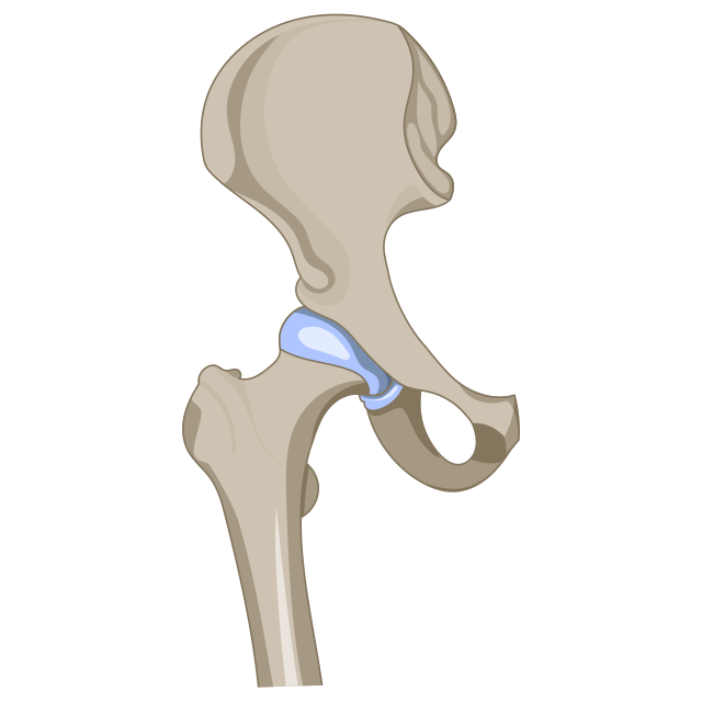
The hip joint is a synovial joint with articulation between the femoral head and the acetabulum of the pelvis. The rounded femoral head sits within the cup-shaped acetabulum of pelvis.
Nerve Supply[edit | edit source]
Hilton's law states “The same trunks of nerves, whose branches supply the groups of muscles moving a joint, furnish also a distribution of nerves to the skin over the insertions of the same muscles; and – what at this moment more especially merits our attention – the interior of the joint receives its nerves from the same source.” [1]A joint tends to be innervated by a branch of a motor nerve which also supplies a muscle extending and acting across the joint,another branch of the nerve often supplies the overlying skin.
- The obturator nerve is considered the primary source of innervation to the hip.
- Branches of the femoral and sciatic nerves contribute to its sensory innervation.[2]
- The hip joint receives multiple innervations primarily involving the hip capsule.
- The posterior articular nerve, a branch of the nerve to the quadratus femoris provides the most extensive nerve supply to the hip joint, including the posterior and inferior regions of the capsule and the ischiofemoral ligament.
- Superiorly, the hip capsule is innervated by the superior gluteal nerve.
- Anterior innervation of the capsule is provided by direct branches of the femoral nerve.
- The anteromedial and anteroinferior regions are supplied by the medial articular nerve, which arises from the anterior division of the obturator nerve .
- The ligamentum teres is innervated by the posterior branch of the obturator nerve .[3] [4]
- Acetabular labrum consists of sensory nerve end organs and ramified free nerve endings, suggest that the labrum may provide nociceptive and proprioceptive feedback to and from the hip joint [5]
Arterial Supply[edit | edit source]
Arterial supply of hip joint is by the cruciate and trochanteric anastomoses supply . A branch from the posterior branch of the obturator artery may also be present in the ligamentum teres.
Cruciate anastomosis
- Transverse branch of medial circumflex femoral artery
- Transverse branch of lateral circumflex femoral artery
- Ascending branch of first perforator artery from profunda femoris artery
- Descending branch of inferior gluteal artery
- Obturator artery
Trochanteric anastomosis
- Descending branch of superior gluteal artery
- Ascending branch of medial circumflex femoral artery
- Ascending branch of lateral circumflex femoral artery
- Inferior gluteal artery[6][7]
Clinical Importance[edit | edit source]
Avascular necrosis of the hip is a condition characterized by the death of the femoral head due to disruption of its vascular supply, mainly by Medial Femoral Circumflex Artery. It leads to insidious onset of hip pain, usually with movement, and can be the result of a variety of factors. Some of the most common causes can be remembered by the mnemonic “ASEPTIC”:
- Alcohol/AIDS
- Steroids, sickle cell disease, SLE
- Erlenmeyer flask (Gaucher disease)
- Pancreatitis
- Trauma
- Idiopathic (Legg-Calve-Perthes disease), infection, iatrogenic
- Caisson disease[8]
Neurologic injury usually is associated with traumatic dislocation and fracture-dislocation of the hip. The sciatic nerve, usually the peroneal branch, is most often injured, and this complication can be seen after all types of posterior fracture-dislocations and simple posterior dislocations. The sciatic nerve can be acutely lacerated, stretched, or compressed, or later encased in heterotopic ossification. Neurologic examination at the time of injury often is difficult but is extremely important. Rehabilitation of patients with sciatic nerve injury must begin as early as possible and should focus on the prevention of an equinus foot deformity. [10]
Intramuscular injection is one of the complications and the sciatic nerve is the most frequently affected nerve, especially in children, the elderly and underweight patients. The neurological presentation may range from minor transient pain to severe sensory disturbance and motor loss with poor recovery. Management of nerve injection injury includes drug treatment of pain, physiotherapy, use of assistive devices and surgical exploration. Early recognition of nerve injection injury and appropriate management are crucial in order to reduce neurological deficit and to maximize recovery. If the injection has to be administered into the gluteal muscle, the ventrogluteal region (gluteal triangle) has a more favourable safety profile than the dorsogluteal region (the upper outer quadrant of the buttock).[11]
References[edit | edit source]
- ↑ John Hilton
- ↑ Hip Anatomy
- ↑ Robbins CE (1998) Anatomy and biomechanics, In: The Hip Handbook edited by Fagerson TL (Eds.); Boston, MA: Butterworth-Heinemann, p. 1-37.
- ↑ Harty M (1984) The anatomy of the hip joint. In: Surgery of the Hip Joint, (2nd edn); edited by Tronzo R, Springer-Verlag, New York, p. 49-74.
- ↑ The nerve endings of the acetabular labrum Y T Kim , H Azuma
- ↑ Hip Joint: Embryology, Anatomy and Biomechanics Volume 12 - Issue 3 Ahmed Zaghloul1* and Elalfy M Mohamed2
- ↑ Hip arthroscopy for lateral cam morphology: how important are the vessels? Austin E Wininger, Lindsay E Barter, Nickolas Boutris, Luis F Pulido, Thomas J Ellis, Shane J Nho, Joshua D Harris
- ↑ Anatomy, Abdomen and Pelvis, Hip Arteries James A. Jordan; Bracken Burns.
- ↑ Avascular Necrosis, Blood Supply Femoral Head- Everything You Need To Know - Dr. Nabil Ebraheim. Avaialble from :https://www.youtube.com/watch?v=Qe0MxGuZC78
- ↑ Nerve injury in traumatic dislocation of the hip
- ↑ Sciatic nerve injection injury
- ↑ Glute injection - Everything You Need To Know - Dr. Nabil Ebraheim. Available from:https://www.youtube.com/watch?v=AxKEJQg6lB8


