Femoral Stress Fracture: Difference between revisions
(added Sports Medicine category) |
No edit summary |
||
| (36 intermediate revisions by 6 users not shown) | |||
| Line 2: | Line 2: | ||
'''Original Editors ''' - [[User:Matthias Verstraelen|Matthias Verstraelen]] as part of the [[Vrije Universiteit Brussel Evidence-based Practice Project|Vrije Universiteit Brussel's Evidence-based Practice project]] | '''Original Editors ''' - [[User:Matthias Verstraelen|Matthias Verstraelen]] as part of the [[Vrije Universiteit Brussel Evidence-based Practice Project|Vrije Universiteit Brussel's Evidence-based Practice project]] | ||
'''Top Contributors''' - {{Special:Contributors/{{FULLPAGENAME}}}} | '''Top Contributors''' - {{Special:Contributors/{{FULLPAGENAME}}}} | ||
</div> | </div> | ||
== | == Introduction == | ||
[[File:Right-femoral-stress-fracture.png|thumb|Bone scan: R FSSF]] | |||
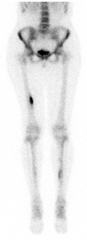
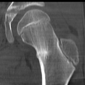
Femoral [[Stress Fractures|stress fractures]] occur in two different regions namely: | |||
# Femoral shaft stress fracture (FSSF): overuse injury in which abnormal stresses are placed on [[Bone Cortical And Cancellous|cancellous bone]] of the [[Femur|femoral]] shaft resulting in microfractures. Most common in young athletic individuals.<ref name=":1">orthobullets Femoral Stress Fractures Available;https://www.orthobullets.com/knee-and-sports/3111/femoral-shaft-stress-fractures (accessed 12.12.2022)</ref> | |||
# Femoral Neck Stress Fracture (FNSF): caused by repetitive loading of the femoral neck that leads to either compression side (inferior-medial neck) or tension side (superior-lateral neck) stress fractures. Most commonly occur in young athletes and military personal . <ref>Orthobullets Femoral Neck Stress Fractures Available:https://www.orthobullets.com/knee-and-sports/3110/femoral-neck-stress-fractures (accessed 12.12.2022)</ref> | |||
Femoral stress fractures can be hard to diagnose. Symptoms are often mild at first, similar to a strained muscle. When the patient doesn’t adapt his or her training, certain stress fractures could lead to complications, even to the point of complete [[Femoral Fractures|femoral fractures]] of the head or shaft <ref>Patel D, Roth M, Kapil N. Stress fractures: diagnosis, treatment and prevention. American Family Physician Jan. 2011; 83: 39-46. Level of evidence: 1A</ref><ref>Zadpoor A, Nikooyan A. The relationship between lower-extremity stress fractures and the ground reaction force: a systematic review. Clinical Biomechanics 2011; 26: 23 -28. Level of evidence: 1B</ref>. | |||
== Etiology == | |||
Occurs through fissure propagation in bone. Repetitive loads, exceeding the threshold of intrinsic bone healing either due to: repetitive stress on normal bone fatigue fracture); repetitive stress on abnormal bone ([[Insufficiency Fracture|insufficiency fracture]]).<ref name=":1" /> | |||
== | == Epidemiology == | ||
The risk factors are as follows: <ref name="Patel">Patel D, Roth M, Kapil N. Stress fractures: diagnosis, treatment and prevention. American Family Physician Jan. 2011; 83: 39-46. (Level of evidence 1A)</ref><ref name="Stress" /><ref name="Zadpoor" /><ref name="Anand">Anand A, Raviraj A, Kodikal G. Subchondral stress fractures of femoral head in healthy adult. Indian J Orthop. 2010 Oct-Dec; 44(4): 458-460. Level of evidence: 3B</ref> | # FNSF make up approximately 11% of stress injuries in athletes. The patient complains of hip or groin pain which is worse with [[weight bearing]] and [[Range of Motion|range of motion]] especially internal rotation. There are 2 types of FNSF: Tension-type FNSF involve the superior-lateral aspect of the neck and are at highest risk for complete fracture; thus, these should be detected early; Compression-type fractures are seen in younger athletes and involve the inferior-medial femoral neck. A trial of non-surgical management can be attempted for patients without a visible fracture line on radiographs in compression type injuries. This injury is common in runners. | ||
# FSSF: Well documented in the literature, and in one study among military recruits, they represented 22.5% of all stress fractures. Patients typically complain of poorly localised, insidious leg pain often mistaken for muscle injury. An exam is often non-focal, although the “[[Fulcrum Test|fulcrum test]]” test can be used by providers to localise the affected pain and suggest the diagnosis. If there is no evidence of a cortical break on imaging, a non-surgical approach can be attempted.<ref>Kiel J, Kaiser K. [https://www.ncbi.nlm.nih.gov/books/NBK507835/ Stress reaction and fractures.] InStatPearls [Internet] 2019 Jun 4. StatPearls Publishing. Available from: https://www.ncbi.nlm.nih.gov/books/NBK507835/ (last accessed 2.12.2019)</ref> | |||
== Risk Factors == | |||
[[File:Femoral-neck-stress-fracture.jpeg|thumb|CT: Femoral-neck-stress-fracture]] | |||
The risk factors are as follows: <ref name="Patel">Patel D, Roth M, Kapil N. Stress fractures: diagnosis, treatment and prevention. American Family Physician Jan. 2011; 83: 39-46. (Level of evidence 1A)</ref><ref name="Stress">Stress Fractures, information from your family doctor. Americain Family Physician Jan. 2011. Level of evidence: 5</ref><ref name="Zadpoor">Zadpoor A, Nikooyan A. The relationship between lower-extremity stress fractures and the ground reaction force: a systematic review. Clinical Biomechanics 2011; 26: 23 -28. (level of evidence 3A)</ref><ref name="Anand">Anand A, Raviraj A, Kodikal G. Subchondral stress fractures of femoral head in healthy adult. Indian J Orthop. 2010 Oct-Dec; 44(4): 458-460. Level of evidence: 3B</ref> | |||
*High-intensity training | *High-intensity training | ||
| Line 34: | Line 34: | ||
*< 3 times exercising/week | *< 3 times exercising/week | ||
*> 10 alcoholic drinks/week | *> 10 alcoholic drinks/week | ||
*Genetic factors <ref name="Korvala">Korvala J, Hartikka H, Pihlajamäki H, Solovieva S, Ruohola J-P, Sahi T, Barral S, Ott J, Ala-Kokko L, Männikkö M. Genetic predisposition for femoral neck stress fractures in military conscripts. BMC Genetics 2010, 11: 95. Level of evidence: 2B</ref> | *Genetic factors <ref name="Korvala">Korvala J, Hartikka H, Pihlajamäki H, Solovieva S, Ruohola J-P, Sahi T, Barral S, Ott J, Ala-Kokko L, Männikkö M. Genetic predisposition for femoral neck stress fractures in military conscripts. BMC Genetics 2010, 11: 95. Level of evidence: 2B</ref> | ||
*Change of surfaces (indoor track, frozen field) | *Change of surfaces (indoor track, frozen field) | ||
*Biomechanical imbalance (leg length, foot arch, forefoot varus, stance of foot and ankle) | *Biomechanical imbalance (leg length, foot arch, forefoot varus, stance of foot and ankle) | ||
=== Signs and Symptoms === | |||
*Local pain and oedema | |||
*Point tenderness on palpation | |||
*Local pain and | |||
*Point tenderness on | |||
*Local swelling | *Local swelling | ||
*Antalgic gait | *Antalgic gait | ||
| Line 51: | Line 46: | ||
*Pain increases during activity | *Pain increases during activity | ||
*Groin pain | *Groin pain | ||
*Bone marrow | *Bone marrow oedema<ref name="Anand" /><ref name="Niva">Niva M, Mattila V, Kiuru M, Pihlajamäki H. Bone stress Injuries are common in female military trainees. Clin Orthop Relat Res (2009) 467: 2962-2969. Level of evidence: 3A</ref><ref name="Ivkovic">Ivkovic A, Bojanic I, Pecina M. Stress fractures of the femoral shaft in athletes: a new treatment algorithm. Br J Sports Med 2006; 40: 518-520. (Level of evidence 2A)</ref><ref name="Schultz">Schultz, Houglum, Perrin. Third edition, examination of musculoskeletal injuries p.401. Human Kinetics. Level of evidence: 5</ref> | ||
=== Outcome Measures === | |||
# The [[Hop Test|hop test]] and tuning fork test could be used as diagnostic test but there is a lack of recent evidence for their validity. | |||
# Another test is the “fist” test, the therapist create a bilateral pressure on the anterior side of the femur starting at the distal part and moving to the proximal one. | |||
# The most valid test for the diagnosis is the [[Fulcrum Test|fulcrum-test]], while the therapist pushes to the dorsum of the knee <ref name="Ivkovic" />. | |||
=== Differential Diagnosis === | |||
NOF: [[Hip Osteoarthritis|Early osteoarthritis]] [[Hip Labral Disorders|Hip labral tears]] Chondral defects of hip [[Quadriceps Muscle Strain|Rectus strain]] [[Avascular necrosis of the femoral head]] | |||
< | === Diagnostic Procedures === | ||
4 modalities used in different phases of diagnosis and treatment <ref name="Patel" /><ref name="Korvala" /> | |||
#[[X-Rays|plain radiography,]] | |||
# bone scan | |||
#[[MRI Scans|MRI]] (has the highest sensitivity and specificity <ref name="Patel" /> | |||
#[[Ultrasound Scans|ultrasonography]] <br> | |||
== Physical Therapy Management | === Physical Therapy Management === | ||
FNSF | |||
# Conservative Treatment: Patient should be limited weight-bearing with crutches until they are completely free of pain. This normally takes between 6 to 8 weeks but can be up to 14 weeks. During this time, weight-bearing through the injured side can be gradually increased from non-weight-bearing to toe-touch weight bearing to partial weight-bearing, as pain allows.<ref>Robertson GA, Wood AM. [https://www.ncbi.nlm.nih.gov/pmc/articles/PMC6226070/ Femoral neck stress fractures in sport: a current concepts review]. Sports medicine international open. 2017 Feb;1(02):E58-68. Available from: https://www.ncbi.nlm.nih.gov/pmc/articles/PMC6226070/ (last accessed 2.12.2019)</ref> Upper limb conditioning can be initiated. Hydrotherapy can be undertaken, wearing an inflatable jacket for support. Lower-limb athletic activity should be commenced only when there is clear evidence of fracture union, both radiologically and clinically . Activity is normally commenced in a graduated manner, around 12 weeks, specifically focussing on strengthening and range-of-motion exercises around the hip. Patient should begin with a gentle running programme, which should be increased in intensity over 6 to 8 weeks, ensuring the patient remains pain-free throughout. Return to full sport can normally be achieved between 3 and 6 months after injury, though this can require up to a year if not longer. | |||
# Surgical intervention: Post-operatively, the patient should remain non- to toe-touch weight-bearing with crutches for 6 weeks, followed by partial weight-bearing with crutches for a further 6 weeks . After this, weight-bearing is permitted as tolerated. Rehabilitation can then follow the above guideline for conservative management. | |||
A triple-phase bone scan is recommended for an early diagnosis. It is very important to perform an adequate evaluation, patient history, and have a high index of suspicion. This will enable the practitioner to justify having a bone scan performed and thereby decrease the incidence of undiagnosed asymptomatic femoral shaft stress fractures. <ref name=":0">Mark Casterline, M. A. (March 1996). Femoral Stress Fracture. ''Journal Of Athletic Training'', 55-56 (level of evidence 4). | * Ivkovic et al. designed a new treatment algorithm for FNSF. Four phases have to be fulfilled to start normal training and each phase is evaluated by a hop or fulcrum test. 1. Symptomatic, where the patient has to walk with crutches; 2nd, is the asymptomatic one where patient are allowed to walk normally and to start swimming and exercises the upper extremity; 3rd, ‘basic’ phase the patient can perform exercises of lower and upper extremities; 4th, ‘resuming phase’, the athlete is allowed to gradually start normal training <ref name="Ivkovic" />. No recurrence of injury after treatment and follow-up for 48-96 months <ref name="Ivkovic" />.The treatment algorithm is free available in the article from Ivkovic et al.: "Stress fractures of the femoral shaft in athletes: a new treatment algorithm." | ||
* A triple-phase [[Medical Imaging|bone scan]] is recommended for an early diagnosis. It is very important to perform an adequate evaluation, patient history, and have a high index of suspicion. This will enable the practitioner to justify having a bone scan performed and thereby decrease the incidence of undiagnosed asymptomatic femoral shaft stress fractures. <ref name=":0">Mark Casterline, M. A. (March 1996). Femoral Stress Fracture. ''Journal Of Athletic Training'', 55-56 (level of evidence 4). | |||
</ref> | </ref> | ||
* An early diagnosis is needed. Often, x-rays are not going to detect these injuries. Therefore, we must go through the appropriate referral channels to have a triple-phase bone scan ordered. One needs to maintain a high level of suspicion, especially if the athlete is experiencing persistent pain that shows no improvement with treatment.<ref name=":0" /> | |||
FSSF | |||
# Nonoperative: rest, activity modification, protected weight bearing. Indications- most femoral shaft stress fractures. Restrict weight bearing until the fracture heals and incorporate cross-training into running programs | |||
# Operative: locked intramedullary reconstruction nail. Indications: prophylactic fixation, patients with low bone mass or patients >60 years old; fracture completion or displacement<ref name=":1" /> | |||
== | === Prevention === | ||
Includes: | |||
# Modify their training schedules | |||
# Wear shock-absorbing shoe inserts. | |||
# Use insoles if indicated. Lowers the incidence because the improves [[biomechanics]], less fatigue and limit the impact on the ground. The size of these insoles can range in different types to support the forefoot and/or the toes <ref name="Zadpoor" /><ref name="Snyder">Snyder R, De Angelis J, Koester M., Spindler K, Dunn W. Does shoe insole modification prevent stress fractures? A systematic review. HSSJ (2009) 5: 92-98. (Level of evidence 2B)</ref>. | |||
# Calcium and vitamin D supplementation could play a role in the prevention but their data are controversial <ref name="Snyder" />. | |||
# Leg muscle stretching during warm-up has no significant effect on prevention for femoral stress fractures<ref name="Patel" />. | |||
== References == | == References == | ||
| Line 86: | Line 95: | ||
<br> | <br> | ||
[[Category: | [[Category:Conditions]] | ||
[[Category:Bones]] | [[Category:Bones]] | ||
[[Category:Hip]] | [[Category:Hip]] | ||
| Line 94: | Line 103: | ||
[[Category:Primary Contact]] | [[Category:Primary Contact]] | ||
[[Category:Sports Medicine]] | [[Category:Sports Medicine]] | ||
[[Category:Bone - Conditions]] | |||
[[Category:Hip - Conditions]] | |||
[[Category:Knee - Conditions]] | |||
[[Category:Fractures]] | |||
Revision as of 06:36, 12 December 2022
Original Editors - Matthias Verstraelen as part of the Vrije Universiteit Brussel's Evidence-based Practice project
Top Contributors - Matthias Verstraelen, Lucinda hampton, Kim Jackson, Redisha Jakibanjar, Admin, Daniele Barilla, Adam Vallely Farrell, Wanda van Niekerk, Daphne Jackson, Elise Audiens, Claire Knott, Mohit Chand, Laura Ritchie, Evan Thomas, Naomi O'Reilly and WikiSysop
Introduction[edit | edit source]
Femoral stress fractures occur in two different regions namely:
- Femoral shaft stress fracture (FSSF): overuse injury in which abnormal stresses are placed on cancellous bone of the femoral shaft resulting in microfractures. Most common in young athletic individuals.[1]
- Femoral Neck Stress Fracture (FNSF): caused by repetitive loading of the femoral neck that leads to either compression side (inferior-medial neck) or tension side (superior-lateral neck) stress fractures. Most commonly occur in young athletes and military personal . [2]
Femoral stress fractures can be hard to diagnose. Symptoms are often mild at first, similar to a strained muscle. When the patient doesn’t adapt his or her training, certain stress fractures could lead to complications, even to the point of complete femoral fractures of the head or shaft [3][4].
Etiology[edit | edit source]
Occurs through fissure propagation in bone. Repetitive loads, exceeding the threshold of intrinsic bone healing either due to: repetitive stress on normal bone fatigue fracture); repetitive stress on abnormal bone (insufficiency fracture).[1]
Epidemiology[edit | edit source]
- FNSF make up approximately 11% of stress injuries in athletes. The patient complains of hip or groin pain which is worse with weight bearing and range of motion especially internal rotation. There are 2 types of FNSF: Tension-type FNSF involve the superior-lateral aspect of the neck and are at highest risk for complete fracture; thus, these should be detected early; Compression-type fractures are seen in younger athletes and involve the inferior-medial femoral neck. A trial of non-surgical management can be attempted for patients without a visible fracture line on radiographs in compression type injuries. This injury is common in runners.
- FSSF: Well documented in the literature, and in one study among military recruits, they represented 22.5% of all stress fractures. Patients typically complain of poorly localised, insidious leg pain often mistaken for muscle injury. An exam is often non-focal, although the “fulcrum test” test can be used by providers to localise the affected pain and suggest the diagnosis. If there is no evidence of a cortical break on imaging, a non-surgical approach can be attempted.[5]
Risk Factors[edit | edit source]
The risk factors are as follows: [6][7][8][9]
- High-intensity training
- Recreational runners
- Track and field, basketball, soccer, dance
- Women
- Poor nutrition and lifestyle activities
- Lower 25-hydroxyvitamin D
- Female athlete triad
- (history of) smoking
- < 3 times exercising/week
- > 10 alcoholic drinks/week
- Genetic factors [10]
- Change of surfaces (indoor track, frozen field)
- Biomechanical imbalance (leg length, foot arch, forefoot varus, stance of foot and ankle)
Signs and Symptoms[edit | edit source]
- Local pain and oedema
- Point tenderness on palpation
- Local swelling
- Antalgic gait
- Painful and limited passive and active ROM of hip and/or knee (flexion, internal rotation, extension)
- Pain increases during activity
- Groin pain
- Bone marrow oedema[9][11][12][13]
Outcome Measures[edit | edit source]
- The hop test and tuning fork test could be used as diagnostic test but there is a lack of recent evidence for their validity.
- Another test is the “fist” test, the therapist create a bilateral pressure on the anterior side of the femur starting at the distal part and moving to the proximal one.
- The most valid test for the diagnosis is the fulcrum-test, while the therapist pushes to the dorsum of the knee [12].
Differential Diagnosis[edit | edit source]
NOF: Early osteoarthritis Hip labral tears Chondral defects of hip Rectus strain Avascular necrosis of the femoral head
Diagnostic Procedures[edit | edit source]
4 modalities used in different phases of diagnosis and treatment [6][10]
- plain radiography,
- bone scan
- MRI (has the highest sensitivity and specificity [6]
- ultrasonography
Physical Therapy Management[edit | edit source]
FNSF
- Conservative Treatment: Patient should be limited weight-bearing with crutches until they are completely free of pain. This normally takes between 6 to 8 weeks but can be up to 14 weeks. During this time, weight-bearing through the injured side can be gradually increased from non-weight-bearing to toe-touch weight bearing to partial weight-bearing, as pain allows.[14] Upper limb conditioning can be initiated. Hydrotherapy can be undertaken, wearing an inflatable jacket for support. Lower-limb athletic activity should be commenced only when there is clear evidence of fracture union, both radiologically and clinically . Activity is normally commenced in a graduated manner, around 12 weeks, specifically focussing on strengthening and range-of-motion exercises around the hip. Patient should begin with a gentle running programme, which should be increased in intensity over 6 to 8 weeks, ensuring the patient remains pain-free throughout. Return to full sport can normally be achieved between 3 and 6 months after injury, though this can require up to a year if not longer.
- Surgical intervention: Post-operatively, the patient should remain non- to toe-touch weight-bearing with crutches for 6 weeks, followed by partial weight-bearing with crutches for a further 6 weeks . After this, weight-bearing is permitted as tolerated. Rehabilitation can then follow the above guideline for conservative management.
- Ivkovic et al. designed a new treatment algorithm for FNSF. Four phases have to be fulfilled to start normal training and each phase is evaluated by a hop or fulcrum test. 1. Symptomatic, where the patient has to walk with crutches; 2nd, is the asymptomatic one where patient are allowed to walk normally and to start swimming and exercises the upper extremity; 3rd, ‘basic’ phase the patient can perform exercises of lower and upper extremities; 4th, ‘resuming phase’, the athlete is allowed to gradually start normal training [12]. No recurrence of injury after treatment and follow-up for 48-96 months [12].The treatment algorithm is free available in the article from Ivkovic et al.: "Stress fractures of the femoral shaft in athletes: a new treatment algorithm."
- A triple-phase bone scan is recommended for an early diagnosis. It is very important to perform an adequate evaluation, patient history, and have a high index of suspicion. This will enable the practitioner to justify having a bone scan performed and thereby decrease the incidence of undiagnosed asymptomatic femoral shaft stress fractures. [15]
- An early diagnosis is needed. Often, x-rays are not going to detect these injuries. Therefore, we must go through the appropriate referral channels to have a triple-phase bone scan ordered. One needs to maintain a high level of suspicion, especially if the athlete is experiencing persistent pain that shows no improvement with treatment.[15]
FSSF
- Nonoperative: rest, activity modification, protected weight bearing. Indications- most femoral shaft stress fractures. Restrict weight bearing until the fracture heals and incorporate cross-training into running programs
- Operative: locked intramedullary reconstruction nail. Indications: prophylactic fixation, patients with low bone mass or patients >60 years old; fracture completion or displacement[1]
Prevention[edit | edit source]
Includes:
- Modify their training schedules
- Wear shock-absorbing shoe inserts.
- Use insoles if indicated. Lowers the incidence because the improves biomechanics, less fatigue and limit the impact on the ground. The size of these insoles can range in different types to support the forefoot and/or the toes [8][16].
- Calcium and vitamin D supplementation could play a role in the prevention but their data are controversial [16].
- Leg muscle stretching during warm-up has no significant effect on prevention for femoral stress fractures[6].
References[edit | edit source]
- ↑ 1.0 1.1 1.2 orthobullets Femoral Stress Fractures Available;https://www.orthobullets.com/knee-and-sports/3111/femoral-shaft-stress-fractures (accessed 12.12.2022)
- ↑ Orthobullets Femoral Neck Stress Fractures Available:https://www.orthobullets.com/knee-and-sports/3110/femoral-neck-stress-fractures (accessed 12.12.2022)
- ↑ Patel D, Roth M, Kapil N. Stress fractures: diagnosis, treatment and prevention. American Family Physician Jan. 2011; 83: 39-46. Level of evidence: 1A
- ↑ Zadpoor A, Nikooyan A. The relationship between lower-extremity stress fractures and the ground reaction force: a systematic review. Clinical Biomechanics 2011; 26: 23 -28. Level of evidence: 1B
- ↑ Kiel J, Kaiser K. Stress reaction and fractures. InStatPearls [Internet] 2019 Jun 4. StatPearls Publishing. Available from: https://www.ncbi.nlm.nih.gov/books/NBK507835/ (last accessed 2.12.2019)
- ↑ 6.0 6.1 6.2 6.3 Patel D, Roth M, Kapil N. Stress fractures: diagnosis, treatment and prevention. American Family Physician Jan. 2011; 83: 39-46. (Level of evidence 1A)
- ↑ Stress Fractures, information from your family doctor. Americain Family Physician Jan. 2011. Level of evidence: 5
- ↑ 8.0 8.1 Zadpoor A, Nikooyan A. The relationship between lower-extremity stress fractures and the ground reaction force: a systematic review. Clinical Biomechanics 2011; 26: 23 -28. (level of evidence 3A)
- ↑ 9.0 9.1 Anand A, Raviraj A, Kodikal G. Subchondral stress fractures of femoral head in healthy adult. Indian J Orthop. 2010 Oct-Dec; 44(4): 458-460. Level of evidence: 3B
- ↑ 10.0 10.1 Korvala J, Hartikka H, Pihlajamäki H, Solovieva S, Ruohola J-P, Sahi T, Barral S, Ott J, Ala-Kokko L, Männikkö M. Genetic predisposition for femoral neck stress fractures in military conscripts. BMC Genetics 2010, 11: 95. Level of evidence: 2B
- ↑ Niva M, Mattila V, Kiuru M, Pihlajamäki H. Bone stress Injuries are common in female military trainees. Clin Orthop Relat Res (2009) 467: 2962-2969. Level of evidence: 3A
- ↑ 12.0 12.1 12.2 12.3 Ivkovic A, Bojanic I, Pecina M. Stress fractures of the femoral shaft in athletes: a new treatment algorithm. Br J Sports Med 2006; 40: 518-520. (Level of evidence 2A)
- ↑ Schultz, Houglum, Perrin. Third edition, examination of musculoskeletal injuries p.401. Human Kinetics. Level of evidence: 5
- ↑ Robertson GA, Wood AM. Femoral neck stress fractures in sport: a current concepts review. Sports medicine international open. 2017 Feb;1(02):E58-68. Available from: https://www.ncbi.nlm.nih.gov/pmc/articles/PMC6226070/ (last accessed 2.12.2019)
- ↑ 15.0 15.1 Mark Casterline, M. A. (March 1996). Femoral Stress Fracture. Journal Of Athletic Training, 55-56 (level of evidence 4).
- ↑ 16.0 16.1 Snyder R, De Angelis J, Koester M., Spindler K, Dunn W. Does shoe insole modification prevent stress fractures? A systematic review. HSSJ (2009) 5: 92-98. (Level of evidence 2B)


