Diverticulitis: Difference between revisions
No edit summary |
(Added under review tempalate) |
||
| Line 1: | Line 1: | ||
<div class="editorbox">'''Original Editors '''- [[Pathophysiology of Complex Patient Problems|Cate Hurst and Katie Countryman from Bellarmine University's Pathophysiology of Complex Patient Problems project.]] | <div class="editorbox">'''Original Editors '''- [[Pathophysiology of Complex Patient Problems|Cate Hurst and Katie Countryman from Bellarmine University's Pathophysiology of Complex Patient Problems project.]] | ||
'''Top Contributors''' - {{Special:Contributors/{{FULLPAGENAME}}}} | '''Top Contributors''' - {{Special:Contributors/{{FULLPAGENAME}}}} | ||
</div> | </div>This article or area is currently under review and may only be partially complete. Please come back soon to see the finished work! ({{REVISIONDAY}}/{{REVISIONMONTH}}/{{REVISIONYEAR}}) | ||
== Introduction == | == Introduction == | ||
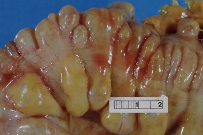
[[Image:Diverticula.jpg|400x300px|alt=|thumb|Showing multiple pouches (diverticula) colon]]Colonic diverticulitis is a possible adverse result of colonic diverticulosis. Colonic diverticulosis is when multiple tiny pouches, or diverticula, are seen in the colon. Diverticulosis is mainly asymptomatic whilst acute diverticulitis is a potentially life-threatening illness.<ref name=":0">Radiopedia [https://radiopaedia.org/articles/colonic-diverticulosis?lang=gb Colonic diverticulosis] Available:https://radiopaedia.org/articles/colonic-diverticulosis?lang=gb (accessed 22.1.20230</ref> | [[Image:Diverticula.jpg|400x300px|alt=|thumb|Showing multiple pouches (diverticula) colon]]Colonic diverticulitis is a possible adverse result of colonic diverticulosis. Colonic diverticulosis is when multiple tiny pouches, or diverticula, are seen in the colon. Diverticulosis is mainly asymptomatic whilst acute diverticulitis is a potentially life-threatening illness.<ref name=":0">Radiopedia [https://radiopaedia.org/articles/colonic-diverticulosis?lang=gb Colonic diverticulosis] Available:https://radiopaedia.org/articles/colonic-diverticulosis?lang=gb (accessed 22.1.20230</ref> | ||
Revision as of 23:36, 12 March 2024
Top Contributors - Katie Countryman, Cate Hurst, Khloud Shreif, Elaine Lonnemann, Lucinda hampton, Kim Jackson, WikiSysop, 127.0.0.1, George Prudden and Karen Wilson
This article or area is currently under review and may only be partially complete. Please come back soon to see the finished work! (12/03/2024)
Introduction[edit | edit source]
Colonic diverticulitis is a possible adverse result of colonic diverticulosis. Colonic diverticulosis is when multiple tiny pouches, or diverticula, are seen in the colon. Diverticulosis is mainly asymptomatic whilst acute diverticulitis is a potentially life-threatening illness.[1]
The most common symptoms of diverticulitis include severe left lower quadrant abdominal pain, marked changes in bowel habits, fever, and nausea. Possible complications include perforation of bowels, abscess formation, fistula formation, obstruction, and bleeding.
Diverticulitis diagnosis is typically confirmed with the presence of constitutional symptoms, bloody stools, elevated white blood cell count, and with the use of imaging studies.[2] Depending on the severity of the condition, diverticulitis can be treated with rest, changes in diet or antibiotics, and in severe cases may require surgery.[3]
Epidemiolgy/ Etiology[edit | edit source]
Diverticulitis is a complication of diverticulosis, and the demographics of the condition are much alike. Elderly patients are most at risk and 4% of people with diverticulosis go on to develop diverticulitis.[1]
Risk Factors include: Increased age; Constipation; Sedentary lifestyle; Obesity; Smoking; NSAIDS; Red meat[4]
Colonic diverticular development may involve involve bowel wall abnormality, increased intraluminal pressure, and lack of dietary fibre. Diverticulitis is the result of obstruction of the neck of the diverticulum (outpouch), with consequential inflammation, perforation, and infection. A walled of region of soft tissue may later progress to abscess formation and generalised peritonitis.[1]
Characteristics/Clinical Presentation[edit | edit source]
The presentation and signs and symptoms can vary for each individual patient. Although many of the patients have the same side effects, they are usually experienced at different intensities and at various times. Some of the most common signs and symptoms that are present with the diverticulitis diagnosis include the following[2]:
- Sudden abdominal pain, usually in the left lower quadrant abdomen,
- Palpable mass
- Irregular bowel movements
- Bowel sounds absent or decreased
- Flatulence
- Fever
- Nausea/ Vomiting
- Bloody stools
- Increased frequency of urination[4]
- Complication may be lower gastrointestinal haemorrhage ("diverticular haemorrhage")[1]
Treatment[edit | edit source]
Treatment depends on a range of factors, in particular comorbidities and stage of the disease.
- Localised disease: conservative management with intravenous antibiotics and rehydration usually is enough.
- Surgery may be required if a patient has a complication (abscess, fistula formation, bowel obstruction), has had multiple episodes of uncomplicated diverticulitis, or is immune-compromised. Surgery may be recommended or it may require emergency surgery[1]. There are two main types of surgery[3]: Primary bowel resection/laparoscopic procedure; Bowel resection with colostomy[4].
Prevention of diverticulitis is from a variety of lifestyle changes. Adherence to a high-fiber diet, decreased red meat intake, prevention of constipation with an adequate balance of fluids and fiber intake, cessation of smoking, and regular exercise during remission may decrease the risk of diverticulitis.[4]
Diagnosis[edit | edit source]
Diverticulitis is typically diagnosed during an acute attack due to complaints of severe abdominal pain. Diverticulitis diagnosis is typically confirmed with the presence of constitutional symptoms, bloody stools, elevated white blood cell count, and with the use of imaging studies.[2]
Due to the prevalence of abdominal pain in a number of conditions, the physician may order a number of tests to rule out other causes of abdominal pain and associated symptoms.[3]
Physical Therapy Management[edit | edit source]
As a physical therapist, the optimal goal is to help a patient return moving in a functional way. Being active helps decrease the chances of developing diverticulitis because movement helps promote proper bowel movement. Therapists can help patients with proper exercise, strengthening, and positioning to help them get the best and safest movement possible. Patients with diverticulitis must be cautious about doing activities that increase the pressure on their abdomen so further herniation does not happen[2].
Exercise can be seen as a protective mechanism because it is promoting movement to the body, but also the different systems that could be affected from a sedentary lifestyle[2]. Depending on the symptoms that the patients present with, it is up to the therapist to do appropriate screening or testing to identify what is involved and what is causing the issues.
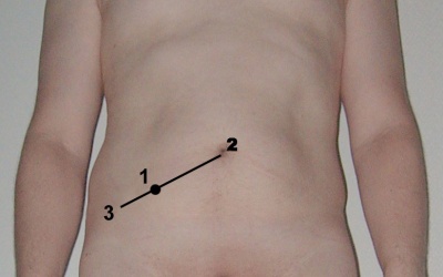
A common area of pain is the left lower quadrant, including referred pain to low back or thigh from an abscess[5]. For example, the obturator test, manual muscle testing and palpation of the illiopsoas ,or McBurney’s point palpation can be done to look at positive or negative testing of referred pain to the thigh[2][5].
References[edit | edit source]
- ↑ 1.0 1.1 1.2 1.3 1.4 Radiopedia Colonic diverticulosis Available:https://radiopaedia.org/articles/colonic-diverticulosis?lang=gb (accessed 22.1.20230
- ↑ 2.0 2.1 2.2 2.3 2.4 2.5 Goodman CC, Snyder TE. Differential diagnosis for physical therapists: screening for referral. 4th ed. St. Louis: Saunders Elsevier, 2007.
- ↑ 3.0 3.1 3.2 MCS. Diverticulitis Disease and Condition [Internet]. - Mayo Clinic. 2014 [cited 2016Mar31]. Retrieved from: http://www.mayoclinic.org/diseases-conditions/diverticulitis/basics/definition/con-20033495
- ↑ 4.0 4.1 4.2 4.3 Goodman CC, Fuller KS. Pathology: implications for the physical therapist. 3rd ed. St. Louis: Saunders Elsevier, 2009.
- ↑ 5.0 5.1 Hammond N. Left Lower-Quadrant Pain: Guidelines from the American College of Radiology Appropriateness Criteria. American Family Physician. 2010;82(7):766-770.


