Current Management of Carpal Tunnel Syndrome: Difference between revisions
No edit summary |
Kim Jackson (talk | contribs) m (Text replacement - "brachial plexus" to "brachial plexus") |
||
| (17 intermediate revisions by 4 users not shown) | |||
| Line 1: | Line 1: | ||
<div class=" | <div class="editorbox"> | ||
'''Original Editor '''- [[User:Wanda van Niekerk|Wanda van Niekerk]] based on the course by [https://members.physio-pedia.com/instructor/loren-szmiga/ Loren Szmiga] | |||
'''Top Contributors''' - {{Special:Contributors/{{FULLPAGENAME}}}} | |||
</div> | </div> | ||
< | == Introduction == | ||
[[Carpal Tunnel Syndrome|Carpal tunnel syndrome]] (CTS) is a common neurological disorder. The cases of CTS are on the rise as its incidence in the working population can reach 8%.<ref name=":10">Joshi A, Patel K, Mohamed A, Oak S, Zhang MH, Hsiung H, Zhang A, Patel UK. [https://www.ncbi.nlm.nih.gov/pmc/articles/PMC9389835/pdf/cureus-0014-00000027053.pdf Carpal Tunnel Syndrome: Pathophysiology and Comprehensive Guidelines for Clinical Evaluation and Treatment]. Cureus. 2022 Jul 20;14(7):e27053.</ref> Carpal tunnel syndrome is considered an occupational hazard, and production workers, material movers, and office administrative staff are at-risk occupations. Activities that involve excessive engagement of wrist flexion or prolonged wrist movements can lead to median nerve injury. <ref name=":10" /> Treatment options include conservative measures or surgery. | |||
This article discusses the anatomy and pathophysiology of CTS along with treatment considerations. | |||
== What is Carpal Tunnel Syndrome == | == What is Carpal Tunnel Syndrome == | ||
Carpal tunnel syndrome is an entrapment or compression of the median nerve at the wrist as it passes through the carpal tunnel.<ref name=":3">Padua L, Coraci D, Erra C, Pazzaglia C, Paolasso I, Loreti C, Caliandro P, Hobson-Webb LD. Carpal tunnel syndrome: clinical features, diagnosis, and management. The Lancet Neurology. 2016 Nov 1;15(12):1273-84.</ref> It is the most common compressive neuropathy and is more common in females.<ref>Ostergaard PJ, Meyer MA, Earp BE. [https://www.ncbi.nlm.nih.gov/pmc/articles/PMC7174467/pdf/12178_2020_Article_9616.pdf Non-operative treatment of carpal tunnel syndrome.] Current reviews in musculoskeletal medicine. 2020 Apr;13(2):141-7.</ref> | Carpal tunnel syndrome is an entrapment or compression of the [[Median Nerve|median nerve]] at the wrist as it passes through the carpal tunnel.<ref name=":3">Padua L, Coraci D, Erra C, Pazzaglia C, Paolasso I, Loreti C, Caliandro P, Hobson-Webb LD. Carpal tunnel syndrome: clinical features, diagnosis, and management. The Lancet Neurology. 2016 Nov 1;15(12):1273-84.</ref> It is the most common compressive neuropathy and is more common in females.<ref>Ostergaard PJ, Meyer MA, Earp BE. [https://www.ncbi.nlm.nih.gov/pmc/articles/PMC7174467/pdf/12178_2020_Article_9616.pdf Non-operative treatment of carpal tunnel syndrome.] Current reviews in musculoskeletal medicine. 2020 Apr;13(2):141-7.</ref> Symptoms occur in the thumb, index finger, middle finger and the radial half of the ring finger. Early symptoms include: | ||
* Pain | * Pain | ||
* Numbness and tingling | * Numbness and tingling | ||
* | * Paraesthesia | ||
* Can also lead to burning symptoms | * Can also lead to burning symptoms | ||
| Line 22: | Line 23: | ||
* The carpal tunnel is a U-shaped, osteofibrous canal | * The carpal tunnel is a U-shaped, osteofibrous canal | ||
* The floor of the tunnel is formed by the carpal bones and the roof by the flexor retinaculum | * The floor of the tunnel is formed by the carpal bones, and the roof by the flexor retinaculum | ||
* The tunnel is located deep to the flexor retinaculum/ transverse carpal ligament, between the tubercles of the scaphoid and trapezoid on the lateral side and the pisiform and hook of hamate on the medial side | * The tunnel is located deep to the [[flexor retinaculum]] / transverse carpal ligament, between the tubercles of the [[scaphoid]] and [[trapezoid]] on the lateral side and the [[pisiform]] and hook of [[hamate]] on the medial side | ||
* The four main structures passing through the tunnel are: | * The four main structures passing through the tunnel are: | ||
** Four tendons of flexor digitorum superficialis | ** Four tendons of [[Flexor Digitorum Superficialis|flexor digitorum superficialis]] | ||
** Four tendons of flexor digitorum profundus | ** Four tendons of [[Flexor Digitorum Profundus|flexor digitorum profundus]] | ||
** One tendon of the flexor pollicis longus | ** One tendon of the [[Flexor Pollicis Longus|flexor pollicis longus]] | ||
** [[File:Brachial Plexus shutterstock 445800688 RESIZED.jpg|thumb|Path of the Median Nerve]]Median nerve | ** [[File:Brachial Plexus shutterstock 445800688 RESIZED.jpg|thumb|Path of the Median Nerve]][[Median Nerve|Median nerve]] | ||
*** Path of the median nerve: | *** Path of the median nerve: | ||
**** Begins in the axillary region with the root of median nerves situated in the anterior rami of C5-T1 | **** Begins in the axillary region with the root of median nerves situated in the anterior rami of C5-T1 | ||
**** The median nerve is formed by fascicles of the medial and lateral cords of the brachial plexus | **** The median nerve is formed by fascicles of the medial and lateral cords of the [[Brachial Plexus|brachial plexus]] | ||
**** Runs distally in the arm next to the brachial artery until the middle of the arm | **** Runs distally in the arm next to the brachial artery until the middle of the arm and descends into the cubital fossa (anterior elbow) | ||
**** Principal nerve supply to the anterior compartment of the forearm | **** Principal nerve supply to the anterior compartment of the forearm | ||
**** The muscular branch in the forearm supplies all the superficial and intermediate layers of the forearm flexors, except for flexor carpi ulnaris | **** The muscular branch in the forearm supplies all the superficial and intermediate layers of the forearm flexors, except for [[Flexor Carpi Ulnaris Muscle|flexor carpi ulnaris]]. These muscles are: | ||
***** Pronator teres | *****[[Pronator Teres|Pronator teres]] | ||
***** Palmaris longus | ***** [[Palmaris Longus|Palmaris longus]] | ||
***** Flexor digitorum superficialis | ***** [[Flexor Digitorum Superficialis|Flexor digitorum superficialis]] | ||
***** Flexor carpi radialis | ***** [[Flexor Carpi Radialis|Flexor carpi radialis]] | ||
**** The terminal branch of the median nerve enters the hand through the carpal tunnel, along with the tendons of flexor digitorum profundus, flexor digitorum superficialis and flexor pollicis longus | **** The terminal branch of the median nerve enters the hand through the carpal tunnel, along with the tendons of flexor digitorum profundus, flexor digitorum superficialis and flexor pollicis longus | ||
**** Distal to the carpal tunnel the nerve supplies five intrinsic muscles in the thenar part | **** Distal to the carpal tunnel, the nerve supplies five intrinsic muscles in the thenar part | ||
**** The median nerve supplies sensation to the skin on: | **** The median nerve supplies sensation to the skin on: | ||
***** | ***** The entire palmar surface | ||
***** | ***** The sides of the first three digits | ||
***** | ***** The lateral half of the fourth digit and | ||
***** | ***** The dorsal aspects of the distal halves of these digits | ||
**** Innervation to the thenar eminence includes flexor pollicis brevis, opponens pollicis and abductor pollicis brevis | **** Innervation to the [[Thenar and Hypothenar Muscles Of The Hand|thenar eminence]] includes flexor pollicis brevis, opponens pollicis and abductor pollicis brevis | ||
== Aetiology == | == Aetiology == | ||
| Line 57: | Line 58: | ||
* History of repetitive wrist movement or exposure to vibrations or forceful angular motions such as typing, gaming, machine work | * History of repetitive wrist movement or exposure to vibrations or forceful angular motions such as typing, gaming, machine work | ||
* Specific conditions may also be associated with an increased risk for the development of carpal tunnel syndrome (CTS). These can include: | * Specific conditions may also be associated with an increased risk for the development of carpal tunnel syndrome (CTS). These can include: | ||
** Diabetes | ** [[Diabetes]]. The hyperglycemic conditions cause tendon inflammation and prevent tendons from gliding past one another | ||
** Pregnancy | ** Pregnancy. Fluid retention during pregnancy may increase pressure in the carpal tunnel. | ||
** Obesity | ** Obesity | ||
** Rheumatoid arthritis | ** [[Rheumatoid Arthritis|Rheumatoid arthritis]]. Inflammatory conditions can lead to synovial hyperplasia, which reduces the space in the carpal tunnel. | ||
** Fall on an outstretched hand (FOOSH) – this can displace the lunate bone which can cause pressure in the carpal tunnel | ** Fall on an outstretched hand (FOOSH) – this can displace the [[lunate]] bone, which can cause pressure in the carpal tunnel | ||
* CTS is more commonly seen in females, | * CTS is more commonly seen in females, usually between 36 and 60. Females may be more susceptible than males to compression of the median nerve because of a smaller relative cross-sectional area of the carpal tunnel. | ||
== Pathophysiology == | == Pathophysiology == | ||
| Line 71: | Line 72: | ||
* Osteoarthritis | * Osteoarthritis | ||
* Trauma | * Trauma | ||
* Acromegaly | * [[Acromegaly]] | ||
All of these place pressure on the median nerve. | All of these place pressure on the median nerve. | ||
It is hypothesised that the compression of the median nerve leads to the development of local | It is hypothesised that the compression of the median nerve leads to the development of local ischaemia, and this may cause demyelination of the nerve, resulting in clinical symptoms. Normal pressure in the carpal tunnel varies between 2 – 10 mmHg. Repetitive wrist motion causes fluctuations in carpal tunnel pressure. Wrist extension can result in a 10-fold increase in pressure, and wrist flexion can result in an 8-fold increase in pressure.<ref name=":1" /> | ||
== Clinical Presentation == | == Clinical Presentation == | ||
| Line 82: | Line 83: | ||
* Symptoms may arise spontaneously, but not commonly<ref name=":2" /> | * Symptoms may arise spontaneously, but not commonly<ref name=":2" /> | ||
* Numbness | * Numbness | ||
* Tingling or pins and needles sensation in the median nerve distribution of the hand (thumb, index finger, middle finger and half of the ring finger) | * Tingling or pins and needles sensation in the median nerve distribution of the hand (thumb, index finger, middle finger and radial half of the ring finger) | ||
* Symptoms are worst at night or early morning (complaints of nocturnal burning pain) and are relieved by shaking of the hand<ref name=":2" /> | * Symptoms are worst at night or early morning (complaints of nocturnal burning pain) and are relieved by shaking of the hand<ref name=":2" /> | ||
* As symptoms worsen, intermittent pain and numbness may be experienced during daytime activities such as driving, lifting, working on the computer | * As symptoms worsen, intermittent pain and numbness may be experienced during daytime activities such as driving, lifting, working on the computer | ||
* Increased symptoms with static gripping of objects such as a phone or steering wheel | * Increased symptoms with static gripping of objects such as a phone or steering wheel | ||
* As symptoms progress, increased tingling and numbness and burning pain in the hand may be reported<ref name=":0" /> | * As symptoms progress, increased tingling and numbness and burning pain in the hand may be reported<ref name=":0" /> | ||
* If symptoms are left untreated, patients can complain of constant pain, swelling | * If symptoms are left untreated, patients can complain of constant pain, hand swelling, difficulties with motor control and weakness and visible atrophy of the thenar eminence<ref name=":0" /> | ||
* Sensory deprivation may also be present, resulting in clumsiness, weakness, loss of grip and pinch strength<ref name=":2" /> | * Sensory deprivation may also be present, resulting in clumsiness, weakness, loss of grip and pinch strength<ref name=":2" /> | ||
| Line 94: | Line 95: | ||
* Pronator teres syndrome | * Pronator teres syndrome | ||
* Anterior | * [[Anterior Interosseous Nerve Syndrome|Anterior interosseous nerve syndrome]] | ||
* Cervicobrachial syndromes | * Cervicobrachial syndromes | ||
* Injury to the digital nerves at the palm of the hand | * Injury to the digital nerves at the palm of the hand | ||
* Carpometacarpal arthritis of the thumb | * Carpometacarpal arthritis of the thumb | ||
* Cervical radiculopathy | * Cervical radiculopathy | ||
* De Quervain’s tenosynovitis | * [[De Quervain's Tenosynovitis|De Quervain’s tenosynovitis]] | ||
* Peripheral neuropathy | * Peripheral neuropathy | ||
* Raynaud syndrome | * [[Raynaud's Phenomenon|Raynaud's syndrome]] | ||
* Ulnar compressive neuropathy<ref>Wipperman J, Goerl K. [http://thepafp.org/website/wp-content/uploads/2017/05/2016-Carpal-Tunnel-Syndrome.pdf Carpal tunnel syndrome: diagnosis and management]. American family physician. 2016 Dec 15;94(12):993-9.</ref> | * Ulnar compressive neuropathy<ref>Wipperman J, Goerl K. [http://thepafp.org/website/wp-content/uploads/2017/05/2016-Carpal-Tunnel-Syndrome.pdf Carpal tunnel syndrome: diagnosis and management]. American family physician. 2016 Dec 15;94(12):993-9.</ref> | ||
| Line 107: | Line 108: | ||
Electrophysical assessment (i.e., nerve conduction studies) can measure and examine median nerve dysfunction. This is useful when diagnosing carpal tunnel syndrome to assess nerve function and quantify damage to the nerve.<ref name=":3" /> | Electrophysical assessment (i.e., nerve conduction studies) can measure and examine median nerve dysfunction. This is useful when diagnosing carpal tunnel syndrome to assess nerve function and quantify damage to the nerve.<ref name=":3" /> | ||
There is a debate in recent literature with traditionalists arguing that nerve conduction studies are the gold standard for confirmation of a carpal tunnel syndrome diagnosis | There is a debate in recent literature, with traditionalists arguing that nerve conduction studies are the gold standard for confirmation of a carpal tunnel syndrome diagnosis and contemporary thinkers arguing that a diagnosis is possible based on clinical symptoms. Furthermore, even amongst traditionalists in favour of nerve conduction tests, there seems to be no consensus on the single best technique to be used.<ref name=":0" /> | ||
Neuromuscular ultrasound is a valuable tool to investigate carpal tunnel syndrome as it provides information on median nerve morphology and the surrounding structures.<ref>Walker FO, Cartwright MS, Alter KE, Visser LH, Hobson-Webb LD, Padua L, Strakowski JA, Preston DC, Boon AJ, Axer H, van Alfen N. Indications for neuromuscular ultrasound: Expert opinion and review of the literature. Clinical Neurophysiology. 2018 Dec 1;129(12):2658-79.</ref> Recent research highlights that (based on expert consensus) combining electrodiagnosis and ultrasound is more effective than using either modality on its own. In cases where electrodiagnostic studies are normal or unable to localise suspected carpal tunnel syndrome, ultrasound can add value.<ref>Pelosi L, Arányi Z, Beekman R, Bland J, Coraci D, Hobson-Webb LD, Padua L, Podnar S, Simon N, van Alfen N, Verhamme C. Expert consensus on the combined investigation of carpal tunnel syndrome with electrodiagnostic tests and neuromuscular ultrasound. Clinical Neurophysiology. 2022 Jan 6.</ref> | Neuromuscular ultrasound is a valuable tool to investigate carpal tunnel syndrome as it provides information on median nerve morphology and the surrounding structures.<ref>Walker FO, Cartwright MS, Alter KE, Visser LH, Hobson-Webb LD, Padua L, Strakowski JA, Preston DC, Boon AJ, Axer H, van Alfen N. Indications for neuromuscular ultrasound: Expert opinion and review of the literature. Clinical Neurophysiology. 2018 Dec 1;129(12):2658-79.</ref> Recent research highlights that (based on expert consensus) combining electrodiagnosis and ultrasound is more effective than using either modality on its own. In cases where electrodiagnostic studies are normal or unable to localise suspected carpal tunnel syndrome, ultrasound can add value.<ref>Pelosi L, Arányi Z, Beekman R, Bland J, Coraci D, Hobson-Webb LD, Padua L, Podnar S, Simon N, van Alfen N, Verhamme C. Expert consensus on the combined investigation of carpal tunnel syndrome with electrodiagnostic tests and neuromuscular ultrasound. Clinical Neurophysiology. 2022 Jan 6.</ref> | ||
| Line 113: | Line 114: | ||
Magnetic Resonance Imaging (MRI) is becoming more popular as a diagnostic tool for carpal tunnel syndrome. It can define the deeper and lateral limits of the carpal tunnel in more detail than ultrasound. It has also been shown to provide objective and accurate information about the anatomy and pathologies of the carpal tunnel.<ref>Vo NQ, Nguyen DD, Hoang NT, Ngo DH, Nguyen TH, Trong BL, Le NT, Thanh TN. [https://www.researchgate.net/profile/Thao-Nguyen-Thanh/publication/360041860_Magnetic_resonance_imaging_as_a_first-choice_imaging_modality_in_carpal_tunnel_syndrome_new_evidence/links/625fbbff9be52845a911d504/Magnetic-resonance-imaging-as-a-first-choice-imaging-modality-in-carpal-tunnel-syndrome-new-evidence.pdf Magnetic resonance imaging as a first-choice imaging modality in carpal tunnel syndrome: new evidence]. Acta Radiologica. 2022 Apr 18:02841851221094227.</ref> | Magnetic Resonance Imaging (MRI) is becoming more popular as a diagnostic tool for carpal tunnel syndrome. It can define the deeper and lateral limits of the carpal tunnel in more detail than ultrasound. It has also been shown to provide objective and accurate information about the anatomy and pathologies of the carpal tunnel.<ref>Vo NQ, Nguyen DD, Hoang NT, Ngo DH, Nguyen TH, Trong BL, Le NT, Thanh TN. [https://www.researchgate.net/profile/Thao-Nguyen-Thanh/publication/360041860_Magnetic_resonance_imaging_as_a_first-choice_imaging_modality_in_carpal_tunnel_syndrome_new_evidence/links/625fbbff9be52845a911d504/Magnetic-resonance-imaging-as-a-first-choice-imaging-modality-in-carpal-tunnel-syndrome-new-evidence.pdf Magnetic resonance imaging as a first-choice imaging modality in carpal tunnel syndrome: new evidence]. Acta Radiologica. 2022 Apr 18:02841851221094227.</ref> | ||
X-ray is recommended to exclude other causes of wrist pain or bony pathology<ref>Chammas M, Boretto J, Burmann LM, Ramos RM, Neto FCS, Silva JB. Carpal tunnel syndrome – part 1 (anatomy, physiology, | X-ray is recommended to exclude other causes of wrist pain or bony pathology<ref>Chammas M, Boretto J, Burmann LM, Ramos RM, Neto FCS, Silva JB. Carpal tunnel syndrome – part 1 (anatomy, physiology, aetiology and diagnosis). Revista brasileira de Ortopedia (English edition) 2014 September-October; 49 (5):429-436.</ref> | ||
== Physical Examination == | == Physical Examination == | ||
The location of | The location of symptoms is key for diagnosis.<ref name=":5">Szmiga, L. Carpal Tunnel Syndrome. Course. Plus. 2022</ref> | ||
* Carpal compression test | * Carpal compression test | ||
| Line 131: | Line 132: | ||
* Reverse Phalen’s test | * Reverse Phalen’s test | ||
** Have the patient fully extend their wrists by placing the palms of both hands together for 30 – 60 seconds | ** Have the patient fully extend their wrists by placing the palms of both hands together for 30 – 60 seconds | ||
** | ** A positive test is when symptoms are reproduced | ||
<div class="row"> | <div class="row"> | ||
<div class="col-md-6"> {{#ev:youtube|EdC5QQV6A8w|250}} <div class="text-right"><ref>Clinical Physio. Phalens Test for Carpal Tunnel Syndrome | Clinical Physio. Available from: https://www.youtube.com/watch?v=EdC5QQV6A8w [last accessed 20/06/2022]</ref></div></div> | <div class="col-md-6"> {{#ev:youtube|EdC5QQV6A8w|250}} <div class="text-right"><ref>Clinical Physio. Phalens Test for Carpal Tunnel Syndrome | Clinical Physio. Available from: https://www.youtube.com/watch?v=EdC5QQV6A8w [last accessed 20/06/2022]</ref></div></div> | ||
| Line 147: | Line 148: | ||
=== Conservative Management === | === Conservative Management === | ||
* Wrist control orthosis – blocks wrist extension and flexion motion which decreases compression of the carpal tunnel<ref name=":6">Karjalanen T, Raatikainen S, Jaatinen K, Lusa V. [https://www.ncbi.nlm.nih.gov/pmc/articles/PMC8877380/pdf/jcm-11-00950.pdf Update on Efficacy of Conservative Treatments for Carpal Tunnel Syndrome.] Journal of | * Wrist control orthosis – blocks wrist extension and flexion motion which decreases the compression of the carpal tunnel<ref name=":6">Karjalanen T, Raatikainen S, Jaatinen K, Lusa V. [https://www.ncbi.nlm.nih.gov/pmc/articles/PMC8877380/pdf/jcm-11-00950.pdf Update on Efficacy of Conservative Treatments for Carpal Tunnel Syndrome.] Journal of Clinical Medicine. 2022 Feb 11;11(4):950.</ref> | ||
* Patients wear | ** Patients wear this orthosis mostly at night<ref name=":6" /> | ||
* Activity modification – educate patients on how to modify daily activities and avoid positions that cause increased compression of the nerve<ref name=":5" /> | * Activity modification – educate patients on how to modify daily activities and avoid positions that cause increased compression of the nerve<ref name=":5" /> | ||
* Ergonomics education<ref>Trillos-Chacón MC, Castillo-M JA, Tolosa-Guzman I, Medina AF, Ballesteros SM. Strategies for | * Ergonomics education<ref>Trillos-Chacón MC, Castillo-M JA, Tolosa-Guzman I, Medina AF, Ballesteros SM. Strategies for preventing carpal tunnel syndrome in the workplace: A systematic review. Applied Ergonomics. 2021 May 1;93:103353.</ref> | ||
* Desk and keyboard height | * Desk and keyboard height | ||
** Elbow wrist and finger alignment | ** Elbow wrist and finger alignment | ||
| Line 160: | Line 161: | ||
** At the treating physician’s discretion | ** At the treating physician’s discretion | ||
* Neurodynamic techniques | * Neurodynamic techniques | ||
** Can improve short-term pain and functional status in individuals with carpal tunnel syndrome<ref name=":7">Núñez de Arenas-Arroyo S, Cavero-Redondo I, Torres-Costoso A, Reina-Gutiérrez S, Álvarez-Bueno C, Martínez-Vizcaíno V. [https://www.researchgate.net/publication/356275601_Short-term_Effects_of_Neurodynamic_Techniques_for_Treating_Carpal_Tunnel_Syndrome_A_Systematic_Review_With_Meta-analysis/link/61af1e7fc11c103836979c9b/download Short-Term Effects of Neurodynamic Techniques for Treating Carpal Tunnel Syndrome: A Systematic Review With Meta-Analysis]. | ** Can improve short-term pain and functional status in individuals with carpal tunnel syndrome<ref name=":7">Núñez de Arenas-Arroyo S, Cavero-Redondo I, Torres-Costoso A, Reina-Gutiérrez S, Álvarez-Bueno C, Martínez-Vizcaíno V. [https://www.researchgate.net/publication/356275601_Short-term_Effects_of_Neurodynamic_Techniques_for_Treating_Carpal_Tunnel_Syndrome_A_Systematic_Review_With_Meta-analysis/link/61af1e7fc11c103836979c9b/download Short-Term Effects of Neurodynamic Techniques for Treating Carpal Tunnel Syndrome: A Systematic Review With Meta-Analysis]. Journal of orthopaedic & sports physical therapy. 2021 Dec;51(12):566-80.</ref> | ||
** Neurodynamic techniques are more effective than electrotherapy interventions in improvement of function and more effective than surgery to improve pain<ref name=":7" /> | ** Neurodynamic techniques are more effective than electrotherapy interventions in the improvement of function and more effective than surgery to improve pain<ref name=":7" /> | ||
** Read the systematic review here: [https://www.researchgate.net/publication/356275601_Short-term_Effects_of_Neurodynamic_Techniques_for_Treating_Carpal_Tunnel_Syndrome_A_Systematic_Review_With_Meta-analysis/link/61af1e7fc11c103836979c9b/download Short-Term Effects of Neurodynamic Techniques for Treating Carpal Tunnel Syndrome: A Systematic Review With Meta-Analysis.]<ref name=":7" /> | ** Read the systematic review here: [https://www.researchgate.net/publication/356275601_Short-term_Effects_of_Neurodynamic_Techniques_for_Treating_Carpal_Tunnel_Syndrome_A_Systematic_Review_With_Meta-analysis/link/61af1e7fc11c103836979c9b/download Short-Term Effects of Neurodynamic Techniques for Treating Carpal Tunnel Syndrome: A Systematic Review With Meta-Analysis.]<ref name=":7" /> | ||
** Read more on Median nerve neurodynamic techniques here: [[Neurodynamic Treatment]] | |||
* Exercise therapy and mobilisation techniques | |||
** Researchers argue that gliding exercises may improve symptoms.<ref name=":8">Martins RS, Siqueira MG. [https://www.scielo.br/j/anp/a/LtyNnDmxCGYQGzk5fzQ6byB/?lang=en Conservative therapeutic management of carpal tunnel syndrome]. Arquivos de neuro-psiquiatria. 2017;75:819-24.</ref> The supporting theory is that gliding exercises may prevent or stretch adhesions around the tendons and median nerve and thus reduce oedema and increase venous return. This may result in reduced pressure in the carpal tunnel.<ref name=":8" /> | |||
** Examples of tendon gliding exercises are shown below in the section on Surgical Management | |||
** There is conflicting evidence on the effectiveness of tendon gliding exercises as conservative management. This may be due to the methodological quality of studies and the fact that the exercise therapy intervention is often applied and assessed in association with other management approaches.<ref>Huisstede BM, Fridén J, Coert JH, Hoogvliet P, European HANDGUIDE Group. Carpal tunnel syndrome: hand surgeons, hand therapists, physical medicine, and rehabilitation physicians agree on a multidisciplinary treatment guideline—results from the European HANDGUIDE Study. Archives of physical medicine and rehabilitation. 2014 Dec 1;95(12):2253-63.</ref> A Cochrane review (2012)<ref name=":9">Page MJ, O'Connor D, Pitt V, Massy‐Westropp N. Exercise and mobilisation interventions for carpal tunnel syndrome. Cochrane Database of Systematic Reviews. 2012(6).</ref>, reported limited evidence to justify the use of exercise and mobilisation techniques as a conservative intervention for Carpal Tunnel Syndrome, but the authors did suggest that decision-making and consideration for the use of exercise and mobilisation techniques as an intervention should be based on the clinician's experience as well as the patient's preference.<ref name=":9" /> | |||
* Read more: [https://www.scielo.br/j/anp/a/LtyNnDmxCGYQGzk5fzQ6byB/?lang=en Conservative therapeutic management of carpal tunnel syndrome]<ref name=":8" /> | |||
=== Surgical Management === | === Surgical Management === | ||
Open | Open or endoscopic surgery, where the transverse ligament is cut, creates space in the carpal tunnel and reduces pressure on the median nerve.<ref>Nazarieh M, Hakakzadeh A, Ghannadi S, Maleklou F, Tavakol Z, Alizadeh Z. [https://brieflands.com/articles/asjsm-102631.html Non-surgical management and post-surgical rehabilitation of carpal tunnel syndrome: An algorithmic approach and practical guideline]. Asian Journal of Sports Medicine. 2020 Sep 30;11(3).</ref><ref>Pace V, Marzano F, Placella G. [https://www.ncbi.nlm.nih.gov/pmc/articles/PMC9850791/pdf/WJO-14-6.pdf Update on surgical procedures for carpal tunnel syndrome: What is the current evidence and practice? What are the future research directions?] World J Orthop. 2023 Jan 18;14(1):6-12</ref> | ||
==== Open Carpal Tunnel Release ==== | |||
* Traditional incision or mini-incision can be used | |||
* Risk of incomplete transverse ligament release with the choice of mini-incision technique | |||
* Median nerve decompression and visualisation | |||
* It is considered a safe procedure, with reports of only a few cases of wound infections | |||
==== Endoscopic Carpal Tunnel Release ==== | |||
* Single-portal and two-portal techniques are used | |||
* Two-portal techniques are considered the lower risk for complications as they allow better visualisation of the neurovascular structures | |||
* Small post-surgical scars | |||
* Quick recovery time and return to work | |||
==== Post-Surgical Rehabilitation ==== | ==== Post-Surgical Rehabilitation ==== | ||
* Exercises are aimed at reducing stiffness | * Exercises are aimed at reducing stiffness | ||
* Therapy is short-term as recovery time is quick after surgery<ref name=":5" /> | |||
* Finger abduction and adduction | * Finger abduction and adduction | ||
** Gets intrinsic muscles working | ** Gets intrinsic muscles working | ||
| Line 179: | Line 201: | ||
** Straight fist with fingers touching the palm of the hand | ** Straight fist with fingers touching the palm of the hand | ||
** Tabletop position | ** Tabletop position | ||
** Back to straight hand | ** Back to a straight hand | ||
[[File:Finger Tendon glides.png|thumb|450x450px|Finger Tendon Glide Exercises. [(A) straight; (B) hook; (C) fist; (D) straight fist; (E) table top.]<ref>Sheereen FJ, Sarkar B, Sahay P, Shaphe MA, Alghadir AH, Iqbal A, Ali T, Ahmad F. Comparison of Two Manual Therapy Programs, including Tendon Gliding Exercises as a Common Adjunct, While Managing the Participants with Chronic Carpal Tunnel Syndrome. Pain Research and Management. 2022 Jun 8;2022.</ref>|alt=|center]] | [[File:Finger Tendon glides.png|thumb|450x450px|Finger Tendon Glide Exercises. [(A) straight; (B) hook; (C) fist; (D) straight fist; (E) table top.]<ref>Sheereen FJ, Sarkar B, Sahay P, Shaphe MA, Alghadir AH, Iqbal A, Ali T, Ahmad F. Comparison of Two Manual Therapy Programs, including Tendon Gliding Exercises as a Common Adjunct, While Managing the Participants with Chronic Carpal Tunnel Syndrome. Pain Research and Management. 2022 Jun 8;2022.</ref>|alt=|center]] | ||
{{#ev:youtube|_myr1Pl0l4o|300}}<ref>Rehab My Patient. Finger Tendon Glide Exercise. Available from: https://www.youtube.com/watch?v=_myr1Pl0l4o [last accessed 20/06/2022]</ref> | {{#ev:youtube|_myr1Pl0l4o|300}}<ref>Rehab My Patient. Finger Tendon Glide Exercise. Available from: https://www.youtube.com/watch?v=_myr1Pl0l4o [last accessed 20/06/2022]</ref> | ||
| Line 189: | Line 211: | ||
* Digit blocking | * Digit blocking | ||
** If patient experiences stiffness after surgery, digit blocking may help to increase motion<ref name=":5" /> | ** If a patient experiences stiffness after surgery, digit blocking may help to increase motion<ref name=":5" /> | ||
** For example if the patient has stiffness in the proximal interphalangeal (PIP) or distal interphalangeal (DIP) joints | ** For example, if the patient has stiffness in the proximal interphalangeal (PIP) or distal interphalangeal (DIP) joints | ||
*** Patient blocks the joint below with the other hand and performs movement | *** Patient blocks the joint below with the other hand and performs movement | ||
*** This forces motion to go through the stiff joint | *** This forces motion to go through the stiff joint | ||
| Line 208: | Line 230: | ||
* Prolonged duration of symptoms without treatment leads to irreversible changes and thenar muscle atrophy | * Prolonged duration of symptoms without treatment leads to irreversible changes and thenar muscle atrophy | ||
* Speedy and correct treatment is | * Speedy and correct treatment is therefore crucial | ||
* Have a look at this article: [https://www.jospt.org/doi/epdf/10.2519/jospt.2019.0501 Carpal Tunnel Syndrome: A Summary of Clinical Practice Guideline Recommendations-Using the Evidence to Guide Physical Therapist Practice | * Have a look at this article: [https://www.jospt.org/doi/epdf/10.2519/jospt.2019.0501 Carpal Tunnel Syndrome: A Summary of Clinical Practice Guideline Recommendations-Using the Evidence to Guide Physical Therapist Practice]<ref>Carpal Tunnel Syndrome. [https://www.jospt.org/doi/epdf/10.2519/jospt.2019.0501 A Summary of Clinical Practice Guideline Recommendations-Using the Evidence to Guide Physical Therapist Practice. (2019)]. The Journal of orthopaedic and sports physical therapy.;49(5):359-60.</ref> | ||
== References == | == References == | ||
<references /> | <references /> | ||
[[Category:Plus Content]] | |||
[[Category:Course Pages]] | |||
[[Category:Hand - Conditions]] | |||
[[Category:Nerves]] | |||
Latest revision as of 19:02, 8 March 2024
Original Editor - Wanda van Niekerk based on the course by Loren Szmiga
Top Contributors - Wanda van Niekerk, Ewa Jaraczewska, Kim Jackson and Jess Bell
Introduction[edit | edit source]
Carpal tunnel syndrome (CTS) is a common neurological disorder. The cases of CTS are on the rise as its incidence in the working population can reach 8%.[1] Carpal tunnel syndrome is considered an occupational hazard, and production workers, material movers, and office administrative staff are at-risk occupations. Activities that involve excessive engagement of wrist flexion or prolonged wrist movements can lead to median nerve injury. [1] Treatment options include conservative measures or surgery.
This article discusses the anatomy and pathophysiology of CTS along with treatment considerations.
What is Carpal Tunnel Syndrome[edit | edit source]
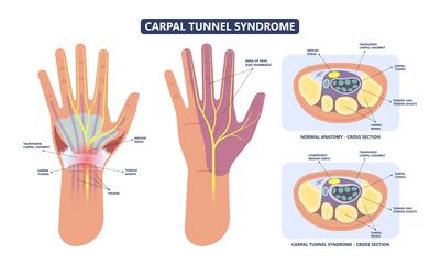
Carpal tunnel syndrome is an entrapment or compression of the median nerve at the wrist as it passes through the carpal tunnel.[2] It is the most common compressive neuropathy and is more common in females.[3] Symptoms occur in the thumb, index finger, middle finger and the radial half of the ring finger. Early symptoms include:
- Pain
- Numbness and tingling
- Paraesthesia
- Can also lead to burning symptoms
Clinically Relevant Anatomy[edit | edit source]
Some clinically relevant anatomical structures include[4]:
- The carpal tunnel is a U-shaped, osteofibrous canal
- The floor of the tunnel is formed by the carpal bones, and the roof by the flexor retinaculum
- The tunnel is located deep to the flexor retinaculum / transverse carpal ligament, between the tubercles of the scaphoid and trapezoid on the lateral side and the pisiform and hook of hamate on the medial side
- The four main structures passing through the tunnel are:
- Four tendons of flexor digitorum superficialis
- Four tendons of flexor digitorum profundus
- One tendon of the flexor pollicis longus
- Median nerve
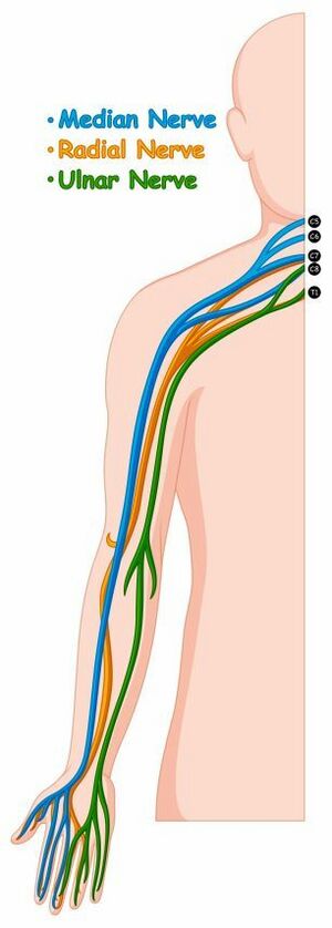
- Path of the median nerve:
- Begins in the axillary region with the root of median nerves situated in the anterior rami of C5-T1
- The median nerve is formed by fascicles of the medial and lateral cords of the brachial plexus
- Runs distally in the arm next to the brachial artery until the middle of the arm and descends into the cubital fossa (anterior elbow)
- Principal nerve supply to the anterior compartment of the forearm
- The muscular branch in the forearm supplies all the superficial and intermediate layers of the forearm flexors, except for flexor carpi ulnaris. These muscles are:
- The terminal branch of the median nerve enters the hand through the carpal tunnel, along with the tendons of flexor digitorum profundus, flexor digitorum superficialis and flexor pollicis longus
- Distal to the carpal tunnel, the nerve supplies five intrinsic muscles in the thenar part
- The median nerve supplies sensation to the skin on:
- The entire palmar surface
- The sides of the first three digits
- The lateral half of the fourth digit and
- The dorsal aspects of the distal halves of these digits
- Innervation to the thenar eminence includes flexor pollicis brevis, opponens pollicis and abductor pollicis brevis
- Path of the median nerve:
Aetiology[edit | edit source]
Increased pressure in the carpal tunnel and compression of the median nerve is the main cause of carpal tunnel syndrome. The aetiology of carpal tunnel syndrome can be related to[5]:
- Work
- Lifestyle
- Injury
- Genetic predisposition
- History of repetitive wrist movement or exposure to vibrations or forceful angular motions such as typing, gaming, machine work
- Specific conditions may also be associated with an increased risk for the development of carpal tunnel syndrome (CTS). These can include:
- Diabetes. The hyperglycemic conditions cause tendon inflammation and prevent tendons from gliding past one another
- Pregnancy. Fluid retention during pregnancy may increase pressure in the carpal tunnel.
- Obesity
- Rheumatoid arthritis. Inflammatory conditions can lead to synovial hyperplasia, which reduces the space in the carpal tunnel.
- Fall on an outstretched hand (FOOSH) – this can displace the lunate bone, which can cause pressure in the carpal tunnel
- CTS is more commonly seen in females, usually between 36 and 60. Females may be more susceptible than males to compression of the median nerve because of a smaller relative cross-sectional area of the carpal tunnel.
Pathophysiology[edit | edit source]
Increased interstitial pressure in the carpal tunnel due to various causes such as:
- Mechanical overuse
- Osteoarthritis
- Trauma
- Acromegaly
All of these place pressure on the median nerve.
It is hypothesised that the compression of the median nerve leads to the development of local ischaemia, and this may cause demyelination of the nerve, resulting in clinical symptoms. Normal pressure in the carpal tunnel varies between 2 – 10 mmHg. Repetitive wrist motion causes fluctuations in carpal tunnel pressure. Wrist extension can result in a 10-fold increase in pressure, and wrist flexion can result in an 8-fold increase in pressure.[5]
Clinical Presentation[edit | edit source]
- Symptoms can develop gradually over months, years or decades[6]
- Symptoms may arise spontaneously, but not commonly[6]
- Numbness
- Tingling or pins and needles sensation in the median nerve distribution of the hand (thumb, index finger, middle finger and radial half of the ring finger)
- Symptoms are worst at night or early morning (complaints of nocturnal burning pain) and are relieved by shaking of the hand[6]
- As symptoms worsen, intermittent pain and numbness may be experienced during daytime activities such as driving, lifting, working on the computer
- Increased symptoms with static gripping of objects such as a phone or steering wheel
- As symptoms progress, increased tingling and numbness and burning pain in the hand may be reported[4]
- If symptoms are left untreated, patients can complain of constant pain, hand swelling, difficulties with motor control and weakness and visible atrophy of the thenar eminence[4]
- Sensory deprivation may also be present, resulting in clumsiness, weakness, loss of grip and pinch strength[6]
Differential Diagnosis[edit | edit source]
The process of differential diagnosis should consider all conditions which could potentially cause dysfunction of the median nerve, the brachial plexus, C5 to C8 nerve root systems and the central nervous system.[7] Conditions to consider can include[7]:
- Pronator teres syndrome
- Anterior interosseous nerve syndrome
- Cervicobrachial syndromes
- Injury to the digital nerves at the palm of the hand
- Carpometacarpal arthritis of the thumb
- Cervical radiculopathy
- De Quervain’s tenosynovitis
- Peripheral neuropathy
- Raynaud's syndrome
- Ulnar compressive neuropathy[8]
Diagnosis of Carpal Tunnel Syndrome[edit | edit source]
Electrophysical assessment (i.e., nerve conduction studies) can measure and examine median nerve dysfunction. This is useful when diagnosing carpal tunnel syndrome to assess nerve function and quantify damage to the nerve.[2]
There is a debate in recent literature, with traditionalists arguing that nerve conduction studies are the gold standard for confirmation of a carpal tunnel syndrome diagnosis and contemporary thinkers arguing that a diagnosis is possible based on clinical symptoms. Furthermore, even amongst traditionalists in favour of nerve conduction tests, there seems to be no consensus on the single best technique to be used.[4]
Neuromuscular ultrasound is a valuable tool to investigate carpal tunnel syndrome as it provides information on median nerve morphology and the surrounding structures.[9] Recent research highlights that (based on expert consensus) combining electrodiagnosis and ultrasound is more effective than using either modality on its own. In cases where electrodiagnostic studies are normal or unable to localise suspected carpal tunnel syndrome, ultrasound can add value.[10]
Magnetic Resonance Imaging (MRI) is becoming more popular as a diagnostic tool for carpal tunnel syndrome. It can define the deeper and lateral limits of the carpal tunnel in more detail than ultrasound. It has also been shown to provide objective and accurate information about the anatomy and pathologies of the carpal tunnel.[11]
X-ray is recommended to exclude other causes of wrist pain or bony pathology[12]
Physical Examination[edit | edit source]
The location of symptoms is key for diagnosis.[13]
- Carpal compression test
- Apply firm pressure directly over the carpal tunnel for 30 seconds.
- The test is positive when paraesthesia, pain or other symptoms are reproduced
- Read more here: Carpal Compression Test
Carpal Compression Test video provided by Clinically Relevant
- Phalen’s test
- Have the patient fully flex their wrists by placing the dorsal surfaces of both hands together for 30 – 60 seconds
- A positive test is when symptoms (numbness, tingling, pain) are reproduced
- Read more here: Phalen's Test
- Reverse Phalen’s test
- Have the patient fully extend their wrists by placing the palms of both hands together for 30 – 60 seconds
- A positive test is when symptoms are reproduced
- Tinel’s sign
- Test is performed by lightly tapping over the median nerve for 30 seconds to reproduce symptoms
- Read more here: Tinel's Sign
- Read more on other physical examination tests here: Physical Examination of Carpal Tunnel Syndrome
Management of Carpal Tunnel Syndrome[edit | edit source]
Conservative Management[edit | edit source]
- Wrist control orthosis – blocks wrist extension and flexion motion which decreases the compression of the carpal tunnel[18]
- Patients wear this orthosis mostly at night[18]
- Activity modification – educate patients on how to modify daily activities and avoid positions that cause increased compression of the nerve[13]
- Ergonomics education[19]
- Desk and keyboard height
- Elbow wrist and finger alignment
- Medication
- Non-steroidal anti-inflammatory medication
- Oral steroids
- Corticosteroid injections[18]
- At the treating physician’s discretion
- Neurodynamic techniques
- Can improve short-term pain and functional status in individuals with carpal tunnel syndrome[20]
- Neurodynamic techniques are more effective than electrotherapy interventions in the improvement of function and more effective than surgery to improve pain[20]
- Read the systematic review here: Short-Term Effects of Neurodynamic Techniques for Treating Carpal Tunnel Syndrome: A Systematic Review With Meta-Analysis.[20]
- Read more on Median nerve neurodynamic techniques here: Neurodynamic Treatment
- Exercise therapy and mobilisation techniques
- Researchers argue that gliding exercises may improve symptoms.[21] The supporting theory is that gliding exercises may prevent or stretch adhesions around the tendons and median nerve and thus reduce oedema and increase venous return. This may result in reduced pressure in the carpal tunnel.[21]
- Examples of tendon gliding exercises are shown below in the section on Surgical Management
- There is conflicting evidence on the effectiveness of tendon gliding exercises as conservative management. This may be due to the methodological quality of studies and the fact that the exercise therapy intervention is often applied and assessed in association with other management approaches.[22] A Cochrane review (2012)[23], reported limited evidence to justify the use of exercise and mobilisation techniques as a conservative intervention for Carpal Tunnel Syndrome, but the authors did suggest that decision-making and consideration for the use of exercise and mobilisation techniques as an intervention should be based on the clinician's experience as well as the patient's preference.[23]
- Read more: Conservative therapeutic management of carpal tunnel syndrome[21]
Surgical Management[edit | edit source]
Open or endoscopic surgery, where the transverse ligament is cut, creates space in the carpal tunnel and reduces pressure on the median nerve.[24][25]
Open Carpal Tunnel Release[edit | edit source]
- Traditional incision or mini-incision can be used
- Risk of incomplete transverse ligament release with the choice of mini-incision technique
- Median nerve decompression and visualisation
- It is considered a safe procedure, with reports of only a few cases of wound infections
Endoscopic Carpal Tunnel Release[edit | edit source]
- Single-portal and two-portal techniques are used
- Two-portal techniques are considered the lower risk for complications as they allow better visualisation of the neurovascular structures
- Small post-surgical scars
- Quick recovery time and return to work
Post-Surgical Rehabilitation[edit | edit source]
- Exercises are aimed at reducing stiffness
- Therapy is short-term as recovery time is quick after surgery[13]
- Finger abduction and adduction
- Gets intrinsic muscles working
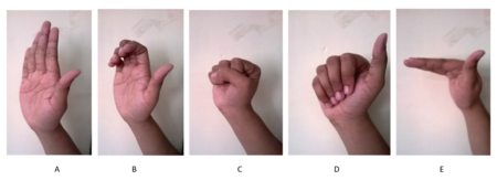
- Tendon glides[26]
- Promotes excursion of the flexor digitorum profundus and flexor digitorum superficialis[13]
- Hand straight
- Hook fist
- Full fist
- Straight fist with fingers touching the palm of the hand
- Tabletop position
- Back to a straight hand

- Dosage of tendon glide exercises[13]
- Patient repeats tendon glides three to five times
- Stiffness will determine how many times a day patients should perform tendon glides
- Very stiff after surgery – perform tendon glides six to eight times a day
- Minimal stiffness – perform tendon glides one to three times a day
- Digit blocking
- If a patient experiences stiffness after surgery, digit blocking may help to increase motion[13]
- For example, if the patient has stiffness in the proximal interphalangeal (PIP) or distal interphalangeal (DIP) joints
- Patient blocks the joint below with the other hand and performs movement
- This forces motion to go through the stiff joint
- Patient repeats this 5 times, holding it for 5 seconds
- This exercise can be done with individual digits or all at once, depending on which joints are lacking in motion
- Thumb opposition
- Allows for movement of flexor pollicis longus
- Patient touching their thumb to fifth finger – thumb opposition
Final Thoughts[edit | edit source]
- Prolonged duration of symptoms without treatment leads to irreversible changes and thenar muscle atrophy
- Speedy and correct treatment is therefore crucial
- Have a look at this article: Carpal Tunnel Syndrome: A Summary of Clinical Practice Guideline Recommendations-Using the Evidence to Guide Physical Therapist Practice[32]
References[edit | edit source]
- ↑ 1.0 1.1 Joshi A, Patel K, Mohamed A, Oak S, Zhang MH, Hsiung H, Zhang A, Patel UK. Carpal Tunnel Syndrome: Pathophysiology and Comprehensive Guidelines for Clinical Evaluation and Treatment. Cureus. 2022 Jul 20;14(7):e27053.
- ↑ 2.0 2.1 Padua L, Coraci D, Erra C, Pazzaglia C, Paolasso I, Loreti C, Caliandro P, Hobson-Webb LD. Carpal tunnel syndrome: clinical features, diagnosis, and management. The Lancet Neurology. 2016 Nov 1;15(12):1273-84.
- ↑ Ostergaard PJ, Meyer MA, Earp BE. Non-operative treatment of carpal tunnel syndrome. Current reviews in musculoskeletal medicine. 2020 Apr;13(2):141-7.
- ↑ 4.0 4.1 4.2 4.3 Urits I, Gress K, Charipova K, Orhurhu V, Kaye AD, Viswanath O. Recent advances in the understanding and management of carpal tunnel syndrome: a comprehensive review. Current pain and headache reports. 2019 Oct;23(10):1-8.
- ↑ 5.0 5.1 Osiak K, Elnazir P, Walocha JA, Pasternak A. Carpal tunnel syndrome: state-of-the-art review. Folia Morphologica. 2021 Nov 9.
- ↑ 6.0 6.1 6.2 6.3 Wang L. Guiding treatment for carpal tunnel syndrome. Physical Medicine and Rehabilitation Clinics. 2018 Nov 1;29(4):751-60.
- ↑ 7.0 7.1 Physiopedia. Carpal Tunnel Syndrome.
- ↑ Wipperman J, Goerl K. Carpal tunnel syndrome: diagnosis and management. American family physician. 2016 Dec 15;94(12):993-9.
- ↑ Walker FO, Cartwright MS, Alter KE, Visser LH, Hobson-Webb LD, Padua L, Strakowski JA, Preston DC, Boon AJ, Axer H, van Alfen N. Indications for neuromuscular ultrasound: Expert opinion and review of the literature. Clinical Neurophysiology. 2018 Dec 1;129(12):2658-79.
- ↑ Pelosi L, Arányi Z, Beekman R, Bland J, Coraci D, Hobson-Webb LD, Padua L, Podnar S, Simon N, van Alfen N, Verhamme C. Expert consensus on the combined investigation of carpal tunnel syndrome with electrodiagnostic tests and neuromuscular ultrasound. Clinical Neurophysiology. 2022 Jan 6.
- ↑ Vo NQ, Nguyen DD, Hoang NT, Ngo DH, Nguyen TH, Trong BL, Le NT, Thanh TN. Magnetic resonance imaging as a first-choice imaging modality in carpal tunnel syndrome: new evidence. Acta Radiologica. 2022 Apr 18:02841851221094227.
- ↑ Chammas M, Boretto J, Burmann LM, Ramos RM, Neto FCS, Silva JB. Carpal tunnel syndrome – part 1 (anatomy, physiology, aetiology and diagnosis). Revista brasileira de Ortopedia (English edition) 2014 September-October; 49 (5):429-436.
- ↑ 13.0 13.1 13.2 13.3 13.4 13.5 Szmiga, L. Carpal Tunnel Syndrome. Course. Plus. 2022
- ↑ Clinical Physio. Phalens Test for Carpal Tunnel Syndrome | Clinical Physio. Available from: https://www.youtube.com/watch?v=EdC5QQV6A8w [last accessed 20/06/2022]
- ↑ Clinical Physio. Reverse Phalens Test for Carpal Tunnel Syndrome | Clinical Physio. Available from: https://www.youtube.com/watch?v=5Fc8oDOPOmY [last accessed 20/6/2022]
- ↑ Tim Fraticelli. PT Progress. Tinel's Sign for Carpal Tunnel Syndrome. Available from: https://www.youtube.com/watch?v=UgV71aUIgLo [last accessed 20/06/2022]
- ↑ BMJ Learning. Tests for carpal tunnel syndrome. Available from: https://www.youtube.com/watch?v=3JPJcnFaPhU[last accessed 20/06/2022]
- ↑ 18.0 18.1 18.2 Karjalanen T, Raatikainen S, Jaatinen K, Lusa V. Update on Efficacy of Conservative Treatments for Carpal Tunnel Syndrome. Journal of Clinical Medicine. 2022 Feb 11;11(4):950.
- ↑ Trillos-Chacón MC, Castillo-M JA, Tolosa-Guzman I, Medina AF, Ballesteros SM. Strategies for preventing carpal tunnel syndrome in the workplace: A systematic review. Applied Ergonomics. 2021 May 1;93:103353.
- ↑ 20.0 20.1 20.2 Núñez de Arenas-Arroyo S, Cavero-Redondo I, Torres-Costoso A, Reina-Gutiérrez S, Álvarez-Bueno C, Martínez-Vizcaíno V. Short-Term Effects of Neurodynamic Techniques for Treating Carpal Tunnel Syndrome: A Systematic Review With Meta-Analysis. Journal of orthopaedic & sports physical therapy. 2021 Dec;51(12):566-80.
- ↑ 21.0 21.1 21.2 Martins RS, Siqueira MG. Conservative therapeutic management of carpal tunnel syndrome. Arquivos de neuro-psiquiatria. 2017;75:819-24.
- ↑ Huisstede BM, Fridén J, Coert JH, Hoogvliet P, European HANDGUIDE Group. Carpal tunnel syndrome: hand surgeons, hand therapists, physical medicine, and rehabilitation physicians agree on a multidisciplinary treatment guideline—results from the European HANDGUIDE Study. Archives of physical medicine and rehabilitation. 2014 Dec 1;95(12):2253-63.
- ↑ 23.0 23.1 Page MJ, O'Connor D, Pitt V, Massy‐Westropp N. Exercise and mobilisation interventions for carpal tunnel syndrome. Cochrane Database of Systematic Reviews. 2012(6).
- ↑ Nazarieh M, Hakakzadeh A, Ghannadi S, Maleklou F, Tavakol Z, Alizadeh Z. Non-surgical management and post-surgical rehabilitation of carpal tunnel syndrome: An algorithmic approach and practical guideline. Asian Journal of Sports Medicine. 2020 Sep 30;11(3).
- ↑ Pace V, Marzano F, Placella G. Update on surgical procedures for carpal tunnel syndrome: What is the current evidence and practice? What are the future research directions? World J Orthop. 2023 Jan 18;14(1):6-12
- ↑ Sheereen FJ, Sarkar B, Sahay P, Shaphe MA, Alghadir AH, Iqbal A, Ali T, Ahmad F. Comparison of Two Manual Therapy Programs, including Tendon Gliding Exercises as a Common Adjunct, While Managing the Participants with Chronic Carpal Tunnel Syndrome. Pain Research and Management. 2022 Jun 8;2022.
- ↑ Sheereen FJ, Sarkar B, Sahay P, Shaphe MA, Alghadir AH, Iqbal A, Ali T, Ahmad F. Comparison of Two Manual Therapy Programs, including Tendon Gliding Exercises as a Common Adjunct, While Managing the Participants with Chronic Carpal Tunnel Syndrome. Pain Research and Management. 2022 Jun 8;2022.
- ↑ Rehab My Patient. Finger Tendon Glide Exercise. Available from: https://www.youtube.com/watch?v=_myr1Pl0l4o [last accessed 20/06/2022]
- ↑ Rehab My Patient. Stiff finger exercise. Available from: https://www.youtube.com/watch?v=hvrD9FSzlik [last accessed 20/06/2022]
- ↑ Rehab My Patient. Finger pain exercise. Available from: https://www.youtube.com/watch?v=5qRb3ht3zsA[last accessed 20/06/2022]
- ↑ Rehab Mhttps://www.youtube.com/watch?v=BSyip8aUsqIy Patient. Thumb opposition proprioception. Available from: http://www.youtube.com/watch?v=dMH0bHeiRNg [last accessed 20/06/2022]
- ↑ Carpal Tunnel Syndrome. A Summary of Clinical Practice Guideline Recommendations-Using the Evidence to Guide Physical Therapist Practice. (2019). The Journal of orthopaedic and sports physical therapy.;49(5):359-60.


