When refering to evidence in academic writing, you should always try to reference the primary (original) source. That is usually the journal article where the information was first stated. In most cases Physiopedia articles are a secondary source and so should not be used as references. Physiopedia articles are best used to find the original sources of information (see the references list at the bottom of the article).
If you believe that this Physiopedia article is the primary source for the information you are refering to, you can use the button below to access a related citation statement.
The vertebral artery is a major artery in the neck[1]. It branches from the subclavian artery, where it arises from the posterosuperior portion of the subclavian artery.
Course
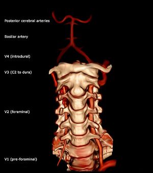
It ascends though the foramina of the transverse processes of the cervical vertebrae, usually starting at C6 but entering as high as C4[2]. It winds behind the superior articular process of the atlas. It enters the cranium through the foramen magnum where it unites with the opposite vertebral artery to form the basilar artery (at the lower border of the pons (part of the brainstem))[3].
Divisions
The vertebral artery can be divided into four divisions:
The first division runs posterocranial between the longus colli and the scalenus anterior.[3] The first division is also called the ‘pre-foraminal division’.[1]
The second division runs cranial through the foramina in the cervical transverse processes of the cervical vertebrae C2.[3] The second division is also called the foraminal division[1].
The third division is defined as the part that rises from C2. It rises from the latter foramen on the medial side of the rectus capitis lateralis, and curves behind the superior articular process of the atlas. Then, it lies in the
groove on the upper surface of the posterior arch of the atlas, and enters the vertebral canal by passing beneath the posterior atlantoöccipital membrane[3].
The fourth part pierces the dura mater and inclines medial to the front of the medulla oblongata.[3]
Injuries to the vertebral artery can be classified as traumatic or spontaneous.
Traumatic injuries usually result from blunt injury to the head and neck but may be due to penetrating trauma. Examples include: motor vehicle crashes; falls; strangulation; or it may be compressed by osteophyte formation or injury to the facet joint;
Spontaneous injuries result from intrinsic weakness of the vessel wall. For example dissection of the vessel caused by an underlying vascular or connective tissue disorder[4]. Examples include: artherosclerotic changes and other vascular risk facotrs (e.g. hypertension, high cholesterol, smoking, diabetes) may contribute dissection or to altered blood flow in the arteries.
Neck movements and stress to VA
The vertebral and carotid artieries are stressed primarily by rotation, extension and traction, but other movements may also stretch the artery. As little as 20% of rotation and extension have been shown to significantly decrease vertebral artery blood flow[2].
The greatest stresses are placed on the verterbal arteries in 4 places:
on entry to the C6 transverse process
within the bony canals of the vertebral transverse processes
between C1 and C2
between C1 and entry of the arteries into the skull.
The most common mechanism for non-penetrating injury to the vertebral artery is extension with or without side flexion or rotation[2].