Synovial Joints
Original Editor - Lucinda hampton
Top Contributors - Lucinda hampton and Joao Costa
Introduction[edit | edit source]

Histologically the three joints in the body are fibrous, cartilaginous, and synovial. Synovial joints are the most common type of joint in the body (see image 1). These joints are termed diarthroses, meaning they are freely mobile.[1]
A key structural characteristic for a synovial joint that is not seen at fibrous or cartilaginous joints is the presence of a joint cavity. The joint cavity contains synovial fluid, secreted by the synovial membrane (synovium), which lines the articular capsule. This fluid-filled space is the site at which the articulating surfaces of the bones contact each other. Hyaline cartilage forms the articular cartilage, covering the entire articulating surface of each bone. The articular cartilage and the synovial membrane are continuous. A few synovial joints of the body have a fibrocartilage structure located between the articulating bones. This is called an articular disc, which is generally small and oval-shaped, or a meniscus, which is larger and C-shaped.[2][3].
Synovial joints are often further classified by the type of movements they permit. There are six such classifications: hinge (elbow), saddle (carpometacarpal joint), planar (acromioclavicular joint), pivot (atlantoaxial joint), condyloid (metacarpophalangeal joint), and ball and socket (hip joint).[2]
Features of all Synovial Joints[edit | edit source]
- Articular capsule with synovial membrane
- Synovial cavity containing synovial fluid
- Hyaline articular cartilage
Additional features within some Synovial Joints[edit | edit source]
- Fibrocartilaginous discs eg. menisci within the knee joint
- Intra-capsular ligaments eg. cruciate ligaments within the knee joint
- Intra-capsular tendons eg. popliteus tendon within the knee joint
- Intra-articular tendons eg. long head of biceps tendon within the shoulder joint

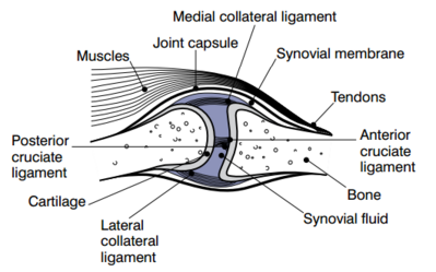
Image 2: In a Synovial joint, the ends of bones are encased in smooth cartilage. Together, they are protected by a joint capsule lined with a synovial membrane that produces synovial fluid. The capsule and fluid protect the cartilage, muscles, and connective tissues.
Additional features surrounding some Synovial Joints[edit | edit source]
- Fat pads
- Bursae
- Extra-capsular ligaments
- Tendons
- Sesamoid bones
Types of synovial joints[edit | edit source]
ball-and-socket joint eg. hip joint
hinge joint eg. elbow joint
condyloid joint eg. radiocarpal joint
saddle joint eg. first carpometocarpal joint
pivot joint eg. medial atlantoaxial joint
plane joint eg. intercarpal joints
Nerve Supply[edit | edit source]
Sensory and autonomic fibers innervate synovial joints:
- The autonomic nerves are vasomotor in function, controlling the dilation or constriction of blood vessels.
- The sensory nerves of the articular capsule and ligaments (articular nerves) provide proprioceptive feedback from Ruffini endings and Pacinian corpuscles. Proprioception of the joint permits reflex control of posture, locomotion, and movement. Free nerve endings convey pain sensation that is diffuse and poorly localized. The articular cartilage has no nerve supply.
Two general principles apply to synovial joint innervation:
- Hilton’s law states: Articular nerves supplying a joint are branches of the nerves that supply the muscles responsible for moving that joint. Hence irritation of articular nerves causes a reflex spasm of the muscles which position the joint for the greatest comfort. These nerves also supply the overlying skin, providing a mechanism for referred pain from joint to skin.
- Gardner’s observation: When part of the articular capsule that is tightened, by contraction of a group of muscles, they receives nerve supply from the same nerves that innervate the antagonist muscles. This relationship provides local reflex arcs that stabilize the joint[2].
Sub Heading 3[edit | edit source]
Resources[edit | edit source]
- bulleted list
- x
or
- numbered list
- x
References[edit | edit source]
- ↑ Radiopedia Synovial Joints Available: https://radiopaedia.org/articles/synovial-joints(accessed 20.6.2021)
- ↑ 2.0 2.1 2.2 Juneja P, Hubbard JB. Anatomy, Joints. 2018 Available: https://www.ncbi.nlm.nih.gov/books/NBK507893/ (accessed 20.6.2021)
- ↑ OSE Synovial joints Available:https://open.oregonstate.education/aandp/chapter/9-4-synovial-joints/ (accessed 20.6.2021)

