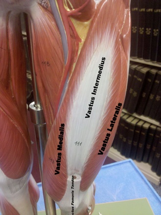
Vastus Intermedius
Original Editor - User:Uchechukwu Chukwuemeka
Top Contributors - Uchechukwu Chukwuemeka, Kim Jackson, Abbey Wright and Lucinda hampton
Description[edit | edit source]
Vastus Intermedius is located centrally, underneath the Rectus femoris[1] at the anterior compartment of the thigh and on each side of it, is situated the Vastus medialis and Vastus Lateralis respectively[2]. It is one of the muscles that made up the quadriceps Femoris muscle. Tensor of Vastus Intermedius is a new muscle that is part of the Quadriceps[3].
Origin[edit | edit source]
It originates from the upper two-thirds of anterior and lateral surfaces of the femur and the intermuscular septum[4].
Insertion[edit | edit source]
It joins the Quadriceps femoris tendon to form the deep part of the tendon and then insert into the lateral margins of the patella[5].
Function[edit | edit source]
Together with other muscles that are part of the Quadriceps femoris, it facilitates knee extension[6][7].
Blood supply[edit | edit source]
The descending branch of the lateral circumference femoral artery supplies this muscle[1].
Innervation[edit | edit source]
The Vastus Intermedius muscle is innervated by a branch of the Femoral nerve, originating from lumbar nerve 2, 3, and 4 nerve roots[4][5].
Assessment[edit | edit source]
Assessment of the muscles individually cannot be done; so, the test for knee extensor integrity is used to assess for it's power.
Palpation[edit | edit source]
The Vastus Intermedius is difficult to palpate. It is the least superficial muscle in the thigh's anterior compartment muscles; thus, it cannot be isolated for stretching and/or massage.
Resources[edit | edit source]
References[edit | edit source]
- ↑ 1.0 1.1 Drake, RL, Vogl, W, Mitchell, AW, Gray, H. Gray's anatomy for Students 2nd ed. Philadelphia : Churchill Livingstone/Elsevier, 2010
- ↑ Grob, K, Manestar, M, Filgueira, L, Kuster, MS, Gilbey, H, Ackland, T. The interaction between the vastus medialis and vastus intermedius and its influence on the extensor apparatus of the knee joint. Knee surgery, Sport Traumatology, Arthroscopy, 2018; 26(3):727-738. doi: 10.1007/s00167-016-4396-3.
- ↑ Veeramani, R, Gnanasekaran, D. Morphometric study of tensor of vastus intermedius in South Indian population. Anatomy of Cell Biology, 2017; 50(1): 7–11. doi: 10.5115/acb.2017.50.1.7
- ↑ 4.0 4.1 Drake, RL, Vogl, W, Mitchell, AW, Gray, H. Gray's anatomy for Students 2nd ed. Philadelphia : Churchill Livingstone/Elsevier, 2010
- ↑ 5.0 5.1 Miller, A, Heckert, KD, Davis, BA.The 3-Minute Musculoskeletal & Peripheral Nerve Exam. New York: Demos Medical Publishing. 2009; p.116-117
- ↑ Moore, KL, Dalley, AF, Agur, AM. Clinically oriented anatomy. 7th ed. Baltimore, MD: Lippincott Williams & Wilkins, 2014
- ↑ Hislop, HJ, Montgomery,J. Daniels and Worthingham's Muscle Testing: Techniques of Manual Examination. 8th ed. Missouri: Saunders Elsevier, 2007; p201-204


