Biomechanics of the Thorax: Difference between revisions
Sonal Joshi (talk | contribs) (Added details to table on articulations) |
Sonal Joshi (talk | contribs) (added new details to function of thorax) |
||
| Line 29: | Line 29: | ||
It is like a compartment sealed by various structures from each side. | It is like a compartment sealed by various structures from each side. | ||
* They can be described as<ref name=":0" />, | * They can be described as<ref name=":0" />, (IMAGE) | ||
{| class="wikitable" | {| class="wikitable" | ||
| Line 61: | Line 61: | ||
* Refer the [[Thoracic Anatomy]] for further details of [https://www.physio-pedia.com/Thoracic_Vertebrae Thoracic vertebrae] & the joint which they form. | * Refer the [[Thoracic Anatomy]] for further details of [https://www.physio-pedia.com/Thoracic_Vertebrae Thoracic vertebrae] & the joint which they form. | ||
==== Articulations ==== | ==== Articulations<ref name=":1">Pamela K. Levangie, Cynthia C. Norkin, 2005, Joint structure and function: A comprehensive analysis, 4th. Edn, Philadelphia, FA Davis publishers.</ref> (IMAGE) ==== | ||
{| class="wikitable" | {| class="wikitable" | ||
|+ | |+ | ||
| Line 97: | Line 97: | ||
|5. | |5. | ||
|Costotransverse | |Costotransverse | ||
| | |Costal tubercle of rib 1 to 10 and costal facet on | ||
| | |||
transverse process of corresponding vertebrae | |||
|<nowiki>- Synovial type of joint</nowiki> | |||
|- | |- | ||
|6. | |6. | ||
|Costochondral | |Costochondral | ||
| | |1 to 10th rib with costal cartilage | ||
| | |<nowiki>- Synchondrosis type of joint</nowiki> | ||
- Have no ligamentous support | |||
|- | |- | ||
|7. | |7. | ||
|Costosternal | |Costosternal | ||
| | |Costal cartilage of ribs 1 to 7 with sternum | ||
| | |<nowiki>-Joints with 1, 6 and 7 ribs</nowiki> | ||
are synchondroses type. | |||
-Joints with 2 to 5 ribs are | |||
synovial type. | |||
|- | |- | ||
|8. | |8. | ||
|Interchondral | |Interchondral | ||
| | |7 to 10 costal cartilage with cartilage above it | ||
| | |<nowiki>- Synovial type of joint</nowiki> | ||
|} | |} | ||
| Line 119: | Line 129: | ||
==== Kinematics ==== | ==== Kinematics ==== | ||
The motion of the ribs in conjunction with sternum and thoracic vertebrae helps produce the movements of respiration. These consist of inspiration and expiration. | The motion of the ribs in conjunction with sternum and thoracic vertebrae helps produce the movements of respiration. These consist of inspiration and expiration. Thoracic kinematics consists of understanding the changes in intrathoracic volume during ventilation. These occur due to complex, synchronized activity of the ribs along with sternum and primary muscles of respiration. It is determined by<ref name=":1" /> | ||
==== Muscles involved in | * Type & angles of articulations | ||
* movement of manubrium sternum | |||
* elasticity of costal cartilage | |||
The kinematics involved in the thorax can be further classified into<ref name=":0" />, | |||
* Changes in vertical diameter | |||
* Changes in anterior-posterior diameter (Pump handle motion) | |||
* Changes in transverse diameter (Bucket handle motion) | |||
===== Changes in Vertical diameter<ref name=":0" /> ===== | |||
* This is mainly due to excursion of Diaphragm muscle. | |||
* During inspiration : Diaphragm contracts -> lowering of the dome of diaphragm -> increase in vertical diameter of thorax. (VIDEO) | |||
* During expiration : Diaphragm relaxes -> dome recoils upward to resting position -> decrease in vertical diameter of thorax. | |||
===== Changes in A-P and transverse diameter<ref name=":1" /> ===== | |||
* There is a single axis of motion for 1 to 10 ribs through the center of their costovertebral(CV) and costotransverse(CT) joints. This influences the motion of these ribs during the process of respiration | |||
* The two main motions occurring in thorax during ventilation can be explained as below, | |||
{| class="wikitable" | |||
|+(VIDEO) | |||
! | |||
!Changes in Anterior-Posterior diameter | |||
!Changes in Transverse diameter | |||
|- | |||
|Mainly motion of | |||
|Upper ribs | |||
|Lower ribs | |||
|- | |||
|common Axis of motion oriented | |||
|nearly in frontal plane | |||
|nearly in sagittal plane | |||
|- | |||
|Thoracic motion occurs | |||
|in sagittal plane | |||
|in frontal plane | |||
|- | |||
|During Inspiration | |||
| | |||
* Upper ribs elevate | |||
* Motion is of anterior part of the ribs | |||
* It pushes sternum ventrally and superiorly | |||
* Total motion of sternum and upper ribs together increases in anterior-posterior diameter of thorax | |||
* Motion of sternum resembles pump handle movement, hence the name. | |||
| | |||
* Lower ribs elevate | |||
* Motion is more in the lateral part of ribs due to its more angled shape and indirect attachment of the ribs | |||
* Total motion of lower ribs together increases the transverse diameter of thorax | |||
* Motion of the lower ribs resembles bucket handle movement, hence the name. | |||
|} | |||
==== Muscles involved in respiration ==== | |||
* To bring about the intrathoracic volume changes during respiration, [https://www.physio-pedia.com/Muscles_of_Respiration?utm_source=physiopedia&utm_medium=search&utm_campaign=ongoing_internal muscles of respiration] play a crucial role. The muscles can be divided into primary and accessory depending on type of breathing i.e. quite or forced, in which they are used. | |||
* The detailed function of each muscle can be accessed by clicking on the muscle name mentioned below. Each has been linked to independent physiopedia pages dedicated to explaining them. | |||
===== Primary Muscles ===== | ===== Primary Muscles ===== | ||
* Diaphragm | * [https://www.physio-pedia.com/How_We_Breathe#cite_ref-:7_16-4 Diaphragm] | ||
* Intercoastal muscles | * [https://www.physio-pedia.com/Muscles_of_Respiration?utm_source=physiopedia&utm_medium=search&utm_campaign=ongoing_internal Intercoastal muscles] | ||
* Scalene | * [[Scalene]] | ||
===== Accessory Muscles ===== | ===== Accessory Muscles ===== | ||
* Sternocleidomastoid | * [[Sternocleidomastoid]] | ||
* Trapezius | * [[Trapezius]] | ||
* Pectoralis major | * [[Pectoralis major]] | ||
* Pectoralis minor | * Pectoralis minor | ||
* Subclavius | * Subclavius | ||
| Line 142: | Line 209: | ||
* Transverse thoracis | * Transverse thoracis | ||
=== Developmental Differences with Age === | === Developmental Differences with Age<ref name=":1" /> === | ||
==== In Newborn ==== | |||
* The chest wall is cartilaginous and therefore more compliant to external pressures applied during birth. | |||
* The muscles act more as stabilizers vs mobilizers in adults. | |||
* The complete ossification of ribs does not happen several months after birth. | |||
* There is more horizontal alignment of ribs as compared to adults. | |||
* There is very little motion of ribcage during tidal breathing. | |||
* The fibers of diaphragm are 20% fatigue-resistant vs 50% in adult. | |||
* Accessory muscle use gradually increases as infant gains head control. | |||
* The anterior part of rib cage becomes oblique as toddler is able to sit and stand. This promotes development of bucket handle motion. | |||
==== In Elderly ==== | |||
* Articulations of chest wall undergo fibrosis, leading to reduced thoracic mobility. | |||
* Narrowing of airways, decrease in elastic recoil and increase in lung compliance. | |||
* Increased kyphosis causes reduced mobility of ribcage. | |||
* Muscles of ventilation have fewer muscle fibers, lower oxidative capacity and decrease in number of fast twitch type II fibers. Therefore, ventilation is more energy consuming with age. | |||
* The resting position of diaphragm is less domed due to reduced abdominal tone. | |||
== References == | == References == | ||
Revision as of 22:20, 4 May 2022
Original Editor - User Name
Top Contributors - Sonal Joshi and Manisha Shrestha
This article is currently under review and may not be up to date. Please come back soon to see the finished work! (15/12/2021)
Introduction[edit | edit source]
Biomechanics is an area of science that uses principles of physics to measure and study, how forces interact with and affect living body[1]. These forces could be Internal i.e. within the body or External i.e. outside the body.
To understand the biomechanics of thorax we will need to study the basic anatomy, joint articulations, muscles associated in the thorax along with kinematics of the rib cage.
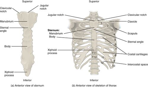
The thorax is formed by the thoracic vertebrae ,the ribs and the sternum.
It has two main functions,
- Providing anchor for muscle attachments
- Major role of ventilation
Structure of Thorax[edit | edit source]
The main three structures mentioned above work in a coordinated motion in thorax to produce respiration.
Anatomy[edit | edit source]
- The rib cage is a system of various bones and muscle. Bones involved are sternum, 12 pairs of Ribs, 12 Thoracic vertebrae.
It is like a compartment sealed by various structures from each side.
- They can be described as[1], (IMAGE)
| Side of compartment | Contents |
|---|---|
| Posterior-laterally | - Thoracic vertebrae
- Ribs - Intercoastal muscles and membrane |
| Anteriorly | - Coastal cartilages
- Sternum -Intercoastal muscles and membrane |
| Superiorly | - Upper ribs and clavicle
- Cervical fascia surrounding esophagus and trachea |
| Inferiorly | -Diaphragm muscle |
- The sternum consists of manubrium, body and xiphoid process. Its details can be referred to on page : sternum.
- The ribs are from T1 to T12 region
- Refer the Thoracic Anatomy for further details of Thoracic vertebrae & the joint which they form.
Articulations[2] (IMAGE)[edit | edit source]
| Joint | Bones involved in the joint articulation | Special features | |
|---|---|---|---|
| 1. | Manubriosternal | Manubrium and superior part of body of Sternum | - Synchondrosis type of joint
- Also called Angle of Louis |
| 2. | Xiphisternal | Xiphoid process and inferior part of body of Sternum | - Synchondrosis type of joint
- Ossifies by 40 to 50 yrs. of age |
| 3. | Typical Costovertebral (CV) | Head of ribs 2 to 9, two adjacent vertebral bodies and
interposed intervertebral disk |
- Synovial type of joint |
| 4. | Atypical Costovertebral | Head of ribs 1, 10 to 12 and corresponding vertebral
body |
- More mobile than
typical CV joint |
| 5. | Costotransverse | Costal tubercle of rib 1 to 10 and costal facet on
transverse process of corresponding vertebrae |
- Synovial type of joint |
| 6. | Costochondral | 1 to 10th rib with costal cartilage | - Synchondrosis type of joint
- Have no ligamentous support |
| 7. | Costosternal | Costal cartilage of ribs 1 to 7 with sternum | -Joints with 1, 6 and 7 ribs
are synchondroses type. -Joints with 2 to 5 ribs are synovial type. |
| 8. | Interchondral | 7 to 10 costal cartilage with cartilage above it | - Synovial type of joint |
Function of Thorax[edit | edit source]
Kinematics[edit | edit source]
The motion of the ribs in conjunction with sternum and thoracic vertebrae helps produce the movements of respiration. These consist of inspiration and expiration. Thoracic kinematics consists of understanding the changes in intrathoracic volume during ventilation. These occur due to complex, synchronized activity of the ribs along with sternum and primary muscles of respiration. It is determined by[2]
- Type & angles of articulations
- movement of manubrium sternum
- elasticity of costal cartilage
The kinematics involved in the thorax can be further classified into[1],
- Changes in vertical diameter
- Changes in anterior-posterior diameter (Pump handle motion)
- Changes in transverse diameter (Bucket handle motion)
Changes in Vertical diameter[1][edit | edit source]
- This is mainly due to excursion of Diaphragm muscle.
- During inspiration : Diaphragm contracts -> lowering of the dome of diaphragm -> increase in vertical diameter of thorax. (VIDEO)
- During expiration : Diaphragm relaxes -> dome recoils upward to resting position -> decrease in vertical diameter of thorax.
Changes in A-P and transverse diameter[2][edit | edit source]
- There is a single axis of motion for 1 to 10 ribs through the center of their costovertebral(CV) and costotransverse(CT) joints. This influences the motion of these ribs during the process of respiration
- The two main motions occurring in thorax during ventilation can be explained as below,
| Changes in Anterior-Posterior diameter | Changes in Transverse diameter | |
|---|---|---|
| Mainly motion of | Upper ribs | Lower ribs |
| common Axis of motion oriented | nearly in frontal plane | nearly in sagittal plane |
| Thoracic motion occurs | in sagittal plane | in frontal plane |
| During Inspiration |
|
|
Muscles involved in respiration[edit | edit source]
- To bring about the intrathoracic volume changes during respiration, muscles of respiration play a crucial role. The muscles can be divided into primary and accessory depending on type of breathing i.e. quite or forced, in which they are used.
- The detailed function of each muscle can be accessed by clicking on the muscle name mentioned below. Each has been linked to independent physiopedia pages dedicated to explaining them.
Primary Muscles[edit | edit source]
Accessory Muscles[edit | edit source]
- Sternocleidomastoid
- Trapezius
- Pectoralis major
- Pectoralis minor
- Subclavius
- Levatores costarum
- Serratus posterior superior
- Serratus posterior inferior
- Abdominal muscles
- Transverse thoracis
Developmental Differences with Age[2][edit | edit source]
In Newborn[edit | edit source]
- The chest wall is cartilaginous and therefore more compliant to external pressures applied during birth.
- The muscles act more as stabilizers vs mobilizers in adults.
- The complete ossification of ribs does not happen several months after birth.
- There is more horizontal alignment of ribs as compared to adults.
- There is very little motion of ribcage during tidal breathing.
- The fibers of diaphragm are 20% fatigue-resistant vs 50% in adult.
- Accessory muscle use gradually increases as infant gains head control.
- The anterior part of rib cage becomes oblique as toddler is able to sit and stand. This promotes development of bucket handle motion.
In Elderly[edit | edit source]
- Articulations of chest wall undergo fibrosis, leading to reduced thoracic mobility.
- Narrowing of airways, decrease in elastic recoil and increase in lung compliance.
- Increased kyphosis causes reduced mobility of ribcage.
- Muscles of ventilation have fewer muscle fibers, lower oxidative capacity and decrease in number of fast twitch type II fibers. Therefore, ventilation is more energy consuming with age.
- The resting position of diaphragm is less domed due to reduced abdominal tone.
References[edit | edit source]
- ↑ 1.0 1.1 1.2 1.3 Neumann DA. Kinesiology of the musculoskeletal system-e-book: foundations for rehabilitation. Elsevier Health Sciences; 2016 Nov 3.
- ↑ 2.0 2.1 2.2 2.3 Pamela K. Levangie, Cynthia C. Norkin, 2005, Joint structure and function: A comprehensive analysis, 4th. Edn, Philadelphia, FA Davis publishers.


