CREST Syndrome: Difference between revisions
Aya Alhindi (talk | contribs) No edit summary |
Aya Alhindi (talk | contribs) No edit summary |
||
| Line 10: | Line 10: | ||
== Description == | == Description == | ||
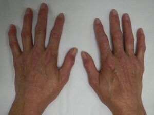
[[File:CREST Syndrome.jpeg|thumb|300x300px| CREST syndrome (calcinosis and sclerodactyly)]] | [[File:CREST Syndrome.jpeg|thumb|300x300px| CREST syndrome (calcinosis and sclerodactyly)]] | ||
CREST syndrome (also known as Cutaneous systemic sclerosis) is an autoimmune disease that has been defined as a subtype of progressive systemic sclerosis with limited skin involvement. <ref>Meyer O. CREST syndrome. Ann Med Interne (Paris) [Internet]. 2002;153(3):183–8. Available from: <nowiki>https://europepmc.org/article/med/12218901</nowiki></ref>The word "CREST " is an acronym for the clinical features that are seen in a patient with this disease: | CREST syndrome (also known as Cutaneous systemic sclerosis) is an autoimmune disease that has been defined as a subtype of progressive systemic sclerosis (SSc) with limited skin involvement. <ref>Meyer O. CREST syndrome. Ann Med Interne (Paris) [Internet]. 2002;153(3):183–8. Available from: <nowiki>https://europepmc.org/article/med/12218901</nowiki></ref>The word "CREST " is an acronym for the clinical features that are seen in a patient with this disease: | ||
* Calcinosis- when calcium salts are deposited into the skin and subcutaneous tissue.<ref>Le C, Bedocs PM. Calcinosis Cutis. StatPearls Publishing; 2023.</ref> | * Calcinosis- when calcium salts are deposited into the skin and subcutaneous tissue.<ref>Le C, Bedocs PM. Calcinosis Cutis. StatPearls Publishing; 2023.</ref> | ||
| Line 21: | Line 21: | ||
Wide variation in prevalence of systemic sclerosis was observed, with slightly higher estimates reported in North America (13.5–44.3 per 100,000 individuals) compared to Europe (7.2–33.9 per 100,000 individuals), which may be a true reflection of epidemiological variation or an artifact of clinical data analyses.<ref name=":0">Bergamasco A, Hartmann N, Wallace L, Verpillat P. Epidemiology of systemic sclerosis and systemic sclerosis-associated interstitial lung disease. Clin Epidemiol [Internet]. 2019;11:257–73. Available from: http://dx.doi.org/10.2147/clep.s191418</ref> The apparent increase in both incidence and prevalence over the last 50 years is most likely due to improved classification, earlier diagnosis, and survival. CREST syndrome may account for 22-25% of all occurrences of systemic sclerosis, according to serum antibody investigations; however, epidemiologic research specifically looking at CREST syndrome are missing.<ref>Wangkaew S, Euathrongchit J, Wattanawittawas P, Kasitanon N, Louthrenoo W. Incidence and predictors of interstitial lung disease (ILD) in Thai patients with early systemic sclerosis: Inception cohort study. Mod Rheumatol [Internet]. 2016;26(4):588–93. Available from: https://academic.oup.com/mr/article-pdf/26/4/588/39351804/mr0588.pdf</ref> SSc diagnosis was reported to occur at the ages of 33.5-59.8 years in Europe and 46.1-49.1 years in North America, and to occur more commonly in women (female:male ratio of 3.8-11.5:1 in Europe and 4.6-15:1 in North America). Women have continuously greater prevalence and incidence rates, indicating a clinically significant difference in the occurrence of SSc across genders.<ref name=":0" /> | Wide variation in prevalence of systemic sclerosis was observed, with slightly higher estimates reported in North America (13.5–44.3 per 100,000 individuals) compared to Europe (7.2–33.9 per 100,000 individuals), which may be a true reflection of epidemiological variation or an artifact of clinical data analyses.<ref name=":0">Bergamasco A, Hartmann N, Wallace L, Verpillat P. Epidemiology of systemic sclerosis and systemic sclerosis-associated interstitial lung disease. Clin Epidemiol [Internet]. 2019;11:257–73. Available from: http://dx.doi.org/10.2147/clep.s191418</ref> The apparent increase in both incidence and prevalence over the last 50 years is most likely due to improved classification, earlier diagnosis, and survival. CREST syndrome may account for 22-25% of all occurrences of systemic sclerosis, according to serum antibody investigations; however, epidemiologic research specifically looking at CREST syndrome are missing.<ref>Wangkaew S, Euathrongchit J, Wattanawittawas P, Kasitanon N, Louthrenoo W. Incidence and predictors of interstitial lung disease (ILD) in Thai patients with early systemic sclerosis: Inception cohort study. Mod Rheumatol [Internet]. 2016;26(4):588–93. Available from: https://academic.oup.com/mr/article-pdf/26/4/588/39351804/mr0588.pdf</ref> SSc diagnosis was reported to occur at the ages of 33.5-59.8 years in Europe and 46.1-49.1 years in North America, and to occur more commonly in women (female:male ratio of 3.8-11.5:1 in Europe and 4.6-15:1 in North America). Women have continuously greater prevalence and incidence rates, indicating a clinically significant difference in the occurrence of SSc across genders.<ref name=":0" /> | ||
== | == Pathophysiology == | ||
The pathogenesis of SSc is a self-amplifying process that begins with microvascular/endothelial damage, then progresses to an immunological response and [[Inflammation Acute and Chronic|inflammation]], and lastly to diffuse [[fibrosis]].<ref>Cutolo M, Soldano S, Smith V. Pathophysiology of systemic sclerosis: current understanding and new insights. Expert Rev Clin Immunol [Internet]. 2019;15(7):753–64. Available from: http://dx.doi.org/10.1080/1744666x.2019.1614915</ref>Routine histology can be used to visualise the key pathophysiological events of SSc. The endothelial cells enlarge first, followed by a lympho-histiocytic inflammatory infiltration around the damaged blood vessels. Later, a dense extracellular matrix deposition with activated myofibroblasts and homogenised collagen bundles occurs.<ref>Rosendahl A-H, Schönborn K, Krieg T. Pathophysiology of systemic sclerosis (scleroderma). Kaohsiung J Med Sci [Internet]. 2022;38(3):187–95. Available from: http://dx.doi.org/10.1002/kjm2.12505</ref> | |||
Although the primary cause of CREST syndrome is unknown, it is reasonable to speculate that vascular endothelial cell abnormalities induce mononuclear infiltration, and that the resulting changes in TH1 and/or TH2 cell and cytokine balance result in abnormal fibroblast activity and increased collagen deposition. | |||
== | == Diagnosis == | ||
In the absence of a diagnostic test proving the absence or presence of SSc, the diagnosis is based on a combination of clinical and laboratory findings. According to the latest classification scheme from 2013, SSc is confirmed by:<ref>van den Hoogen F, Khanna D, Fransen J, Johnson SR, Baron M, Tyndall A, et al. 2013 classification criteria for systemic sclerosis: an American College of Rheumatology/European League against Rheumatism collaborative initiative. Arthritis and rheumatism [Internet]. 2013 [cited 2023 Sep 8];65(11). Available from: https://pubmed.ncbi.nlm.nih.gov/24122180/</ref> | |||
'''Major Criteria:''' | |||
* skin thickening of the fingers of both hands extending proximal to the metacarpophalangeal joints (MCP) is sufficient to classify a subject as having SSc. | |||
* skin thickening sparing the fingers’ are classified as not having SSc. | |||
'''Minor criteria:''' | |||
# Skin thickening of the fingers -Puffy fingers and Sclerodactyly of the fingers (distal to the metacarpophalangeal joints but proximal to the proximal interphalangeal joints) | |||
# Fingertip lesions -Digital tip ulcers and Fingertip pitting scars | |||
# Telangiectasia | |||
# Abnormal nailfold capillaries | |||
# Pulmonary arterial hypertension and/or interstitial lung disease | |||
# Raynaud’s phenomenon | |||
# SSc-related autoantibodies: | |||
* Anticentromere. | |||
* Anti–topoisomerase I [anti–Scl-70]. | |||
* Anti–RNA polymerase III. | |||
== Etiology == | |||
== Medical Management == | == Medical Management == | ||
Revision as of 20:12, 8 September 2023
This article or area is currently under construction and may only be partially complete. Please come back soon to see the finished work! (8/09/2023)
Original Editor - User Name
Top Contributors - Aya Alhindi, Kim Jackson and Khloud Shreif
Description[edit | edit source]
CREST syndrome (also known as Cutaneous systemic sclerosis) is an autoimmune disease that has been defined as a subtype of progressive systemic sclerosis (SSc) with limited skin involvement. [1]The word "CREST " is an acronym for the clinical features that are seen in a patient with this disease:
- Calcinosis- when calcium salts are deposited into the skin and subcutaneous tissue.[2]
- Raynaud's phenomenon.
- Esophageal dysmotility-which can cause difficulty in swallowing.
- Sclerodactyly- scleroderma in which the fingers become thin and shiny with sclerotic skin at the tip due to subcutaneous and intracutaneous calcinosis and diffused fibrosis of the collagen.[3]
- Telangiectasia-small widened blood vessels on the skin.[4]
Epidemiology[edit | edit source]
Wide variation in prevalence of systemic sclerosis was observed, with slightly higher estimates reported in North America (13.5–44.3 per 100,000 individuals) compared to Europe (7.2–33.9 per 100,000 individuals), which may be a true reflection of epidemiological variation or an artifact of clinical data analyses.[5] The apparent increase in both incidence and prevalence over the last 50 years is most likely due to improved classification, earlier diagnosis, and survival. CREST syndrome may account for 22-25% of all occurrences of systemic sclerosis, according to serum antibody investigations; however, epidemiologic research specifically looking at CREST syndrome are missing.[6] SSc diagnosis was reported to occur at the ages of 33.5-59.8 years in Europe and 46.1-49.1 years in North America, and to occur more commonly in women (female:male ratio of 3.8-11.5:1 in Europe and 4.6-15:1 in North America). Women have continuously greater prevalence and incidence rates, indicating a clinically significant difference in the occurrence of SSc across genders.[5]
Pathophysiology[edit | edit source]
The pathogenesis of SSc is a self-amplifying process that begins with microvascular/endothelial damage, then progresses to an immunological response and inflammation, and lastly to diffuse fibrosis.[7]Routine histology can be used to visualise the key pathophysiological events of SSc. The endothelial cells enlarge first, followed by a lympho-histiocytic inflammatory infiltration around the damaged blood vessels. Later, a dense extracellular matrix deposition with activated myofibroblasts and homogenised collagen bundles occurs.[8]
Although the primary cause of CREST syndrome is unknown, it is reasonable to speculate that vascular endothelial cell abnormalities induce mononuclear infiltration, and that the resulting changes in TH1 and/or TH2 cell and cytokine balance result in abnormal fibroblast activity and increased collagen deposition.
Diagnosis[edit | edit source]
In the absence of a diagnostic test proving the absence or presence of SSc, the diagnosis is based on a combination of clinical and laboratory findings. According to the latest classification scheme from 2013, SSc is confirmed by:[9]
Major Criteria:
- skin thickening of the fingers of both hands extending proximal to the metacarpophalangeal joints (MCP) is sufficient to classify a subject as having SSc.
- skin thickening sparing the fingers’ are classified as not having SSc.
Minor criteria:
- Skin thickening of the fingers -Puffy fingers and Sclerodactyly of the fingers (distal to the metacarpophalangeal joints but proximal to the proximal interphalangeal joints)
- Fingertip lesions -Digital tip ulcers and Fingertip pitting scars
- Telangiectasia
- Abnormal nailfold capillaries
- Pulmonary arterial hypertension and/or interstitial lung disease
- Raynaud’s phenomenon
- SSc-related autoantibodies:
- Anticentromere.
- Anti–topoisomerase I [anti–Scl-70].
- Anti–RNA polymerase III.
Etiology[edit | edit source]
Medical Management[edit | edit source]
Physical Therapy Management[edit | edit source]
References[edit | edit source]
- ↑ Meyer O. CREST syndrome. Ann Med Interne (Paris) [Internet]. 2002;153(3):183–8. Available from: https://europepmc.org/article/med/12218901
- ↑ Le C, Bedocs PM. Calcinosis Cutis. StatPearls Publishing; 2023.
- ↑ Nelson FRT, Blauvelt CT. The Hand and wrist. In: Nelson FRT, Blauvelt CT, editors. A Manual of Orthopaedic Terminology. Elsevier; 2015. p. 307–41.
- ↑ Telangiectasia (Spider Veins) [Internet]. Pennmedicine.org. [cited 2023 Sep 8]. Available from: https://www.pennmedicine.org/for-patients-and-visitors/patient-information/conditions-treated-a-to-z/telangiectasia-spider-veins
- ↑ 5.0 5.1 Bergamasco A, Hartmann N, Wallace L, Verpillat P. Epidemiology of systemic sclerosis and systemic sclerosis-associated interstitial lung disease. Clin Epidemiol [Internet]. 2019;11:257–73. Available from: http://dx.doi.org/10.2147/clep.s191418
- ↑ Wangkaew S, Euathrongchit J, Wattanawittawas P, Kasitanon N, Louthrenoo W. Incidence and predictors of interstitial lung disease (ILD) in Thai patients with early systemic sclerosis: Inception cohort study. Mod Rheumatol [Internet]. 2016;26(4):588–93. Available from: https://academic.oup.com/mr/article-pdf/26/4/588/39351804/mr0588.pdf
- ↑ Cutolo M, Soldano S, Smith V. Pathophysiology of systemic sclerosis: current understanding and new insights. Expert Rev Clin Immunol [Internet]. 2019;15(7):753–64. Available from: http://dx.doi.org/10.1080/1744666x.2019.1614915
- ↑ Rosendahl A-H, Schönborn K, Krieg T. Pathophysiology of systemic sclerosis (scleroderma). Kaohsiung J Med Sci [Internet]. 2022;38(3):187–95. Available from: http://dx.doi.org/10.1002/kjm2.12505
- ↑ van den Hoogen F, Khanna D, Fransen J, Johnson SR, Baron M, Tyndall A, et al. 2013 classification criteria for systemic sclerosis: an American College of Rheumatology/European League against Rheumatism collaborative initiative. Arthritis and rheumatism [Internet]. 2013 [cited 2023 Sep 8];65(11). Available from: https://pubmed.ncbi.nlm.nih.gov/24122180/


