Functional Anatomy of the Hip-Muscles and Fascia: Difference between revisions
No edit summary |
No edit summary |
||
| Line 7: | Line 7: | ||
</div> | </div> | ||
== Description == | == Description == | ||
One theory assumes that the human body has two muscular systems: local and global. The local muscular system acts close to the joint axis, provides joint compression, and is responsible for | One theory assumes that the human body has two muscular systems: local and global. The local muscular system acts close to the joint axis, provides joint compression, and is responsible for joint stability. Large forces produced by muscles and small changes in their length create joint compression, thus producing active stabilisation of the joint.<ref name=":0" /> The global system contains superficial muscles generating greater torque and a larger moment arm.<ref name=":0">Retchford TH, Crossley KM, Grimaldi A, Kemp JL, Cowan SM. [http://ismni.org/jmni/pdf/51/01RETCHFORD.pdf Can local muscles augment stability in the hip? A narrative literature review]. J Musculoskelet Neuronal Interact. 2013 Mar 1;13(1):1-2.</ref> However, it is the muscle architecture and the line of action that determines the muscles' primary role. | ||
Daily activities require the hip joint to withstand high forces | Daily activities require the hip joint to withstand high forces. This is made possible by the contribution of the individual muscles surrounding the joint.<ref>Correa TA, Crossley KM, Kim HJ, Pandy MG. Contributions of individual muscles to hip joint contact force in normal walking. Journal of biomechanics. 2010 May 28;43(8):1618-22.</ref> Active stability provided by the hip muscles can increase passive stability in both the normal hip, as well as the hip with structural abnormality.<ref name=":0" /> | ||
== Muscles == | == Muscles == | ||
<blockquote>"There are 22 muscles that provide stability around the hip joint to give it that 365-degree mobility. It's a huge responsibility to maintain that stability of the ball and socket" | <blockquote>"There are 22 muscles that provide stability around the hip joint to give it that 365-degree mobility. It's a huge responsibility to maintain that stability of the ball and socket." ''Rina Pandya''</blockquote>[[File:Deep muscles of the gluteal region Primal.png|thumb|External Rotators of the Hip]] | ||
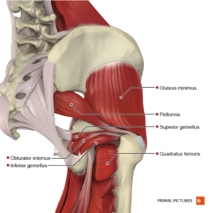
=== External Rotators === | === External Rotators === | ||
===== | ===== Attachments: ===== | ||
[[Quadratus Femoris|Quadratus femoris]]: | [[Quadratus Femoris|Quadratus femoris]]: ischial tuberosity to the intertrochanteric crest of the femur. | ||
[[Obturator Internus|Obturator internus]] and [[Obturator Externus|externus]] : | [[Obturator Internus|Obturator internus]] and [[Obturator Externus|externus]]: obturator membrane and ischiopubic ramus to the greater trochanter (internus) or intertrochanteric fossa of the femur (externus). | ||
Gemelli ([[Gemellus Superior|superior]] and [[Gemellus Inferior|inferior]]): | Gemelli ([[Gemellus Superior|superior]] and [[Gemellus Inferior|inferior]]): ischial spine (superior) or ischial tuberosity (inferior) to the greater trochanter and obturator internus tendon. | ||
[[Piriformis]]: | [[Piriformis]]: anterior surface of the sacrum and sacrotuberous ligament to the greater trochanter. | ||
===== Function: ===== | ===== Function: ===== | ||
* Active stabilisers of the hip joint. | * Active stabilisers of the hip joint. A primary role of these muscles is to stabilise the femoral head in the acetabulum. | ||
* When deep external rotators are resected during hip arthroplasty with a posterior surgical approach there is an increased rate of prosthetic dislocation and functional deficits. With capsular repairs, the dislocation rate is lower. | * When deep external rotators are resected during hip arthroplasty with a posterior surgical approach, there is an increased rate of prosthetic dislocation and functional deficits. With capsular repairs, the dislocation rate is lower. | ||
* Resisted '''external rotation''' of the hip with '''extension''' activates [[piriformis]], but its line of force is not conducive to enhance joint compression. | * Resisted '''external rotation''' of the hip with '''extension''' activates [[piriformis]], but its line of force is not conducive to enhance joint compression. | ||
* If the hip joint is tight with external rotation, and the patient is not able to bring | * If the hip joint is tight with external rotation, and the patient is not able to bring their leg up and put their sock on, it could indicate tightness of the piriformis. In addition, the patient may complain of shooting pain in the buttock region.<ref name=":1">Pandya R. Anatomy of the Hip Course. Physioplus. 2022.</ref> | ||
=== Internal Rotators === | === Internal Rotators === | ||
===== | ===== Attachments: ===== | ||
[[File:1122 Gluteal Muscles that Move the Femur a.png|thumb|Tensor Fascia Latae]] | [[File:1122 Gluteal Muscles that Move the Femur a.png|thumb|Tensor Fascia Latae]] | ||
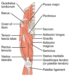
[[Tensor Fascia Lata|Tensor | [[Tensor Fascia Lata|Tensor fascia latae (TFL)]]: the anterior superior iliac spine to iliotibial tract, between the deep and superficial layers of the iliotibial band. | ||
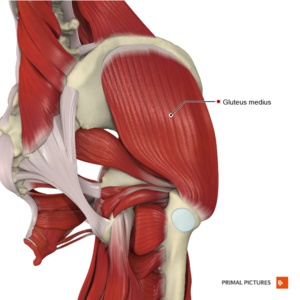
[[Gluteus Medius|Gluteus medius]] | [[Gluteus Medius|Gluteus medius]]: the outer surface of the ilium, between the iliac crest, and the anterior and posterior gluteal lines to the greater trochanter. The muscle has three segments; anterior, posterior, middle or superficial, each with a specific orientation of muscle fibres. | ||
[[Gluteus Minimus|Gluteus minimus]] | [[Gluteus Minimus|Gluteus minimus]]: the outer surface of the ilium, between the anterior and posterior gluteal lines to the greater trochanter. | ||
===== Function: ===== | ===== Function: ===== | ||
* ''TFL'' works in different movement planes: | * ''TFL'' works in different movement planes: it assists with ''hip abduction'' in the frontal plane, performs ''hip flexion'' in the sagittal plane, completes ''internal rotation'' in the transverse plane together with anterior gluteus medius and gluteus minimus.<ref>Besomi Molina M. [https://espace.library.uq.edu.au/data/UQ_faa2512/s4448975_phd_thesis.pdf?dsi_version=c89c7bc7d3c2aa1f7baf5f94dd139f58&Expires=1645651232&Key-Pair-Id=APKAJKNBJ4MJBJNC6NLQ&Signature=Z-bcaV2dAbyx7rLyKjlyLGsaWRtC7f8UPHqVsNzbHloHrH~Xf75lrhKEM1wOf8aQlqLgxGLRm1RZLMP6mX7ABwYdWG1cB-VAR-7pFSx8Fp-JFBTjqx5iRnEar9ohdaqyDsgWTB2pWNIoJXKze1Ja3RvBpoGrIDTBrRzFYVB9oOPsWNQu08jpyWv8DjvuN-FYc~heC2~anGXwldgmEYmAnrVbdimPUreWAnhsR8lobvn25zOTnM7HSFCy5LuMDwwxKRl-Gn3vyG2Q8cFVfb3INpF0fjO6lJtZGEqBFOefqoypD5-8msaFq51kgAkfpQVD3ncWDN3YhOBBWTy4Jw2S5A__ Towards the investigation of the tensor fascia lata muscle and iliotibial band function in runners: the relevance of the why and the how]. The University of Queensland, Australia. A thesis submitted for the degree of Doctor of Philosophy at The University of Queensland in 2020.</ref> | ||
* ''TFL'' limited elasticity | * ''TFL'' has limited elasticity, which limits the bulging of the thigh muscles and, thus, helps keep them contained so they can contract efficiently.<ref name=":1" /> | ||
* ''Gluteus medius'' and '' | * ''Gluteus medius'' and g''luteus minimus'' are primary abductors and ''assist with internal rotation.'' | ||
[[File: Intermediate muscles of the gluteal region Primal.png|thumb|Hip Abductor]] | [[File: Intermediate muscles of the gluteal region Primal.png|thumb|Hip Abductor]] | ||
=== Abductors === | === Abductors === | ||
[[Gluteus Minimus|Gluteus minimus]] | |||
[[Gluteus Medius|Gluteus medius]] | |||
[[Gluteus Medius|Gluteus medius]] | |||
[[Tensor Fascia Lata|Tensor Fascia Latae]] | [[Tensor Fascia Lata|Tensor Fascia Latae]] | ||
[[Piriformis]] | [[Piriformis]] | ||
See above for attachments. | |||
===== Function: ===== | ===== Function: ===== | ||
'' | The functions of g''luteus minimus'' include: | ||
* Stabilisation of the hip and pelvis through modulation of the joint capsule | * Stabilisation of the hip and pelvis through modulation of the joint capsule. | ||
* Stabilisation of the femoral head in the acetabulum | * Stabilisation of the femoral head in the acetabulum. | ||
* Rotation and flexion of the hip | * Rotation and flexion of the hip. | ||
* Prevention of anterior dislocation and migration of the femoral head in a superior and medial direction | * Prevention of anterior dislocation and migration of the femoral head in a superior and medial direction. | ||
* Proprioceptive role. | * Proprioceptive role. | ||
''Gluteus medius'' | ''Gluteus medius'': | ||
* | * Is a primary abductor of the hip. | ||
* | * Stabilises the pelvis and hip. | ||
* | * Prevents the pelvis from adduction in a single-leg stance. | ||
* | * Is an important stabiliser of the pelvis on the hip as it contracts prior to and after foot contact regardless of the walking speed.<ref name=":0" /> | ||
''Piriformis'': | ''Piriformis'': | ||
| Line 82: | Line 82: | ||
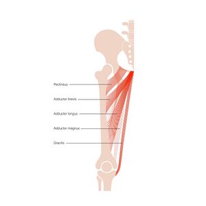
=== Adductors === | === Adductors === | ||
===== | ===== Attachments: ===== | ||
[[Adductor Longus|Adductor longus]]: pubic bone between the crest and symphysis to linea aspera of the femur | [[Adductor Longus|Adductor longus]]: pubic bone between the crest and symphysis to linea aspera of the femur. | ||
[[Adductor Brevis|Adductor brevis]]: body and inferior ramus of the pubis to linea aspera of the femur | [[Adductor Brevis|Adductor brevis]]: body and inferior ramus of the pubis to linea aspera of the femur. | ||
[[Adductor Magnus|Adductor magnus]]: ischial tuberosity and inferior ramus of the pubis to linea aspera and the adductor tubercle | [[Adductor Magnus|Adductor magnus]]: ischial tuberosity and inferior ramus of the pubis to linea aspera and the adductor tubercle. | ||
[[Gracilis]]: inferior pubic ramus to medial side of the tibial tuberosity | [[Gracilis]]: inferior pubic ramus to medial side of the tibial tuberosity. | ||
[[Pectineus Muscle|Pectineus:]] pectineal line of the pubis and pubic tubercle to pectineal line of the femur | [[Pectineus Muscle|Pectineus:]] pectineal line of the pubis and pubic tubercle to pectineal line of the femur. | ||
===== Function: ===== | ===== Function: ===== | ||
* | * The hip adductors contribute to static balance performance.<ref>Porto JM, Freire Junior RC, Bocarde L, Fernandes JA, Marques NR, Rodrigues NC, de Abreu DC. Contribution of hip abductor–adductor muscles on the static and dynamic balance of community-dwelling older adults. Ageing clinical and experimental research. 2019 May;31(5):621-7.</ref> | ||
* ''Adductor longus'' provides some ''medial rotation'' | * ''Adductor longus'' provides some ''medial rotation.'' | ||
* ''Adductor | * ''Adductor magnus'' ''extends the hip'' through his attachment on the ischial tuberosity. | ||
* In open chain activation, | * In open chain activation, their primary function is hip adduction. | ||
* In closed chain activation, hip adductors help to ''stabilise the pelvis and lower extremity'' during the stance phase of gait. | * In closed chain activation, hip adductors help to ''stabilise the pelvis and lower extremity'' during the stance phase of gait. | ||
* Secondary roles of '''hip adductors''' include '''hip flexion and rotation'''<ref>Kiel J, Kaiser K. Adductor strain. Available at https://europepmc.org/article/nbk/nbk493166 (last access 23.02.2022)</ref> | * Secondary roles of the '''hip adductors''' include '''hip flexion and rotation'''.<ref>Kiel J, Kaiser K. Adductor strain. Available at https://europepmc.org/article/nbk/nbk493166 (last access 23.02.2022)</ref> | ||
* ''Gracilis'' and ''semitendinosus'' create the conjoined tendons known as the [[Pes Anserinus Bursitis|pes anserinus]].<ref>Walters BB, Varacallo M. [https://europepmc.org/article/nbk/nbk532889 Anatomy, Bony Pelvis and Lower Limb, Thigh Sartorius Muscle]. In: StatPearls. StatPearls Publishing, Treasure Island (FL); 2021</ref> | * ''Gracilis'' and ''semitendinosus'' create the conjoined tendons known as the [[Pes Anserinus Bursitis|pes anserinus]].<ref>Walters BB, Varacallo M. [https://europepmc.org/article/nbk/nbk532889 Anatomy, Bony Pelvis and Lower Limb, Thigh Sartorius Muscle]. In: StatPearls. StatPearls Publishing, Treasure Island (FL); 2021</ref> | ||
[[File:Muscles of the iliac region Primal.png|thumb|Hip Flexors]] | [[File:Muscles of the iliac region Primal.png|thumb|Hip Flexors]] | ||
| Line 105: | Line 105: | ||
=== Flexors === | === Flexors === | ||
===== | ===== Attachments ===== | ||
[[Iliopsoas]] has three portions: | [[Iliopsoas]] has three portions:<ref name=":1" /> | ||
[[Psoas Major|Psoas major]]: transverse processes of vertebrae T12–L5 to lesser trochanter of the femur | * [[Iliacus]]: lateral edge of the sacrum and iliac fossa to lesser trochanter of the femur. | ||
* [[Psoas Major|Psoas major]]: transverse processes of vertebrae T12–L5 to lesser trochanter of the .femur | |||
* [[Psoas Minor|Psoas minor]]: vertebral bodies of T12–L1 to iliopubic ramus. | |||
[[ | [[Rectus Femoris|Rectus femoris]]: anterior-inferior iliac spine, a superior rim of the femoral acetabulum to the base of the patella.[[File:Muscles of the thigh anterior compartment Primal.png|thumb|Hip Flexors]][[Sartorius]]: anterior superior iliac spine to the upper medial side of the tibia. | ||
[[Tensor Fascia Lata|Tensor fascia latae]]: see above. | |||
[[Tensor Fascia Lata|Tensor | |||
===== Function: ===== | ===== Function: ===== | ||
* ''Psoas major'' and '' | * ''Psoas major'' and i''liacus'' are separately innervated. They are active throughout hip flexion. | ||
* ''Iliacus'' and both '' | * ''Iliacus'' and both p''soas muscles'' have a role similar to that of the rotator cuff muscles at the shoulder. They affect hip joint stability by creating tension in musculotendinous units as they pass over the anterior aspect of the hip joint.<ref name=":0" /> | ||
* ''Sartorius'' serves as both a hip and ''knee flexor'' and ''hip external rotator.'' | * ''Sartorius'' serves as both a hip and ''knee flexor'' and ''hip external rotator.'' | ||
* [[Thomas Test|Thomas test]] assesses the length of the hip flexors. The | * The [[Thomas Test|Thomas test]] assesses the length of the hip flexors. The contralateral limb is held in flexion. As flexion of this limb increases, the testing side (if tight) will lift off the bed due to posterior tilting of the lumbar spine. The knee extension on the tested side indicates rectus femoris or sartorius tightness | ||
[[File:Muscles of the thigh posterior compartment Primal.png|thumb|Hip Extensors]] | [[File:Muscles of the thigh posterior compartment Primal.png|thumb|Hip Extensors]] | ||
| Line 190: | Line 188: | ||
[[Category:Course Pages]] | [[Category:Course Pages]] | ||
[[Category:Hip - Muscles]] | [[Category:Hip - Muscles]] | ||
[[Category:Physioplus Content]] | |||
Revision as of 10:53, 4 March 2022
Original Editor - Ewa Jaraczewska
Top Contributors - Ewa Jaraczewska, Jess Bell, Kim Jackson and Lucinda hampton
Description[edit | edit source]
One theory assumes that the human body has two muscular systems: local and global. The local muscular system acts close to the joint axis, provides joint compression, and is responsible for joint stability. Large forces produced by muscles and small changes in their length create joint compression, thus producing active stabilisation of the joint.[1] The global system contains superficial muscles generating greater torque and a larger moment arm.[1] However, it is the muscle architecture and the line of action that determines the muscles' primary role.
Daily activities require the hip joint to withstand high forces. This is made possible by the contribution of the individual muscles surrounding the joint.[2] Active stability provided by the hip muscles can increase passive stability in both the normal hip, as well as the hip with structural abnormality.[1]
Muscles[edit | edit source]
"There are 22 muscles that provide stability around the hip joint to give it that 365-degree mobility. It's a huge responsibility to maintain that stability of the ball and socket." Rina Pandya
External Rotators[edit | edit source]
Attachments:[edit | edit source]
Quadratus femoris: ischial tuberosity to the intertrochanteric crest of the femur.
Obturator internus and externus: obturator membrane and ischiopubic ramus to the greater trochanter (internus) or intertrochanteric fossa of the femur (externus).
Gemelli (superior and inferior): ischial spine (superior) or ischial tuberosity (inferior) to the greater trochanter and obturator internus tendon.
Piriformis: anterior surface of the sacrum and sacrotuberous ligament to the greater trochanter.
Function:[edit | edit source]
- Active stabilisers of the hip joint. A primary role of these muscles is to stabilise the femoral head in the acetabulum.
- When deep external rotators are resected during hip arthroplasty with a posterior surgical approach, there is an increased rate of prosthetic dislocation and functional deficits. With capsular repairs, the dislocation rate is lower.
- Resisted external rotation of the hip with extension activates piriformis, but its line of force is not conducive to enhance joint compression.
- If the hip joint is tight with external rotation, and the patient is not able to bring their leg up and put their sock on, it could indicate tightness of the piriformis. In addition, the patient may complain of shooting pain in the buttock region.[3]
Internal Rotators[edit | edit source]
Attachments:[edit | edit source]
Tensor fascia latae (TFL): the anterior superior iliac spine to iliotibial tract, between the deep and superficial layers of the iliotibial band.
Gluteus medius: the outer surface of the ilium, between the iliac crest, and the anterior and posterior gluteal lines to the greater trochanter. The muscle has three segments; anterior, posterior, middle or superficial, each with a specific orientation of muscle fibres.
Gluteus minimus: the outer surface of the ilium, between the anterior and posterior gluteal lines to the greater trochanter.
Function:[edit | edit source]
- TFL works in different movement planes: it assists with hip abduction in the frontal plane, performs hip flexion in the sagittal plane, completes internal rotation in the transverse plane together with anterior gluteus medius and gluteus minimus.[4]
- TFL has limited elasticity, which limits the bulging of the thigh muscles and, thus, helps keep them contained so they can contract efficiently.[3]
- Gluteus medius and gluteus minimus are primary abductors and assist with internal rotation.
Abductors[edit | edit source]
See above for attachments.
Function:[edit | edit source]
The functions of gluteus minimus include:
- Stabilisation of the hip and pelvis through modulation of the joint capsule.
- Stabilisation of the femoral head in the acetabulum.
- Rotation and flexion of the hip.
- Prevention of anterior dislocation and migration of the femoral head in a superior and medial direction.
- Proprioceptive role.
Gluteus medius:
- Is a primary abductor of the hip.
- Stabilises the pelvis and hip.
- Prevents the pelvis from adduction in a single-leg stance.
- Is an important stabiliser of the pelvis on the hip as it contracts prior to and after foot contact regardless of the walking speed.[1]
Piriformis:
- Externally (laterally) rotates the femur during the hip extension and abducts the femur during hip flexion.[5]
- Acts as an auxiliary muscle and shows coactivation during pelvic floor muscles contracture.[6]
Adductors[edit | edit source]
Attachments:[edit | edit source]
Adductor longus: pubic bone between the crest and symphysis to linea aspera of the femur.
Adductor brevis: body and inferior ramus of the pubis to linea aspera of the femur.
Adductor magnus: ischial tuberosity and inferior ramus of the pubis to linea aspera and the adductor tubercle.
Gracilis: inferior pubic ramus to medial side of the tibial tuberosity.
Pectineus: pectineal line of the pubis and pubic tubercle to pectineal line of the femur.
Function:[edit | edit source]
- The hip adductors contribute to static balance performance.[7]
- Adductor longus provides some medial rotation.
- Adductor magnus extends the hip through his attachment on the ischial tuberosity.
- In open chain activation, their primary function is hip adduction.
- In closed chain activation, hip adductors help to stabilise the pelvis and lower extremity during the stance phase of gait.
- Secondary roles of the hip adductors include hip flexion and rotation.[8]
- Gracilis and semitendinosus create the conjoined tendons known as the pes anserinus.[9]
Flexors[edit | edit source]
Attachments[edit | edit source]
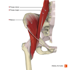
Iliopsoas has three portions:[3]
- Iliacus: lateral edge of the sacrum and iliac fossa to lesser trochanter of the femur.
- Psoas major: transverse processes of vertebrae T12–L5 to lesser trochanter of the .femur
- Psoas minor: vertebral bodies of T12–L1 to iliopubic ramus.
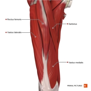
Rectus femoris: anterior-inferior iliac spine, a superior rim of the femoral acetabulum to the base of the patella.
Sartorius: anterior superior iliac spine to the upper medial side of the tibia.
Tensor fascia latae: see above.
Function:[edit | edit source]
- Psoas major and iliacus are separately innervated. They are active throughout hip flexion.
- Iliacus and both psoas muscles have a role similar to that of the rotator cuff muscles at the shoulder. They affect hip joint stability by creating tension in musculotendinous units as they pass over the anterior aspect of the hip joint.[1]
- Sartorius serves as both a hip and knee flexor and hip external rotator.
- The Thomas test assesses the length of the hip flexors. The contralateral limb is held in flexion. As flexion of this limb increases, the testing side (if tight) will lift off the bed due to posterior tilting of the lumbar spine. The knee extension on the tested side indicates rectus femoris or sartorius tightness
Extensors[edit | edit source]
Attachements:[edit | edit source]
Gluteus maximus: ilium, sacrum, coccyx, and the sacrotuberous ligament to gluteal tuberosity of the femur and iliotibial band.
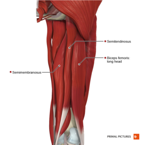
Long head: ischial tuberosity to lateral tibial condyle and head of the fibula.
Short head: upper supra-condylar line and linea aspera to lateral tibial condyle and head of the fibula.
Semimembranosus: ischial tuberosity to superior and medial surface of the tibia
Semitendinosus: ischial tuberosity to the medial condyle of the tibia
Function:[edit | edit source]
- Gluteus maximus is one of the primary hip extensors, it assists with hip external rotation and is the strongest and the biggest muscle in the body
- Upper and lower fibres of the gluteus maximus contribute to abduction and adduction as well. So it is an accessory muscle for abduction/adduction as well
- Gluteus maximus is prone to weakness and inhibition[10]
- Gluteus maximus originates partially from the thoracolumbar fascia which explains why any issue with the lumbar spine or even the thoracolumbar junction produces an anomaly or tightness or weakness of the gluteus max
- The biceps femoris long head is the most affected muscle in the hamstring strain injury.[11]
Inversion of muscular action[edit | edit source]
The muscles of the hip joint can contribute to movement in several different planes depending on the position of the hip, which is caused by a change in the relationship between a muscle’s line of action and the hip’s axis of rotation manifests as a muscle’s secondary function.
For example, the gluteus medius and minimus act as abductors when the hip is extended and as internal rotators when the hip is flexed. The adductor longus acts as a flexor at 50° of hip flexion, but as an extensor at 70.[12]
Fascia[edit | edit source]
The concept of fascia lines further explain the connection between adjacent structures: muscles, tendons, ligaments. This is called a myofascial continuity and it explains unique strains and connections that can occur following the injury, adhesions, postural changes. [13]
The following are the fascial lines and the hip joint muscles connected with these lines:
- Front functional line: adductor longus
- Back functional line: contralateral gluteus maximus
- Superficial Front line: rectus femoris
- Superficial Back line: hamstrings
- Lateral line: gluteus maximus, Tensor Fascia Latae, Iliotibial Track/Hip Abductors
- Spiral Line: Iliotibial track, Tensor Fascia Latae
- Deep Front Line: Iliacus, Psoas, Adductor Brevis and Longus, Adductor Magnus and Minimus
This video further explains the concept of fascia lines:
Examples of fascia connections in the hip joint:[15]
- The fascia of hip adductors continues to the pelvis and influences the urogenital and pelvic diaphragms
- The iliacus fascia is continuous with the deep pelvic fascia
- The fascia of the gluteus medius is continuous with the fascia of the abdominal obliques at the iliac crest
- There is fascial continuity between the obturator internus and the iliacus which further extends into internal obliques and the diaphragm.
Clinical relevance[edit | edit source]
- Piriformis syndrome is described as irritation of the sciatic nerve at the level of the piriformis muscle. It is a combination of symptoms involving the hip, buttock, and upper thigh. Possible causes include trauma, hematoma, excessive sitting, and anatomic variations of the muscle and nerve.
- Gluteus medius and gluteus minimus muscles weakness can present with Trendelenburg gait.
- External myofascial mobilisation approach based on fascial connectivity including ipsilateral latissimus dorsi, ipsilateral thoracolumbar fascia and contralateral gluteus maximus posteriorly, ipsilateral external oblique and contralateral internal oblique, and hip adductor complex anteriorly led to significant symptom improvement in the spastic chronic pelvic pain syndrome.[16]
- The tightness of the psoas can contribute to low back pain.
Resources[edit | edit source]
- ↑ 1.0 1.1 1.2 1.3 1.4 Retchford TH, Crossley KM, Grimaldi A, Kemp JL, Cowan SM. Can local muscles augment stability in the hip? A narrative literature review. J Musculoskelet Neuronal Interact. 2013 Mar 1;13(1):1-2.
- ↑ Correa TA, Crossley KM, Kim HJ, Pandy MG. Contributions of individual muscles to hip joint contact force in normal walking. Journal of biomechanics. 2010 May 28;43(8):1618-22.
- ↑ 3.0 3.1 3.2 Pandya R. Anatomy of the Hip Course. Physioplus. 2022.
- ↑ Besomi Molina M. Towards the investigation of the tensor fascia lata muscle and iliotibial band function in runners: the relevance of the why and the how. The University of Queensland, Australia. A thesis submitted for the degree of Doctor of Philosophy at The University of Queensland in 2020.
- ↑ Chang C, Jeno SH, Varacallo M. Anatomy, bony pelvis and lower limb, piriformis muscle. StatPearls [Internet]. 2020 Nov 12.
- ↑ Wang Z, Zhu Y, Han D, Huang Q, Maruyama H, Onoda K. Effect of hip external rotator muscle contraction on pelvic floor muscle function and the piriformis. International Urogynecology Journal. 2021 Nov 29:1-7.
- ↑ Porto JM, Freire Junior RC, Bocarde L, Fernandes JA, Marques NR, Rodrigues NC, de Abreu DC. Contribution of hip abductor–adductor muscles on the static and dynamic balance of community-dwelling older adults. Ageing clinical and experimental research. 2019 May;31(5):621-7.
- ↑ Kiel J, Kaiser K. Adductor strain. Available at https://europepmc.org/article/nbk/nbk493166 (last access 23.02.2022)
- ↑ Walters BB, Varacallo M. Anatomy, Bony Pelvis and Lower Limb, Thigh Sartorius Muscle. In: StatPearls. StatPearls Publishing, Treasure Island (FL); 2021
- ↑ Buckthorpe M, Stride M, Della Villa F. Assessing and treating gluteus maximus weakness–a clinical commentary. International journal of sports physical therapy. 2019 Jul;14(4):655.
- ↑ Llurda-Almuzara L, Labata-Lezaun N, López-de-Celis C, Aiguadé-Aiguadé R, Romaní-Sánchez S, Rodríguez-Sanz J, Fernández-de-Las-Peñas C, Pérez-Bellmunt A. Biceps femoris activation during hamstring strength exercises: a systematic review. International Journal of Environmental Research and Public Health. 2021 Jan;18(16):8733.
- ↑ Byrne DP, Mulhall KJ, Baker JF. Anatomy & Biomechanics of the Hip. The Open Sports Medicine Journal, 2010, 4: 51-57
- ↑ Myers TW. Anatomy Trains. Second edition. London: Churchill Livingstone, Elsevier; 2011.
- ↑ CatFitGlobal.Myofascial Lines. 2012. Available from: https://www.youtube.com/watch?v=LTt1DN3ozAs&t=64s [last accessed 24/02/2022]
- ↑ Schultz RL, Feitis R. The endless web. Fascial anatomy and physical reality. USA, CA: North Atlantic Books;1996
- ↑ Ajimsha MS, Ismail LA, Al-Mudahka N, Majzoub A. Effectiveness of external myofascial mobilisation in the management of male chronic pelvic pain of muscle spastic type: A retrospective study. Arab J Urol. 2021 Jul 26;19(3):394-400.
