Spinalis: Difference between revisions
No edit summary |
No edit summary |
||
| (4 intermediate revisions by the same user not shown) | |||
| Line 5: | Line 5: | ||
</div> | </div> | ||
== Introduction == | == Introduction == | ||
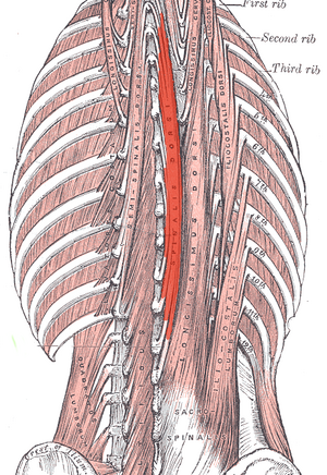
[[File:Spinalis.png|thumb|Musculus spinalis thoracis]] | |||
The spinalis muscle group are part of the the [[Erector Spinae|erector spinae]] (ES) group (the intermediate layer of the intrinsic [[Back Muscles|back muscles]]). The ES is made of three subgroups, with the group divisions occurring by location. | |||
== | # '''Spinalis''' subgroup, most medial | ||
# Longissimus subgroup, between spinalis and iliocostalis | |||
# Iliocostalis subgroup, most lateral | |||
The erector spinae muscles, which spinalis belong to, are the most powerful extensors of the spine.<ref name=":0" /><ref name=":1" /> | |||
== | == Anatomy == | ||
The [[Spinalis Thoracis|spinalis thoracis]] is the major spinalis muscle, arising from the bones of the lower [[Thoracic Vertebrae|thoracic]] and upper [[Lumbar Vertebrae|lumbar vertebral]] spine and inserted into the bones of the upper thoracic vertebral spine. | |||
== | The [[Spinalis Capitis|spinalis capitis]] and [[Spinalis Cervicis|spinalis cervicis]] are variably developed and variably present.<ref name=":0">Radiopedia Erector Spinae Available: https://radiopaedia.org/articles/erector-spinae-group?lang=us<nowiki/>(accessed 4.2.2022)</ref><ref name=":1">Britannica Spinalis Available:https://www.britannica.com/science/spinalis-muscle (accessed 4.2.2022)</ref> | ||
# | The spinalis is innervated by the lateral branches of the posterior rami of the [[Thoracic Spinal Nerves|spinal nerves]]. | ||
# | |||
It receives blood supply via the dorsal branches of the posterior [[Intercostal Muscle Strain|intercostal]] arteries, the deep cervical [[Arteries|artery]], and the muscular branches of the [[Vertebral Artery|vertebral artery]]<ref>Spinalis Anatomy applied Available: https://anatomy.app/encyclopedia/spinalis<nowiki/>(accessed 4.2.2022)</ref>. | |||
== Function == | |||
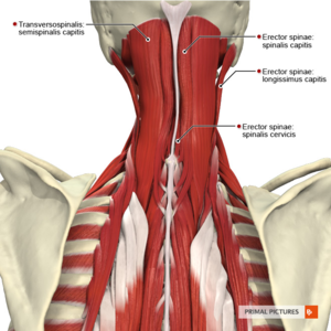
[[File:Muscles of the cervical region intermediate muscles Primal.png|thumb|Spinalis Capitis marked]] | |||
Spinalis works synergistically with the other members of the erector spinae group. Erector spinae muscle group have 3 major functions. | |||
# Unilateral contraction of erector spinae results in lateral flexion (ipsilateral) and rotation of the cervical, thoracic spine and lumbar spines. | |||
# Bilateral contraction of erector spinae causes extension of the head, neck and cervical, thoracic and lumbar vertebrae. | |||
# Helps to stabilize the [[pelvis]] while [[Balance|balancing]] on one leg. During this activity, the contralateral muscle group contracts and prevents the [[Trendelenburg Sign|pelvis from dipping]]<ref>Kenhub Spinalis Available: https://www.kenhub.com/en/library/anatomy/spinalis-muscle<nowiki/>(accessed 4.2.2022)</ref>. | |||
== References == | == References == | ||
<references /> | <references /> | ||
[[Category:Muscles]] | |||
[[Category:Thoracic Spine - Anatomy]] | |||
[[Category:Anatomy]] | |||
Latest revision as of 06:32, 4 February 2022
Original Editor - Lucinda hampton
Top Contributors - Lucinda hampton
Introduction[edit | edit source]
The spinalis muscle group are part of the the erector spinae (ES) group (the intermediate layer of the intrinsic back muscles). The ES is made of three subgroups, with the group divisions occurring by location.
- Spinalis subgroup, most medial
- Longissimus subgroup, between spinalis and iliocostalis
- Iliocostalis subgroup, most lateral
The erector spinae muscles, which spinalis belong to, are the most powerful extensors of the spine.[1][2]
Anatomy[edit | edit source]
The spinalis thoracis is the major spinalis muscle, arising from the bones of the lower thoracic and upper lumbar vertebral spine and inserted into the bones of the upper thoracic vertebral spine.
The spinalis capitis and spinalis cervicis are variably developed and variably present.[1][2]
The spinalis is innervated by the lateral branches of the posterior rami of the spinal nerves.
It receives blood supply via the dorsal branches of the posterior intercostal arteries, the deep cervical artery, and the muscular branches of the vertebral artery[3].
Function[edit | edit source]
Spinalis works synergistically with the other members of the erector spinae group. Erector spinae muscle group have 3 major functions.
- Unilateral contraction of erector spinae results in lateral flexion (ipsilateral) and rotation of the cervical, thoracic spine and lumbar spines.
- Bilateral contraction of erector spinae causes extension of the head, neck and cervical, thoracic and lumbar vertebrae.
- Helps to stabilize the pelvis while balancing on one leg. During this activity, the contralateral muscle group contracts and prevents the pelvis from dipping[4].
References[edit | edit source]
- ↑ 1.0 1.1 Radiopedia Erector Spinae Available: https://radiopaedia.org/articles/erector-spinae-group?lang=us(accessed 4.2.2022)
- ↑ 2.0 2.1 Britannica Spinalis Available:https://www.britannica.com/science/spinalis-muscle (accessed 4.2.2022)
- ↑ Spinalis Anatomy applied Available: https://anatomy.app/encyclopedia/spinalis(accessed 4.2.2022)
- ↑ Kenhub Spinalis Available: https://www.kenhub.com/en/library/anatomy/spinalis-muscle(accessed 4.2.2022)


