Gluteal Tendinopathy: Difference between revisions
No edit summary |
No edit summary |
||
| Line 60: | Line 60: | ||
| {{#ev:youtube|A9pi7_JRgwQ|350}} <ref>What single leg standing assessment can tell you Available from: https://www.youtube.com/watch?v=A9pi7_JRgwQ</ref> | | {{#ev:youtube|A9pi7_JRgwQ|350}} <ref>What single leg standing assessment can tell you Available from: https://www.youtube.com/watch?v=A9pi7_JRgwQ</ref> | ||
|} | |} | ||
{{#ev:youtube|nwBnc3I53QY|350}} | {{#ev:youtube|nwBnc3I53QY|350}}<ref>Resisted External Derotation Test/Gluteal Tendinopathy. Available from: https://www.youtube.com/watch?v=nwBnc3I53QY</ref> | ||
Pain on palpation of the structures over the greater trochanter is considered a cardinal sign in diagnosing Greater Trochanter Pain Syndrome. If the direct compression over this area failed to elicit pain then GT may be excluded and other conditions would be considered<ref>Bird PA, Oakley SP, Shnier R, Kirkham BW. Prospective evaluation of magnetic resonance imaging and physical examination findings in patients with greater trochanteric pain syndrome. Arthritis & Rheumatism: Official Journal of the American College of Rheumatology. 2001 Sep;44(9):2138-45.</ref>. The available literature has not yet provided a clear combination of diagnostic tests to confirm GT diagnosis. Further research is required to reach accurate and objective set of criteria and/or tests. | Pain on palpation of the structures over the greater trochanter is considered a cardinal sign in diagnosing Greater Trochanter Pain Syndrome. If the direct compression over this area failed to elicit pain then GT may be excluded and other conditions would be considered<ref>Bird PA, Oakley SP, Shnier R, Kirkham BW. Prospective evaluation of magnetic resonance imaging and physical examination findings in patients with greater trochanteric pain syndrome. Arthritis & Rheumatism: Official Journal of the American College of Rheumatology. 2001 Sep;44(9):2138-45.</ref>. The available literature has not yet provided a clear combination of diagnostic tests to confirm GT diagnosis. Further research is required to reach accurate and objective set of criteria and/or tests. | ||
| Line 89: | Line 90: | ||
* Sustained postures and stretches | * Sustained postures and stretches | ||
* Minimizing exposure to walking uphill, hopping, running and power walking in the short term. | * Minimizing exposure to walking uphill, hopping, running and power walking in the short term. | ||
This also includes training patients on using proper body mechanics when doing functional activities that require single-leg loading ,such as stairs negotiation, to be performed without excessive hip adduction and minimal trunk lean to minimize high external hip adduction moment. | This also includes training patients on using proper body mechanics when doing functional activities that require single-leg loading ,such as stairs negotiation, to be performed without excessive hip adduction and minimal trunk lean to minimize high external hip adduction moment. | ||
[[File:Activity modifications in GT.jpg|none|thumb|700x700px]] | |||
[[File:Running modifications in GT.jpg|none|thumb|700x700px]] | |||
=== Exercises === | === Exercises === | ||
| Line 100: | Line 104: | ||
To load abductors in frontal plane. subjects in exercise group performed different abduction exercises against spring resistance. | To load abductors in frontal plane. subjects in exercise group performed different abduction exercises against spring resistance. | ||
Motoring load appropriately is recommended by increasing load gradually without pain aggravation. | Motoring load appropriately is recommended by increasing load gradually without pain aggravation<ref name=":1" />. | ||
=== Steroid Injection === | === Steroid Injection === | ||
| Line 107: | Line 111: | ||
=== Surgical Intervention === | === Surgical Intervention === | ||
Removal of trochanteric bursae and ITB release is utilized in patients without gluteal tears. Studies report good to excellent short-medium term outcomes, however, most studies lacked control group and the rationale behind the mechanism of efficacy remains unclear. | Surgery is considered in persistent pain and failed conservative treatment<ref name=":0" />. 90% long-term improvement was reported following GMed tendon repair in 72 patients<ref>Walsh MJ, Walton JR, Walsh NA. Surgical repair of the gluteal tendons: a report of 72 cases. The Journal of arthroplasty. 2011 Dec 1;26(8):1514-9.</ref>. Endoscopic repairs provide an accelerated rehabilitaion and less invasive approach with reduced rates of post-operative infection, scar tissue formation and pain. Patients with larger tears are not suitable for endoscopic techniques. | ||
Removal of trochanteric bursae and ITB release is utilized in patients without gluteal tears. Studies report good to excellent short-medium term outcomes<ref>Craig RA, Gwynne Jones DP, Oakley AP, Dunbar JD. Iliotibial band Z‐lengthening for refractory trochanteric bursitis (greater trochanteric pain syndrome). ANZ journal of surgery. 2007 Nov;77(11):996-8.</ref>, however, most studies lacked control group and the rationale behind the mechanism of efficacy remains unclear<ref name=":0" />. | |||
<br> | <br> | ||
Revision as of 22:50, 20 July 2018
Introduction[edit | edit source]
Gluteal Tendinopathy (GT) is defined as moderate to sever disabling pain over the Greater Trochanter (lateral hip pain). It is often referred to as Greater Trochanter Pain Syndrome (GTPS) and was traditionally diagnosed as Trochanteric Bursitis, however, recent research defines non-inflammatory tendinopathy of the gluteus medius(GMed) and/or gluteus minimus (GMin) muscles to be the main source of lateral hip pain[1].
This condition affects both athletes (particularly runners) and less active people[1]. One of four females over 50 years is likely to be affected by GT[2].
GT has significant impacts on the quality of life, with similar symptoms to those of hip OA. It interferes with sleep (side lying) and common weight bearing tasks[1].
Pathoanatomy/Pathomechanics[edit | edit source]
Tendon structure and loading capacity are influenced by mechanical loading which triggers physiological responses within the tendon. Under normal conditions, the tendon undergoes a cycle of balanced catabolic and anabolic processes. Changes in loading type, intensity or frequency disrupt this harmony. Eccentric contractions in outer ranges (when the muscle is active and the tendon is lengthening simultaneously) represent the greatest form of loading. Failure to adapt to loading, due to rapid increase in intensity and/or frequency with insufficient recovery time, results in a series of catabolic effects which in turn result in altering tenocyte behaviour, reducing load-bearing capacity and predisposing tendons to injury at relatively low tensile loads. A combination of both tensile loading and compression are found to be more damaging than either alone[3].
GMed and GMin tendons are subjected to compression due to several factors:
1- Joint position: the ITB presents a compressive force on gluteal tendons that magnifies as the hip moves into further adduction. Birnbaum et al. [4] reported a change from 4 to 106 N ITB compression as the angle of hip adduction increased from 0 to 40 degrees. Adopting a constant or a repetitive hip adduction during static and dynamic tasks possibly contribute to the development of GT.
Examples of activities and positions:
- standing with one hip in adduction
- sitting with knees together crossed in adduction
- excessive lateral pelvic tilt or shift during dynamic single leg loading tasks.
- Running with a midline or cross-midline foot-ground contact pattern
ITB tension also exert loads on GMed and GMin tendons at higher degrees of flexion through the fascial confluence of the ITB with the gluteal fascia . These findings suggest that combining adduction and flexion, such as in sitting with the knee crossed or adducted, further increases the exerted ITB tension, thus worsens the condition.
2-Muscle Force: an imbalance in controlling frontal plane movement between the trochanteric abductors (GMed and GMin) and the ITB-tensioners (upper abducting portion of gluteus maximus (UGM), tensor fascia lata (TFL) and vastus lateralis (VL) was observed in patients with GT[5][6]. Suggesting an an altered biomechanical force distribution and abnormal mechanical loading on gluteal tendons, however, further studies are needed to confirm this hypothesis.
3-Bony Factors: a biomechanical study using cadaveric modeling showed increased compressive force associated with reduced femoral neck angle[7]. Another study related the severity of GT to lower femoral neck-shaft angle compared with pain-free subjects with hip OA[8]. Lower neck-shaft angle is likely to contribute to greater offset (the difference between the width of the iliac wings and that of the greater trochanters). All these bony factors are suggested to influence ITB compression against gluteal tendons[1].
Females are more likely to develop GT. A study concluded that females, in general, tend to have a relatively smaller GMed insertion on the femur along with shorter moment arm resulting in reduced mechanical efficiency, particularly significant in those with smaller femoral neck shaft angle[9].
Clinical Presentation[edit | edit source]
Lateral hip pain caused by tendinopathy may be challenging to diagnose because of the long list of referred pain possibilities[1].
Pain is the main characteristic of GT, frequently insidious, gradually worsens with time and with different loads and tasks[10]. The pain might be significant after a strong guarding contraction of the adductors during a slip or fall. It gets worse at nigh and sleeping on the affected side worsens the symptoms[11], affecting the quality of sleeping.
Assessing functional limitation and levels of discomfort is important. Patients usually report pain with single-loading tasks such as walking and stairs negotiation. Tasks that require active hip extension, such as sit to stand, are also accompanied with pain and stiffness. The latter is a mutual feature between GT and hip OA, however, patients with OA usually have difficulties manipulating shoes and socks (hip flexion) but this problem is not relevant with GT[1].
Diagnostic Procedures[edit | edit source]
A thorough hip examination is needed basically by obtaining patient's history to understand the nature of the symptoms and rule out Red Flags. Then, the assessor should go into PE with a hypothesis that to be confirmed with clinical tests. The following tests, although have weak diagnostic properties, are commonly used in MSK settings to confirm GT diagnosis:
- Ober's test
- Single stance assessment for 30 sec: recommended over Trendlenberg's test.
- Adduction/External Rotation with resistance
- FABER test: the hip ROM is not limited in GT.
Pain provocation and reproduction of symptoms by loading abductors are the aims of these test. Assessing active abduction in a position of hip adduction may be more useful.Further provocation could be elicited by testing the glueal muscles internal rotation function at a 90 degree hip flexion and maximal external rotation[1]. The traditional Trendelenburg test was useful in diagnosing partial and complete abductor tendon[12] tears at advanced stages of the pathology[1].
| [13] | [14] |
| [15] | [16] |
Pain on palpation of the structures over the greater trochanter is considered a cardinal sign in diagnosing Greater Trochanter Pain Syndrome. If the direct compression over this area failed to elicit pain then GT may be excluded and other conditions would be considered[18]. The available literature has not yet provided a clear combination of diagnostic tests to confirm GT diagnosis. Further research is required to reach accurate and objective set of criteria and/or tests.
Assessing GT could be addressed from a different approach. Functional loading tests as prescribed by Cook and Purdam[19] can be used to assess and track the tendon's response to therapy. Reduced pain on single leg standing and hopping indicates improved loading tolerance of the gluteal tendons. Longer standing time on one leg or increased number of hops to onset of pain reflect improvement. Inadequate eccentric pelvic control in single leg loading tasks indicates a greater hip adduction moment arm and possible gluteal tendons compression. Video analysis of is suggested for athletes to observe pelvic tilt and femoral adduction in running and changing direction[1].
Imaging could be indicated, particularly if there was trauma involved, to exclude sever injury, fracture or other sinister pathology. Ultrasound is superior to MRI in diagnosing bursae, however, MRI is the gold standard in distinguishing direct (soft tissue oedema, tendon thickening, intrasubstance signal abnormality and focal discontinuity or absence of tendon fibres) and indirect signs (fatty atrophy of the GMed and GMin muscles) of GT[1][21].
Differential Diagnosis[edit | edit source]
Femoral-acetabular Impingement (FAI)
Greater Trochanter Pain Syndrome
Management / Interventions
[edit | edit source]
Tendon reloading principles are suggested in managing tendonpathies as a general[1]. A randomized clinical trial by Mellor et al. compared the effectiveness of load management education plus exercise, corticosteroid injection and wait-and see approach. Education and exercise resulted in favorable outcomes in terms of pain and global improvement[2].
Education[edit | edit source]
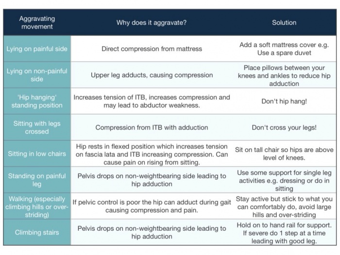
Educating patients on avoiding provocative movements that apply direct pressure on gluteal tendons for such as:
- Direct Pressure: by lying over the affected side
- Hip adduction: prolonged sitting with crossed knee, particularly on low seat, and prolonged weight shifting on one leg (handing on hip).
- Sustained postures and stretches
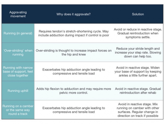
- Minimizing exposure to walking uphill, hopping, running and power walking in the short term.
This also includes training patients on using proper body mechanics when doing functional activities that require single-leg loading ,such as stairs negotiation, to be performed without excessive hip adduction and minimal trunk lean to minimize high external hip adduction moment.
Exercises[edit | edit source]
The exercise program suggested in Mellor et al. study utilized a combination of low load activation of abduction, pelvic control in frontal plane exercises and abductor loading in frontal plane.
To relief pain and activate abductors, isometric exercises were utilized from supine and standing positions using the belt as a resistance.
Bridging variations, squatting and lunges were used for functional Loading progressions and pelvic control training. It is important to integrate functional exercises at different positions to optimize the motor control.
To load abductors in frontal plane. subjects in exercise group performed different abduction exercises against spring resistance.
Motoring load appropriately is recommended by increasing load gradually without pain aggravation[2].
Steroid Injection[edit | edit source]
Corticosteriod injections provided analgesic effect,yet the pain doesn't resolve completely and and often recurrent[1]. Corticosteriods exhibit non-inflammatory effect. on the other hand, GT is reported to be a degenerative condition rather than inflammatory[22]. The pain relieving effect following crticosteriods might interfer with the tendon capacity to respond appropriately to loading[23].
Surgical Intervention[edit | edit source]
Surgery is considered in persistent pain and failed conservative treatment[1]. 90% long-term improvement was reported following GMed tendon repair in 72 patients[24]. Endoscopic repairs provide an accelerated rehabilitaion and less invasive approach with reduced rates of post-operative infection, scar tissue formation and pain. Patients with larger tears are not suitable for endoscopic techniques.
Removal of trochanteric bursae and ITB release is utilized in patients without gluteal tears. Studies report good to excellent short-medium term outcomes[25], however, most studies lacked control group and the rationale behind the mechanism of efficacy remains unclear[1].
Resources
[edit | edit source]
References[edit | edit source]
- ↑ 1.00 1.01 1.02 1.03 1.04 1.05 1.06 1.07 1.08 1.09 1.10 1.11 1.12 1.13 Grimaldi A, Mellor R, Hodges P, Bennell K, Wajswelner H, Vicenzino B. Gluteal tendinopathy: a review of mechanisms, assessment and management. Sports Medicine. 2015 Aug 1;45(8):1107-19.
- ↑ 2.0 2.1 2.2 Mellor, R., Bennell, K., Grimaldi, A., Nicolson, P., Kasza, J., Hodges, P., Wajswelner, H. and Vicenzino, B., 2018. Education plus exercise versus corticosteroid injection use versus a wait and see approach on global outcome and pain from gluteal tendinopathy: prospective, single blinded, randomised clinical trial. bmj, 361, p.k1662.
- ↑ Almekinders LC, Weinhold PS, Maffulli N. Compression etiology in tendinopathy. Clinics in sports medicine. 2003 Oct 1;22(4):703-10.
- ↑ Birnbaum K, Siebert CH, Pandorf T, Schopphoff E, Prescher A, Niethard FU. Anatomical and biomechanical investigations of the iliotibial tract. Surgical and Radiologic Anatomy. 2004 Dec 1;26(6):433-46.
- ↑ Sutter R, Kalberer F, Binkert CA, Graf N, Pfirrmann CW, Gutzeit A. Abductor tendon tears are associated with hypertrophy of the tensor fasciae latae muscle. Skeletal radiology. 2013 May 1;42(5):627-33.
- ↑ Pfirrmann CW, Notzli HP, Dora C, Hodler J, Zanetti M. Abductor tendons and muscles assessed at MR imaging after total hip arthroplasty in asymptomatic and symptomatic patients. Radiology. 2005 Jun;235(3):969-76.
- ↑ Birnbaum K, Prescher A, Niethard FU. Hip centralizing forces of the iliotibial tract within various femoral neck angles. Journal of Pediatric Orthopaedics B. 2010 Mar 1;19(2):140-9.
- ↑ Fearon AM, Stephens S, Cook JL, Smith PN, Neeman T, Cormick W, Scarvell JM. The relationship of femoral neck shaft angle and adiposity to greater trochanteric pain syndrome in women. A case control morphology and anthropometric study. Br J Sports Med. 2012 Sep 1;46(12):888-92.
- ↑ Woyski D, Olinger A, Wright B. Smaller insertion area and inefficient mechanics of the gluteus medius in females. Surgical and Radiologic Anatomy. 2013 Oct 1;35(8):713-9.
- ↑ Lequesne M. From “periarthritis” to hip “rotator cuff” tears. Trochanteric tendinobursitis. Joint Bone Spine. 2006;4(73):344-8.
- ↑ Connell DA, Bass C, Sykes CJ, Young D, Edwards E. Sonographic evaluation of gluteus medius and minimus tendinopathy. European radiology. 2003 Jun 1;13(6):1339-47.
- ↑ Bird PA, Oakley SP, Shnier R, Kirkham BW. Prospective evaluation of magnetic resonance imaging and physical examination findings in patients with greater trochanteric pain syndrome. Arthritis & Rheumatism: Official Journal of the American College of Rheumatology. 2001 Sep;44(9):2138-45.
- ↑ Classic Gluteal Tendinopathy Diagnosis. Available from: https://www.youtube.com/watch?v=216ZAxN4FNc
- ↑ Ober's Test/ITB tightness Available from: https://www.youtube.com/watch?v=Amjv6FzDeLE
- ↑ Patrick's/Faber/Figure four test. Available from: https://www.youtube.com/watch?v=89Qiht82zmg
- ↑ What single leg standing assessment can tell you Available from: https://www.youtube.com/watch?v=A9pi7_JRgwQ
- ↑ Resisted External Derotation Test/Gluteal Tendinopathy. Available from: https://www.youtube.com/watch?v=nwBnc3I53QY
- ↑ Bird PA, Oakley SP, Shnier R, Kirkham BW. Prospective evaluation of magnetic resonance imaging and physical examination findings in patients with greater trochanteric pain syndrome. Arthritis & Rheumatism: Official Journal of the American College of Rheumatology. 2001 Sep;44(9):2138-45.
- ↑ Cook JL, Purdam CR. The challenge of managing tendinopathy in competing athletes. Br J Sports Med. 2013 May 9:bjsports-2012.
- ↑ Video gait running analysis: alignment, issues, rear view. Available from: https://www.youtube.com/watch?v=k1hlY0EMYJw
- ↑ Kong A, Van der Vliet A, Zadow S. MRI and US of gluteal tendinopathy in greater trochanteric pain syndrome. European radiology. 2007 Jul 1;17(7):1772-83.
- ↑ Coombes BK, Bisset L, Vicenzino B. Thermal hyperalgesia distinguishes those with severe pain and disability in unilateral lateral epicondylalgia. The Clinical journal of pain. 2012 Sep 1;28(7):595-601.
- ↑ Coombes BK, Bisset L, Brooks P, Khan A, Vicenzino B. Effect of corticosteroid injection, physiotherapy, or both on clinical outcomes in patients with unilateral lateral epicondylalgia: a randomized controlled trial. Jama. 2013 Feb 6;309(5):461-9.
- ↑ Walsh MJ, Walton JR, Walsh NA. Surgical repair of the gluteal tendons: a report of 72 cases. The Journal of arthroplasty. 2011 Dec 1;26(8):1514-9.
- ↑ Craig RA, Gwynne Jones DP, Oakley AP, Dunbar JD. Iliotibial band Z‐lengthening for refractory trochanteric bursitis (greater trochanteric pain syndrome). ANZ journal of surgery. 2007 Nov;77(11):996-8.


