Vestibulo-Ocular Reflex
Introduction[edit | edit source]
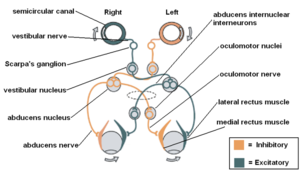
The position of the head in space and quick changes in the direction of head movement are sensed accurately by the vestibular system. When the head is in motion or while walking on the streets, we are able to see the surrounding clearly. The Vestibuloocular reflex (VOR) very well contributes to the maintenance of clear vision. When we move our head to the sides, to maintain a clear vision the eyeballs don’t move along with the head but moves towards the opposite side. These movements of the eyeballs help to fix the gaze on the object and thus we see it clearly[1].
The VOR is one of the reflexes of the vestibular system. Others are Vestibulospinal reflex (VSR) and Vestibulocollic Reflex (VCR). The VOR generates eye movements that enable clear vision while the head is in motion. The VCR acts on the neck musculature to stabilize the head. The VSR generates compensatory body movement to maintain head and postural stability and thereby prevent falls[2]
Abnormalities of the vestibular system result in sensations of dizziness or unsteadiness, which do reach our awareness, as well as problems with focusing our eyes and keeping our balance.
Peripheral processing[edit | edit source]
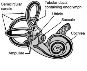
The vestibular portion of the labyrinth Contains five receptors: Three semicircular canals and two otolith organs
Semi Circular canals
- Anterior Semicircular Canal
- Posterior semicircular canal
- Horizontal Semicircular Canal
Otolith Organs
- Utricle
- Saccule
Semicircular Canals
The Semicircular canal (SCC) register the angular acceleration and they are at a right angle to each other. It delivers sensory input about head velocity, and that aids the VOR to generate an eye movement which is equivalent to the velocity of the head movement. Sensory receptors of SCC are situated in the ampulla, and the corresponding ampullary crest which contains vestibular hair cells is projected into the cupula which enables the VOR to generate an eye movement that matches the velocity of the head movement. The angular motion of the head either vertical or horizontal marks either an increase or decrease in hair cell activity and an opposite change in neuronal activity in paired canals. The firing in the neurons returns to its normal state in prolonged motion of the head.
Otolith Organs
The utricle and saccule register forces related to linear acceleration. They respond to linear head motion and static tilt to the gravitational axis. Unlike the SCCs, the Otoliths don’t need hydrodynamic systems to complete its works, it is embedded with calcium carbonate crystals ‘Otoconia’. In an upright individual, the saccule is vertical (parasagittal), whereas the utricle is horizontally oriented (near the plane of the lateral SCC).[3]
The saccule senses linear acceleration in the sagittal plane, associated with a forward pitch of the head. The utricle senses acceleration in the horizontal plane, provoked by a roll (lateral tilt) of the head.[3]
Central Processing[edit | edit source]
The neurons from both SCCs and Otolith organs join the eighth cranial nerve and their cell bodies are in the vestibular ganglion also known as Scarpa’s ganglion. The axons then enter into the pons and most of them go to the floor of the medulla, where the vestibular nuclei are located. A portion of vestibular sensory receptors travels to the cerebellum, reticular formation, thalamus and cerebral cortex.
There are four vestibular nuclei
- Medial vestibular nuclei
- Lateral vestibular nuclei
- Superior vestibular nuclei
- Inferior vestibular nuclei
The medial and superior vestibular nuclei receive input from the SCCs. The output of the medial nucleus is to the medial vestibulospinal tract for controlling neck muscles by its connections to the cervical spinal cord. It also plays a very crucial role in coordinating the interaction between head and eye movement. Neurons from medial and superior vestibular nuclei have ascending connections to motor nuclei of the ocular muscles and thus help in stabilizing the vision during head motion.
The input to the lateral vestibular nuclei is from SCCs, the utricle, the cerebellum and also the spinal cord. The output is to the vestibuloocular tracts and lateral vestibulospinal tracts which activate the antigravity muscles in the neck, trunk and limbs.
The inferior vestibular nucleus receives neurons from SCCs, Otolith organs and cerebellar vermis. The output is to the vestibulospinal tract and vestibuloreticular tract.
In summary, the vestibular system consists of both static and dynamic functions. The static functions are controlled by otolith organs, they monitor the absolute position of the head in the space which is very important in postural control. the dynamic functions are predominantly by the SCCs. It helps in monitoring the head rotations and angular acceleration thereby fixing the gaze through VOR[2]
Clinical Relevance[edit | edit source]
Application of VOR in clinical practice includes Oculocephalic reflex/ doll’s eye reflex, which examines the cranial nerve 3,6,8, (Oculomotor nerve, Abducens nerve, Vestibulocochlear nerve) brainstem nuclei and overall brainstem functions[4]. It is mostly examined in neurological critical care, anaesthetized and also in neonates. The oculocephalic reflex is performed by holding the eyelid open and moving the head from side to side. The reflex seems suppressed in conscious individuals with normal neurological function but is active in comatose patients with gross brain function[5].
- Positive test results indicate an intact brainstem (eyes move opposite to the rotation)
- Negative test results indicate impaired brainstem function (eyes move along with the head movement)[6]
Lesion to the vestibular nerve or vestibular nuclei has been shown to impair the ipsilateral Oculocephalic reflex[7]
Doll's eye reflex in critically ill intensive care patients can solely predict altered mental status after cessation of sedation[8]
Role of Physiotherapist[edit | edit source]
To maintain a clear vision and balance, the brain receives information from the vestibular system, visual system and somatosensory afferents. The information from the vestibular system is very crucial for VOR to work and thus by promoting a clear vision and balance.
Impaired vestibular functions lead to an impaired VOR which makes unclear vision and balance problems. Vestibular Rehabilitation is an evidence-based approach widely used by physiotherapists in improving symptoms like dizziness, vertigo, motion sensitivity, balance and postural control[9]. The VOR is found to be highly plastic and VOR gain can be improved by VOR adaptation exercises which are a part of vestibular rehabilitation[9] [10]
References[edit | edit source]
- ↑ Herdman SJ, Clendaniel RA. Vestibular Rehabilitation, 4th Edition. Philadelphia.FA DavisCompany, 2014
- ↑ 2.0 2.1 Anne Shumway C, Marjorie H. Motor Control. Fourth edition. Baltimore: Lippincott Williams&Wilkins 2012
- ↑ 3.0 3.1 Barber H, Stockwell C. Manual of Electronystagmography. St. Louis, MO: C.V. Mosby; 1976
- ↑ Swathi Somisetty, Das JM. Neuroanatomy, Vestibulo-ocular Reflex (Internet). Nih.gov. StatPearls Publishing; 2022 (cited 2023 Apr 26). Available from: https://www.ncbi.nlm.nih.gov/books/NBK545297/
- ↑ Dishion E, Prasanna Tadi. Doll’s Eyes [Internet]. Nih.gov. StatPearls Publishing; 2022 [cited 2023 Apr 26]. Available from: https://www.ncbi.nlm.nih.gov/books/NBK551716/#:~:text=Definition%2FIntroduction,and%20overall%20gross%20brainstem%20function.
- ↑ Pullen RL. Checking for oculocephalic reflex. Nursing (Internet). 2005 Jun 1 (cited 2023 Apr 26)
- ↑ Foster CA, Foster BD, Spindler J, Harris JP. Functional loss of the horizontal doll's eye reflex following unilateral vestibular lesions. The Laryngoscope. 1994 Apr;104(4):473-8 Available from: https://pubmed.ncbi.nlm.nih.gov/8164488/
- ↑ Tarek Sharshar, Porcher R, Shidasp Siami, Benjamim Rohaut, Bailly-Salin J, Hopkinson NS, et al. Brainstem responses can predict death and delirium in sedated patients in intensive care unit*. Critical Care Medicine [Internet]. 2011 Aug 1 (cited 2023 Apr 26);39(8):1960–7. Available from: https://journals.lww.com/ccmjournal/Abstract/2011/08000/Brainstem_responses_can_predict_death_and_delirium.14.aspx
- ↑ 9.0 9.1 Tonks B. Introduction to Vestibular Rehabilitation Course. Plus. 2021.
- ↑ Muntaseer Mahfuz M, Schubert MC, Figtree WV, Todd CJ, Migliaccio AA. Human vestibulo-ocular reflex adaptation training: time beats quantity. Journal of the Association for Research in Otolaryngology. 2018 Dec 31;19:729-39. https://www.ncbi.nlm.nih.gov/pmc/articles/PMC6249163/


