Matles Test
Original Editors - Nick Libotton as part of the Vrije Universiteit Brussel's Evidence-based Practice project.
Top Contributors - Nick Libotton, Admin, Uchechukwu Chukwuemeka, 127.0.0.1, Kim Jackson and Wanda van Niekerk
Definition/Description [1][edit | edit source]
Definition: The Matles Test is a visual diagnostic test for suspected rupture of the Achilles tendon.
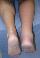
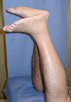
Description: The patient lies prone, actively or passively flexing the knee to 90° with both feet and ankles in a neutral position according to the patient. When an absence of plantar flexion is observed, the test proves positive. The rupture will tend the foot more into dorsal flexion.
Clinically Relevant Anatomy [3][edit | edit source]
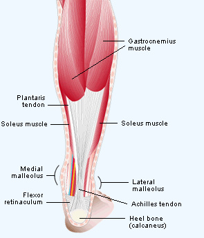
The Achilles Tendon consists of the tendons from 2 major muscles: the Gastrocnemius muscle, a bi-articular muscle that finds its origin on both the lateral and medial epicondyle and the Soleus muscle, which finds it's origin on the dorsal aspect of the tibia and fibula. Sometimes the Plantaris muscle is also present. This is a small muscle in the Popliteal Fossa facing its lateral aspect. It has a very long tendon and inserts together with the Soleus and Gastrocnemius muscles on the back of the heel bone (Calcaneus).
This general insertion is known as the Achilles tendon.
Examination [4][edit | edit source]
If awake, the patient is asked to lie prone and actively flex their knees to 90°. If locally anaesthetized, the examiner passively flexes the knees. The examiner must observe the position of the ankles and feet. An uninjured foot remains in slight plantar flexion. When the patient suffers from an Achilles tendon rupture, the foot will fall into a neutral position or even into dorsiflexion. This is often referred to as 'the angle of dangle'.
This is an observation test, no further palpation or movement is required. See the video below for a demonstration
Key Research [4][edit | edit source]
Along with the calf squeeze test, the Matles test has proven the most accurate to sensitivity compared to other tests, such as the gap palpation test, the Copeland and the O’Brien test. (Level of evidence: C)
It has also shown a high positive predictive value but no significant difference was established between the previously mentioned tests. (Level of evidence: C)
References[edit | edit source]
- ↑ John Kerr. Achilles Tendon Injury: Assessment and management in the emergency department. Advanced Emergency Nursing Journal: July/September 2007 - Volume 29 - Issue 3 - p 249-259. http://www.ncbi.nlm.nih.gov/pubmed/15912711 [1] Level of Evidence: C
- ↑ Pictures found on http://www.foothyperbook.com/trauma/achillesRupture/achillesRuptureClin.htm
- ↑ Schünke M, Schulte E, Schumacher U, Voll M, Wesker K. Prometheus anatomy. Houten: Bohn Stafleu van Loghum, 2005.
- ↑ 4.0 4.1 Nicola Maffulli. The Clinical Diagnosis of Subcutaneous Tear of the Achilles Tendon: A Prospective Study in 174 Patients. The American Journal of Sports Medicine, Vol. 26, No. 2, 1998. http://www.ncbi.nlm.nih.gov/pubmed/9548122 [3] (Level of evidence: C)
- ↑ The Physio Channel. Matles Achilles Tendon Rupture Test Video Demonstration. Available from: http://www.youtube.com/watch?v=KLB-IB7s12s [last accessed 9/9/2023]


