When refering to evidence in academic writing, you should always try to reference the primary (original) source. That is usually the journal article where the information was first stated. In most cases Physiopedia articles are a secondary source and so should not be used as references. Physiopedia articles are best used to find the original sources of information (see the references list at the bottom of the article).
If you believe that this Physiopedia article is the primary source for the information you are refering to, you can use the button below to access a related citation statement.
Biomechanics is an area of science that uses principles of physics to measure and study, how forces interact with and affect living body[1]. These forces could be Internal i.e. within the body or External i.e. outside the body.
To understand the biomechanics of thorax we will need to study the basic anatomy, joint articulations, muscles associated in the thorax along with kinematics of the rib cage.
The thorax is formed by the thoracic vertebrae ,the ribs and the sternum.
Parts of Thorax
It has two main functions,
Providing anchor for muscle attachments
Major role of ventilation
Structure of Thorax
The main three structures mentioned above work in a coordinated motion in thorax to produce respiration.
Anatomy
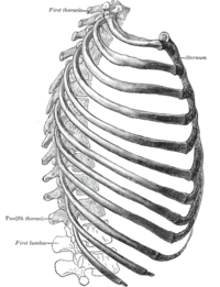
The rib cage is a system of various bones and muscle. Bones involved are sternum, 12 pairs of Ribs, 12 Thoracic vertebrae.
It is like a compartment sealed by various structures from each side.
Refer to the diagram of anterior, posterior and lateral view of thorax to better understand the bones involved in the ribcage.
Head of ribs 1, 10 to 12 and corresponding vertebral
body
- More mobile than
typical CV joint
5.
Costotransverse
Costal tubercle of rib 1 to 10 and costal facet on
transverse process of corresponding vertebrae
- Synovial type of joint
6.
Costochondral
1 to 10th rib with costal cartilage
- Synchondrosis type of joint
- Have no ligamentous support
7.
Costosternal
(Refer diagram below)
Costal cartilage of ribs 1 to 7 with sternum
-Joints with 1, 6 and 7 ribs
are synchondroses type.
-Joints with 2 to 5 ribs are
synovial type.
8.
Interchondral
7 to 10 costal cartilage with cartilage above it
- Synovial type of joint
Function of Thorax
Kinematics
The motion of the ribs in conjunction with sternum and thoracic vertebrae helps produce the movements of respiration. These consist of inspiration and expiration. Thoracic kinematics consists of understanding the changes in intrathoracic volume during ventilation. These occur due to complex, synchronized activity of the ribs along with sternum and primary muscles of respiration. It is determined by[2]
Type & angles of articulations
movement of manubrium sternum
elasticity of costal cartilage
The kinematics involved in the thorax can be further classified into[1],
Changes in vertical diameter
Changes in anterior-posterior diameter (Pump handle motion)
Changes in transverse diameter (Bucket handle motion)
There is a single axis of motion for 1 to 10 ribs through the center of their costovertebral(CV) and costotransverse(CT) joints. This influences the motion of these ribs during the process of respiration
The two main motions occurring in thorax during ventilation can be explained as below,
Refer to the video shown below to understand the kinematics of thorax better,
Total motion of sternum and upper ribs together increases in anterior-posterior diameter of thorax
Changes produced mainly by muscles laterally placed in the ribcage[4]
Motion of sternum resembles pump handlemovement, hence the name.
Lower ribs elevate
Motion is more in the lateral part of ribs due to its more angled shape and indirect attachment of the ribs
Total motion of lower ribs together increases the transverse diameter of thorax
Changes produced mainly by muscles anteriorly & posteriorly placed in the ribcage[4]
Motion of the lower ribs resembles bucket handle movement, hence the name.
Muscles involved in respiration
To bring about the intrathoracic volume changes during respiration, muscles of respiration play a crucial role. The muscles can be divided into primary and accessory depending on type of breathing i.e. quite or forced, in which they are used.
The detailed function of each muscle can be accessed by clicking on the muscle name mentioned below. Each has been linked to independent physiopedia pages dedicated to explaining them.
↑ 1.01.11.21.3Neumann DA. Kinesiology of the musculoskeletal system-e-book: foundations for rehabilitation. Elsevier Health Sciences; 2016 Nov 3.
↑ 2.02.12.2Pamela K. Levangie, Cynthia C. Norkin, 2005, Joint structure and function: A comprehensive analysis, 4th. Edn, Philadelphia, FA Davis publishers.
↑ 4.04.1Loring SH. Action of human respiratory muscles inferred from finite element analysis of rib cage. Journal of Applied Physiology. 1992 Apr 1;72(4):1461-5.
Original Editor - [[User:Manisha Shrestha|Manisha Shrestha] Top Contributors - Sonal Joshi and Manisha Shrestha