
Ankle-Spine Syndrome
Overview
Ankle-Spine Syndrome, also called "Raffet Syndrome II," was first introduced in 2024 by researcher Ahmed Raffet El-Sayed. And is characterized by ipsilateral calf muscle weakness and contralateral lumbar radiculopathy . It is linked to chronic back pain and abnormalities in gait. In the first study that documented the condition, the prevalence of Raffet Syndrome II was found to be 0,6%. Six cases out of 1000 people with calf muscle weakness evaluated were diagnosed with this condition. [1]
Clinical Presentation [1]
- Radicular pain that is sharp and shoots down the lower limb, confined to a small area (5–8 cm).
- Pain increased during dynamic activities (e.g., walking, running, jogging).
- Pain improved during static activities (e.g., standing, sitting) and rest.
- Gait Abnormalities (During mid-stance and terminal stance).
- Ipsilateral Calf Muscle weakness (leading to excessive tibial advancement upon the femur).
- Biomechanical Compensations:
- Ipsilateral pelvic drop compensated by contralateral side bending of the trunk during gait.
- Backward pelvic rotation compensated by contralateral axial rotation of lumber spine during gait which lead to decrease neural frominal dimensions (width, height, area) and increase facet joint compression and nerve root Impingement.[2]
- Chronic low back pain.
- A relevant past history of persistent ankle injuries.
Gait Abnormalities [1][3][4]
Excessive Knee Flexion:
- Occurs during the gait cycle's mid-stance and terminal stance phases as a result of insufficient strength in the calf muscles, specifically the soleus.
- A failure to achieve proper knee extension results from the tibia's excessive advancement over the foot.
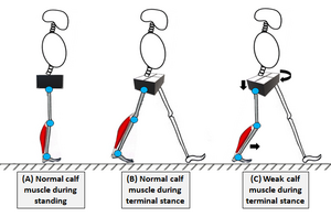
Diagram of the pelvis and calf muscles during walking. (A) A typical standing calf muscle. The position of the pelvis is neutral. (B) The calf muscle in terminal stance is normal. When the knee is fully extended and the heel lifts off the ground, the lower limb is functionally lengthened. (C) The calf muscle is weak in the terminal position.

Delayed or Absent Heel Raise:
- The heel raise begins at start of terminal stance, is delayed or disappeared due to soleus muscle weakness. And lead to inadequate propulsion during gait.
Functional Shortening of the Affected Limb:
- The ipsilateral limb during terminal stance appears relatively shorter due to excessive knee flexion and delayed heel raises.
- The body compensates by excessive pelvic motion.
Pelvic Compensatory Mechanisms:
- Frontal Plane: Excessive pelvic drop toward affected limb to regain limb length.
- Transverse Plane: Excessive backward rotation of the pelvis in affected side.
Shifts in Center of Gravity (COG):
- Excessive forward displacement of COG due to advances the body mass over the hip joint due to knee flexion and tibial advancement.
Exaggerated Heel Rocker:
- Excessive dorsiflexion of the ankle joint (Heel Rocker) is made possible by the weak soleus in mid-stance, which speeds up anterior tibial advancement and knee flexion.
Criteria to Diagnose
The key to diagnose Ankle-Spine Syndrome (Raffet Syndrome II) is present the following in the case[1]:
- Significant calf muscle weakness, particularly the soleus muscle sufficient to induce knee flexion during mid-stance in the gait cycle that cause abnormal pelvic motion.
- Exaggerated decoupling mechanism of the lumbar spine in response to abnormal pelvic motion.
- Presence of an asymptomatic lumbar disc herniation contralateral to affected calf that becomes symptomatic due to biomechanical disruptions.
- A relevant past history of persistent ankle injuries causing unresolved calf muscle weakness.
Outcome Measures[1]
Physiotherapy Management
(Raffet et al., 2024) found that the following methods and approaches were highly successful after 6–8 weeks of interventio[1]:
- Open and closed-chain strengthening exercises targeting the calf muscle.
- Double and single-leg calf raises (with straight and bent knees).
- Wall-sit calf raises.
- Seated calf raises.
- Gait training.
- Lumbopelvic stabilization exercises.
- Core strength.
- Lumbar myofascial release therapy.
- Electrophysical modalities.
References
- ↑ 1.0 1.1 1.2 1.3 1.4 1.5 Raffet, A., Laslett, M., Schamberger, W., Beltagi, A., ElMeligie, M. M., Kentiba, E., Khaled, N., Sayed, H. Y., Omar, A. H., Hawana, M. M., & Fawaz, H. E. (2024, July 11). Ankle spine syndrome “Raffet Syndrome II”. Ipsilateral calf muscle weakness induces contralateral lumbar radiculopathy: 2026, V. 12, No 2. https://hsr-journal.com/index.php/journal/article/view/1048
- ↑ Takahashi, K., Yadav, A. K., Hashimoto, K., Tsubakino, T., Aizawa, T., & Tanaka, Y. (2022). Foraminal stenosis at L5–S1 as an overlooked Pathology of bilateral Radiculopathy: A case series. Journal of Orthopaedic Case Reports, 12(6), 13–18. https://doi.org/10.13107/jocr.2022.v12.i06.2846
- ↑ GAIT Analysis: Normal and pathological function. (2010, June 1). https://pmc.ncbi.nlm.nih.gov/articles/PMC3761742/
- ↑ Neumann D. Kinesiology of the Musculoskeletal System: Foundations for Rehabilitation. Third Edition. Mosby, Elsevier Health Sciences, St. Louis, 2016.