Vastus Lateralis: Difference between revisions
Abbey Wright (talk | contribs) No edit summary |
Joao Costa (talk | contribs) No edit summary |
||
| (7 intermediate revisions by 4 users not shown) | |||
| Line 1: | Line 1: | ||
'''Original Editor ''' [[User:Andeela Hafeez|Andeela Hafeez]] | |||
'''Top Contributors''' - {{Special:Contributors/{{FULLPAGENAME}}}}
| |||
== Description == | == Description == | ||
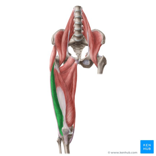
[[File:Vastus lateralis muscle - Kenhub.png|alt=Vastus lateralis muscle (highlighted in green) - anterior view|right|frameless|500x500px|Vastus lateralis muscle (highlighted in green) - anterior view]] | |||
The vastus lateralis muscle is located on the lateral side of the thigh. This muscle is the largest of the [[Quadriceps Muscle|quadriceps]] which includes: [[Rectus Femoris|rectus femoris]], [[Vastus Intermedius|vastus intermedius]], and [[Vastus Medialis|vastus medialis]]. Together, the quadriceps act on the [[knee]] and [[hip]] to promote movement as well as strength and stability. They provide power for and absorb the impact of daily activities such as walking, running, and jumping. | |||
Image: Vastus lateralis muscle (highlighted in green) - anterior view <ref >Vastus lateralis muscle (highlighted in green) - anterior view image - © Kenhub https://www.kenhub.com/en/study/main-muscles-of-lower-limb</ref> | |||
== Anatomy == | == Anatomy == | ||
| Line 23: | Line 23: | ||
=== Blood Supply === | === Blood Supply === | ||
== Function == | == Function == | ||
| Line 48: | Line 47: | ||
*'''Standing ''' | *'''Standing ''' | ||
Stand on one leg and pull the other foot up behind your bottom<br>Keep your knees together and push your hips forwards to increase the stretch <br>Hold for between 10 and 30 seconds | Stand on one leg and pull the other foot up behind your bottom<br>Keep your knees together and push your hips forwards to increase the stretch <br>Hold for between 10 and 30 seconds | ||
| |||
*'''Lying''' | *'''Lying''' | ||
Lay on your front and pull one foot up to meet your buttocks <br>Hold for between 10 and 30 | Lay on your front and pull one foot up to meet your buttocks <br>Hold for between 10 and 30 second | ||
== Clinical relevance == | |||
[[Patellofemoral Pain Syndrome|Patellofemoral pain syndrome]] | |||
=== | |||
Some evidence has shown that in patients with patellofemoral pain syndrome vastus lateralis contracts prematurely when compared with vastus medialis which has been hypothesised to be a cause of knee pain. <ref>Cowan SM, Bennell KL, Hodges PW, Crossley KM, McConnell J. [https://www.sciencedirect.com/science/article/pii/S0003999301747375 Delayed onset of electromyographic activity of vastus medialis obliquus relative to vastus lateralis in subjects with patellofemoral pain syndrome]. Archives of physical medicine and rehabilitation. 2001 Feb 1;82(2):183-9.</ref> In more recent studies this theory has not been clinically proven and vastus lateralis/medialis timing has been shown to be uneven in healthy test subjects. <ref>Chester R, Smith TO, Sweeting D, Dixon J, Wood S, Song F. [https://bmcmusculoskeletdisord.biomedcentral.com/articles/10.1186/1471-2474-9-64 The relative timing of VMO and VL in the aetiology of anterior knee pain: a systematic review and meta-analysis]. BMC musculoskeletal disorders. 2008 Dec;9(1):64.</ref> | |||
== References == | == References == | ||
| Line 75: | Line 62: | ||
[[Category:Anatomy]] | [[Category:Anatomy]] | ||
[[Category:Muscles]] | [[Category:Muscles]] | ||
[[Category:Hip]] | |||
[[Category:Hip - Anatomy]] | |||
[[Category:Hip - Muscles]] | |||
[[Category:Knee]] | |||
[[Category:Knee - Anatomy]] | |||
[[Category:Knee - Muscles]] | |||
Latest revision as of 07:38, 1 April 2022
Original Editor Andeela Hafeez
Top Contributors - Andeela Hafeez, Abbey Wright, Evan Thomas, Joao Costa, WikiSysop, Kim Jackson and Lucinda hampton
Description[edit | edit source]
The vastus lateralis muscle is located on the lateral side of the thigh. This muscle is the largest of the quadriceps which includes: rectus femoris, vastus intermedius, and vastus medialis. Together, the quadriceps act on the knee and hip to promote movement as well as strength and stability. They provide power for and absorb the impact of daily activities such as walking, running, and jumping.
Image: Vastus lateralis muscle (highlighted in green) - anterior view [1]
Anatomy[edit | edit source]
Origin[edit | edit source]
Upper inter-trochanteric line, base of greater trochanter, lateral linea aspera, lateral supracondylar ridge and lateral intermuscular septum[2]
Insertion[edit | edit source]
Lateral quadriceps tendon which attached onto the tibial tubercle.[2]
Nerve Supply[edit | edit source]
Posterior division of femoral nerve (L3,4)
Blood Supply[edit | edit source]
Function[edit | edit source]
Actions[edit | edit source]
1. Extension of the knee
Functional contributions[edit | edit source]
In everyday life, the quadriceps muscle group as a whole allows a person to stand up from sitting, walk up or down stairs along with basic walking and running. These muscles are not active while standing with knees fully extending, but become active during the heel-strike and toe-off phases of gait.[3]
Assessment[edit | edit source]
Palpation[edit | edit source]
In supine:
- Place palpating hand distal to greater trochanter
- Get the patient to actively and isometrically contract quadriceps
- Palpate the contracting muscle focusing on the lateral side to target vastus lateralis
- Continue to palpate distally until the quadriceps tendon
Length Tension Testing / Stretching[edit | edit source]
- Standing
Stand on one leg and pull the other foot up behind your bottom
Keep your knees together and push your hips forwards to increase the stretch
Hold for between 10 and 30 seconds
- Lying
Lay on your front and pull one foot up to meet your buttocks
Hold for between 10 and 30 second
Clinical relevance[edit | edit source]
Some evidence has shown that in patients with patellofemoral pain syndrome vastus lateralis contracts prematurely when compared with vastus medialis which has been hypothesised to be a cause of knee pain. [4] In more recent studies this theory has not been clinically proven and vastus lateralis/medialis timing has been shown to be uneven in healthy test subjects. [5]
References[edit | edit source]
- ↑ Vastus lateralis muscle (highlighted in green) - anterior view image - © Kenhub https://www.kenhub.com/en/study/main-muscles-of-lower-limb
- ↑ 2.0 2.1 Gray HFRS, Gray's Anatomy 15th edition, New York, NY: Barnes & Noble,2010. p396-398
- ↑ Bohm S, Marzilger R, Mersmann F, Santuz A, Arampatzis A. Operating length and velocity of human vastus lateralis muscle during walking and running. Scientific reports. 2018 Mar 22;8(1):5066.
- ↑ Cowan SM, Bennell KL, Hodges PW, Crossley KM, McConnell J. Delayed onset of electromyographic activity of vastus medialis obliquus relative to vastus lateralis in subjects with patellofemoral pain syndrome. Archives of physical medicine and rehabilitation. 2001 Feb 1;82(2):183-9.
- ↑ Chester R, Smith TO, Sweeting D, Dixon J, Wood S, Song F. The relative timing of VMO and VL in the aetiology of anterior knee pain: a systematic review and meta-analysis. BMC musculoskeletal disorders. 2008 Dec;9(1):64.


