Rib Fracture
Introduction[edit | edit source]

Rib fractures occur when a significant enough force directed at the rib causes a break.
- There are a total of 12 pairs of ribs in the thoracic region.
- The first seven ribs attach anteriorly to the sternum and posteriorly to the spinal column.
- Rib numbers 8 through 10 attach similarly but connect to the costal cartilage of the sternum anteriorly.
- Ribs 11 and 12 have the name of “floating” ribs as they only attach posteriorly but do not attach anteriorly.
- Underneath each rib lies the intercostal nerve, artery, and veins which supply to blood supply and innervation.
- The ribs function to protect the underlying organs and structures of the thoracic cavity. Any rib fracture should warrant a thorough evaluation of any concomitant injury, including lungs, heart, kidney, spleen, liver, and neuro-vasculature.[1]
Clinically Relevant Anatomy[edit | edit source]

- The chest comprises of 12 rib bones on each side of the body.
- Each rib attaches to the spine at the back of the body and then travels around to the front of the chest.
The top 7 ribs attach to the sternum, the 8th to 10 ribs attach to the ribs above via cartilage and the 11th and 12th ribs are known as ‘floating’ ribs as they are unattached at the front of the chest[2].
Etiology[edit | edit source]
- Blunt and penetrating trauma: e.g. motor vehicle accidents, falls, assaults - most common injury is blunt thoracic trauma, occurring in 50% of cases
- Pathological fractures eg Osteoporosis, malignancies.
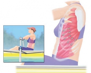
- Stress fractures: occur more commonly in high-level athletes eg rowers, see image R
- Non-accidental injuries in children
- Cardiopulmonary resuscitation (CPR): occurs in 1 in 3 5
- Fetal rib fractures: caused by skeletal dysplasias
- Radiation-induced rib fractures 8-9
- Spontaneous: spontaneous rib fracture[3]

Pathological Process[edit | edit source]

The 4th-10th ribs are the most commonly fractured.
Fractures of the 1st-3rd ribs are associated with high-energy trauma.
When the rib is fractured twice, the term floating rib is used to describe the free fracture fragment, and when three or more contiguous floating ribs are present this is called a flail chest.
Buckle rib fractures can also occur.[3]
Clinical Presentation[edit | edit source]
Typically, patients will provide a history of recent blunt or penetrating thoracic trauma and pain at that site. They may also exhibit decreased ability to perform full inspiration due to pain. The physical exam may reveal chest wall bruising, along with bony tenderness to palpitation or crepitus.
Patients with a broken rib typically experience a sudden onset of chest pain, mid back pain or pain in the side of the ribs at the time of injury that occasionally may radiate into the back, shoulder or neck. Pain is often sharp and intense and may increase during deep breathing, coughing, laughing or sneezing.
Possible presenting symptoms:
- Ache in the ribs that is particularly prominent at night or first thing in the morning (particularly the first few days following injury).
- Pain may increase when lying on the affected side, applying pressure to the rib region or on firmly touching the rib at the site of injury.
- Pain during movements of the upper back such as bending or twisting, or sometimes during certain activities of the upper limb (such as overhead activities, or during heavy pushing, pulling or lifting)[2].
Any vital sign abnormalities such as hypoxia, tachypnea, or significant respiratory distress should undergo further evaluation of other possible injuries such as pneumothorax, hemothorax, cardiac and pulmonary contusions. Lower rib segment injuries should undergo assessment for kidney, liver, and spleen. Any patient with paradoxical chest wall movement or suspicion for multiple rib fractures should be evaluated for flail chest and managed accordingly[1].
Diagnostic Procedures[edit | edit source]
A thorough subjective and objective examination from a physiotherapist is important to assist with diagnosis of a fractured rib
However to determine the likelihood of associated damage to other organs such as the lungs (e.g. a pneumothorax), liver, spleen or kidneys. Investigations such as an X-ray, MRI or CT scan may be performed to confirm diagnosis[2].
- Plain radiograph - may miss up to 50% of rib fractures even with dedicated oblique rib projections
- CT - more sensitive than plain radiography for the detection of rib fractures 1, 3
- Ultrasound - Common application of point of care ultrasonography, used in a complementary manner to conventional radiography in the workup of blunt chest wall trauma and localised chest pain. Ultrasonography is more sensitive and specific than conventional radiography for rib fracture detection in blunt trauma when performed by a trained clinician[3]
Outcome Measures[edit | edit source]
Management / Interventions[edit | edit source]
Rib fractures themselves are treated symptomatically and have a good prognostic outcome.
- For simple, isolated rib fractures, conservative therapy is usually adequate which includes appropriate analgesia, rest, and ice. The use of an incentive spirometer should be encouraged to prevent pulmonary atelectasis and splinting. Intercostal nerve blocks can also be applied to aid in pain control. Rib taping is no longer the recommended treatment as it can impede inspiratory effort[1].
When conservative management fails or for more severe rib fractures, surgical stabilization can be an option.
- Severe rib injuries (e.g. flail chest) may be treated with ORIF, often in the setting of other severe traumatic injuries and in the hope that respiratory function will improve facilitating a shorter ICU stay and quicker recovery.[3]
Associations[edit | edit source]
Rib fractures are often associated with other injuries and the greater the number of rib fractures the more likely are associated injuries:
- brachial plexus or subclavian vessel injuries (1st-3rd rib fractures)
- pneumothorax/haemothorax
- pulmonary laceration
- lung herniation
- liver, kidney and spleen traumatic injuries (10th-12th rib fractures)[3]
Aside from immediate traumatic complications outlined above atelectasis and pneumonia may develop, mainly due to poor respiratory effort secondary to pain, and this increases the morbidity and mortality due to rib fractures
Physiotherapy[edit | edit source]

Physiotherapy treatment can assist patients with this condition and ensure they have a safe return to activity. Treatment may comprise:
- education
- rest from aggravating activities
- dry needling
- protective padding
- exercises to improve posture, flexibility and strength, and to prevent localized lung collapse
- activity modification advice
- taping techniques (e.g. postural taping)
- a graduated return to activity plan
- soft tissue massage
- joint mobilization (usually following completion of fracture healing)
- electrotherapy[2]
- home exercise plan eg postural exercises, deep breathing exercises, thoracic rotation exercises.
References[edit | edit source]
- ↑ 1.0 1.1 1.2 Kuo K, Kim AM. Rib Fracture.Available from:https://www.ncbi.nlm.nih.gov/books/NBK541020/ (last accessed 5.5.2020)
- ↑ 2.0 2.1 2.2 2.3 Physioadvisor.com Rib Fracture Available from:https://www.physioadvisor.com.au/injuries/upper-back-chest/rib-fracture/ (last accessed 5.5.2020)
- ↑ 3.0 3.1 3.2 3.3 3.4 Radiopedia Rib Fractures Available from:https://radiopaedia.org/articles/rib-fractures (last accessed 5.5.2020)

