Gait: Difference between revisions
No edit summary |
No edit summary |
||
| Line 11: | Line 11: | ||
* Gait disorders lead to a loss of personal freedom, falls and injuries and result in a marked reduction in the quality of life<ref name=":0">Pirker W, Katzenschlager R. [https://www.ncbi.nlm.nih.gov/pmc/articles/PMC5318488/ Gait disorders in adults and the elderly.] Wiener Klinische Wochenschrift. 2017 Feb 1;129(3-4):81-95.Available from:https://www.ncbi.nlm.nih.gov/pmc/articles/PMC5318488/ (last accessed 27.6.2020)</ref>. | * Gait disorders lead to a loss of personal freedom, falls and injuries and result in a marked reduction in the quality of life<ref name=":0">Pirker W, Katzenschlager R. [https://www.ncbi.nlm.nih.gov/pmc/articles/PMC5318488/ Gait disorders in adults and the elderly.] Wiener Klinische Wochenschrift. 2017 Feb 1;129(3-4):81-95.Available from:https://www.ncbi.nlm.nih.gov/pmc/articles/PMC5318488/ (last accessed 27.6.2020)</ref>. | ||
This page presents information about: | This page presents information about: | ||
* | * The gait cycle, | ||
* | * Gait analysis | ||
* Gait disorders | * Gait disorders | ||
== Definitions == | == Definitions == | ||
'''Gait''' - the manner or style of walking. | '''Gait''' - the manner or style of walking. | ||
'''Gait Analysis''' - | '''Gait Analysis''' - | ||
'''Gait speed''' | An analysis of each component of the three phases of ambulation is an essential part of the diagnosis of various neurologic disorders and the assessment of patient progress during rehabilitation and recovery from the effects of a neurologic disease, a musculoskeletal injury or disease process, or amputation of a lower limb. | ||
'''Gait speed''' | |||
* The time it takes to walk a specified distance, usually 6 m or less. Slower speeds correlate with an increased risk of mortality in geriatric patients.<ref name=":1">Medical dictionary [https://medical-dictionary.thefreedictionary.com/gait+speed Gait speed] Available from: https://medical-dictionary.thefreedictionary.com/gait+speed (last accessed 28.6.2020)</ref> | |||
* Normal walking speed primarily involves the lower extremities, with the arms and trunk providing stability and balance. | * Normal walking speed primarily involves the lower extremities, with the arms and trunk providing stability and balance. | ||
* | * Faster speeds - body depends on the upper extremities and trunk for propulsion, balance and stability with the lower limb joints producing greater ranges of motion.<ref name="Shultz">Shultz SJ et al. Examination of musculoskeletal injuries. 2nd ed, North Carolina: Human Kinetics, 2005. p55-60.</ref> | ||
'''The gait cycle''' is a repetitive pattern involving steps and strides<ref name="Loudon">Loudon J, et al. The clinical orthopedic assessment guide. 2nd ed. Kansas: Human Kinetics, 2008. p.395-408.</ref> | |||
'''The gait cycle''' is a repetitive pattern involving steps and strides<ref name="Loudon">Loudon J, et al. The clinical orthopedic assessment guide. 2nd ed. Kansas: Human Kinetics, 2008. p.395-408.</ref> | |||
'''A step''' is one single step | '''A step''' is one single step | ||
| Line 44: | Line 43: | ||
Received 25 August 1997; accepted 22 September 1997 Available from: | Received 25 August 1997; accepted 22 September 1997 Available from: | ||
</ref>. | </ref>. | ||
* The gait cycle is one third longer in time, ground reaction force is smaller (so the load is lower)<ref name="Subotnick">Subotnick S. Sports medicine of the lower extremity. Harcourt (USA):Churchill Livingstone, 1999.</ref>. | |||
* The gait cycle is one third longer in time | * Running cycle has one stance phase, shock absorption much larger in comparison, explains why runners have more overload injuries.<ref name="Subotnick" /> | ||
* Running cycle | |||
== The Gait Cycle == | == The Gait Cycle == | ||
| Line 56: | Line 54: | ||
# Regulation of joint forces and moments across synovial joints and skeletal segments. | # Regulation of joint forces and moments across synovial joints and skeletal segments. | ||
# Generation of ground reaction forces. | # Generation of ground reaction forces. | ||
The normal forward step consists of two phases: stance phase; swing phase, | |||
* Stance phase occupies 60% of the gait cycle | * Stance phase occupies 60% of the gait cycle, during which one leg and foot are bearing most or all of the body weight | ||
* Swing phase occupies only 40% of it.<ref name=" | * Swing phase occupies only 40% of it<ref name="Loudon" />, during which the foot is not touching the walking surface and the body weight is borne by the other leg and foot. | ||
The 90 second video below gives the basics of this cycle{{#ev:youtube|DP5-um6SvQI|400}}<ref>Nicole Comninellis The Gait Cycle Animation Available from https://www.youtube.com/watch?time_continue=35&v=DP5-um6SvQI</ref> | * In a complete two-step cycle both feet are in contact with the floor at the same time for about 25 per cent of the time. This part of the cycle is called the double-support phase.Gait cycle phases: the stance phase and the swing phase and involves a combination of open and close chain activities.<ref name="Shultz" /> | ||
The 90 second video below gives the basics of this cycle{{#ev:youtube|DP5-um6SvQI|400}}<ref>Nicole Comninellis The Gait Cycle Animation Available from https://www.youtube.com/watch?time_continue=35&v=DP5-um6SvQI</ref>[[Image:Figure2.jpg|576x576px|right|frameless]]'''Phases of the Gait Cycle (8 phase model):'''<ref name="Loudon" /><ref name="Demos">Demos, Gait analysis, (http://www.ncbi.nlm.nih.gov/books/NBK27235/), 2004.</ref> | |||
[[Image:Figure2.jpg|576x576px|right|frameless]] | |||
#Initial Contact | #Initial Contact | ||
#Loading Response | #Loading Response | ||
| Line 77: | Line 67: | ||
#Mid Swing | #Mid Swing | ||
#Late Swing.<ref name="Berger">Berger W, et al. [https://www.ncbi.nlm.nih.gov/pmc/articles/PMC1193250/ Corrective reactions to stumbling in man: neuronal co-ordination of bilateral leg activity during gait]. J Physiol 1984;357: 109-125.</ref><br> | #Late Swing.<ref name="Berger">Berger W, et al. [https://www.ncbi.nlm.nih.gov/pmc/articles/PMC1193250/ Corrective reactions to stumbling in man: neuronal co-ordination of bilateral leg activity during gait]. J Physiol 1984;357: 109-125.</ref><br> | ||
'''Heel Strike (or initial contact) -''' Short period, begins the moment the foot touches the ground and is the first phase of double support.<ref name="Shultz" /> | '''Heel Strike (or initial contact) -''' Short period, begins the moment the foot touches the ground and is the first phase of double support.<ref name="Shultz" /> | ||
| Line 122: | Line 110: | ||
* Neutral position of the ankle.<ref name="Shultz" /> | * Neutral position of the ankle.<ref name="Shultz" /> | ||
== Anatomical Considerations == | == Gait Cycle - Anatomical Considerations == | ||
* Pelvic region - anterior-posterior displacement, which alternates from left to right. Facilitates anterior movement of the leg (each side anterior-posterior displacement of 4-5°).<ref name="Shultz" /><ref name="Loudon" /><ref name="Demos" /> | |||
* Frontal plane - varus movement in the: foot between heel-strike and foot-flat and between heel-off and toe-off; hip, in lateral movements (when the abductors are too weak, a [[Trendelenburg Gait|Trendelenburg gait]] can be observed).<ref name="Shultz" /><ref name="Demos" /> Valgus movement between foot-flat and heel off in the feet. | |||
* A disorder in any segment of the body can have consequences on the individual's gait pattern.<ref name="Shi">Shi D, et al. [https://www.ncbi.nlm.nih.gov/pubmed/21162970 Effect of anterior cruciate ligament reconstruction on biomechanical features of knee level in walking: a meta analysis.] Chin Med J 2010;123(21):3137-3142.</ref> | |||
== Gait Disorders == | |||
Gait disorders - altered gait pattern due to deformities, weakness or other impairments eg loss of motor control or pain<ref name="Malanga">Malanga G and Delisa J.A. Section One: Clinical Observation. Office of rehabilitation Research and Development No Date. http://www.rehab.research.va.gov/mono/gait/malanga.pdf (accessed 6 February 2010)</ref>. | |||
* [[File:Human falls.jpg|right|frameless]]Prevelence increases with age and the number of people affected will substantially increase in the coming decades due to the expected demographic changes. | * [[File:Human falls.jpg|right|frameless]]Prevelence increases with age and the number of people affected will substantially increase in the coming decades due to the expected demographic changes. | ||
* Lead to a loss of personal freedom and to reduced quality of life. | * Lead to a loss of personal freedom and to reduced quality of life. | ||
| Line 140: | Line 127: | ||
* Any gait disorder should be thoroughly investigated in order to improve patient mobility and independence, to prevent falls and to detect the underlying causes as early as possible. | * Any gait disorder should be thoroughly investigated in order to improve patient mobility and independence, to prevent falls and to detect the underlying causes as early as possible. | ||
* Thorough clinical observation of gait, careful history taking focussed on gait and falls and physical, neurological and orthopedic examinations are basic steps in the categorization of gait disorders and serve as a guide for ancillary investigations and therapeutic interventions. | * Thorough clinical observation of gait, careful history taking focussed on gait and falls and physical, neurological and orthopedic examinations are basic steps in the categorization of gait disorders and serve as a guide for ancillary investigations and therapeutic interventions. | ||
=== Gait Descriptions === | |||
This is not an exhaustive list. | |||
* Antalgic gait a limp adopted so as to avoid pain on weight-bearing structures, characterized by a very short stance phase. | |||
* Ataxic gait an unsteady, uncoordinated walk, with a wide base and the feet thrown out, coming down first on the heel and then on the toes with a double tap.This gait is associated with cerebellar disturbances and can be seen in patients with longstanding alcohol dependency. People with 'Sensory'Disturbances may present with a sensory ataxic gait. Presentation is a wide base of support, high steps, and slapping of feet on the floor in order to gain some sensory feedback. They may also need to rely on observation of foot placement and will often look at the floor during mobility due to a lack of proprioception | |||
=== | * Equine gait a walk accomplished mainly by flexing the hip joint; seen in crossed leg palsy. | ||
* Parkinsonian Gait (seen in parkinson's disease and other neurologic conditions that affect the basal ganglia). Rigidity of joints results in reduced arm swing for balance. A stooped posture and flexed knees are a common presentation. Bradykinesia causes small steps that are shuffling in presentation. There may be occurrences of freezing or short rapid bursts of steps known as ‘festination’ and turning can be difficult. | |||
* Trendelenburg gait, the gait characteristic of paralysis of the gluteus medius muscle, marked by a listing of the trunk toward the affected side at each step. | |||
* Hemiplegic gait a gait involving flexion of the hip because of footdrop and circumduction of the leg. | |||
* | * Steppage gait the gait in footdrop in which the advancing leg is lifted high in order that the toes may clear the ground. It is due to paralysis of the anterior tibial and fibular muscles, and is seen in lesions of the lower motor neuron, such as multiple neuritis, lesions of the anterior motor horn cells, and lesions of the cauda equina. | ||
* Stuttering gait a walking disorder characterized by hesitancy that resembles stuttering; seen in some hysterical or schizophrenic patients as well as in patients with neurologic damage. | |||
* Tabetic gait an ataxic gait in which the feet slap the ground; in daylight the patient can avoid some unsteadiness by watching his feet. | |||
* Waddling gait exaggerated alternation of lateral trunk movements with an exaggerated elevation of the hip, suggesting the gait of a duck; characteristic of muscular dystrophy. | |||
* Diplegic Gait (Spastic gait). Spasticity is normally associated with both lower limbs. Contractures of the adductor muscles can create a ‘scissor’ type gait with a narrowed base of support. Spasticity in the lower half of the legs results in plantarflexed ankles presenting in ‘tiptoe’ walking and often toe dragging. Excessive hip and knee flexion is required to overcome this | |||
* Neuropathies Gaits. High stepping gait to gain floor clearance often due to foot drop<ref name="Shi" /><ref name="Malanga" /><ref name="Washington" /><ref name=":1" /> | |||
'''Musculoskeletal Causes:''' | |||
' | |||
Pathological gait patterns resulting from musculoskeletal are often caused by soft tissue imbalance, joint alignment or bony abnormalities affect the gait pattern as a result<ref name="Malanga" />. | |||
''' | '''Hip Pathology''' | ||
* '''Arthritis''' is a common cause of pathological gait. An arthritic hip has reduced range of movement during swing phase which causes an exaggeration of movement in the opposite limb ‘hip hiking<ref name="Malanga" />. | |||
* '''Excessive Hip Flexion''' can significantly alter gait pattern most commonly due to; • Hip flexion contractures • IT band contractures, • Hip flexor spasticity, • Compensation for excessive knee flexion and ankle DF, • Hip pain • Compensation for excess ankle plantar flexion in mid swing. The deviation of stance phase will occur mainly on the affected side. The result is forward tilt of the trunk and increased demand on the hip extensors or increased lordosis of the spine with anterior pelvic tilt. A person with reduced spinal mobility will adopt a forward flexion position in order to alter their centre of gravity permanently during gait. | |||
* '''Hip Abductor Weakness'''. The abductor muscles stabilise the pelvis to allow the opposite leg to lift during the swing phase. Weak abductor muscles will cause the hip to drop towards the side of the leg swinging forward. This is also known as Trendelenburg gait<ref name="Washington">University of Washington. Pathologic Gait: Musculoskeletal http://courses.washington.edu/anatomy/KinesiologySyllabus/PathGait1Ortho.pdf (accessed 5 February 2015)</ref> | |||
* '''Hip Adductor Contracture.''' During swing phase the leg crosses midline due to the weak adductor muscles, this is known as ‘scissor gait’<ref name="Washington" /> | |||
* '''Weak Hip Extensors''' will cause a person to take a smaller step to lessen the hip flexion required for initial contact, resulting in a lesser force of contraction required from the extensors. Overall gait will be slower to allow time for limb stabilisation. Compensation is increased posterior trunk positioning to maintain alignment of the pelvis in relation to the trunk<ref name="Washington" /> | |||
* '''Hip Flexor Weakness''' results in a smaller step length due to the weakness of the muscle to create the forward motion. Gait will likely be slower and may result in decreased floor clearance of the toes and create a drag | |||
* Knee Pathologies | |||
* '''Weak Quadriceps'''. The quadriceps role is to eccentrically control the knee during flexion through the stance phase. If these muscles are weak the hip extensors will compensate by bringing the limb back into a more extended position, reducing the amount of flexion at the knee during stance phase. Alternatively heel strike will occur earlier increasing the ankle of plantar flexion at the ankle, preventing the forward movement of the tibia, to help stabilise the knee joint<ref name="Washington" />. | |||
* '''Severe Quadriceps Weakness''' or instability at the knee joint will present in hyperextension during the initial contact to stance phase. The knee joint will ‘snap’ back into hyperextension as the bodyweight moves forwards over the limb<ref name="Washington" /> | |||
* '''Knee Flexion Contraction''' will cause a limping type gait pattern. The knee is restricted in extension, meaning heel strike is limited and step length reduced. To compensate the person is likely to ‘toe walk’ during stance phase. Knee flexion contractures of more than 30 degrees will be obvious during normal paced gait. Contractures less then this will be more evident with increased speeds<ref name="Malanga" /><ref name="Washington" />. <br> | |||
'''Ankle Pathologies''' | |||
* '''Ankle Dorsiflexion Weakness''' results in a lack of heel strike and decreased floor clearance. This leads to an increased step height and prolonged swing phase<ref name="Washington" />. | |||
* '''Calf Tightening or Contractures''' due to a period of immobilisation or trauma will cause reduced heel strike due to restricted dorsiflexion. The compensated gait result will be ‘toe walking’ on stance phase, reduced step length and excessive knee and hip flexion during swing phase to ensure floor clearance<ref name="Malanga" />.<br> | |||
'''Foot Pathologies''' | |||
* '''Hallux Rigidus''' results in a lack of dorsiflexion of the great toe. The MPJ uses the windlass effect to raise the arch and stiffen the foot during dorsiflexion of the hallux. This stiffness increases the efficiency of the propulsion portion of the gait cycle. To be efficient in creating stiffness, the hallux should be able to dorsiflex at least 65 degrees. | |||
'''Leg Length Discrepancy''' | |||
* Leg length discrepancy can be as a result of an asymmetrical pelvic, tibia or femur length or for other reasons such as a scoliosis or contractures. The gait pattern will present as a pelvic dip to the shortened side during stance phase with possible ‘toe walking’ on that limb. The opposite leg is likely to increase its knee and hip flexion to reduce its length<ref name="Malanga" />.<br> | |||
'''Antalgic Gait''' | |||
* Antalgic gait due to '''knee pain''' presents with decreased weight bearing on the affected side. The knee remains in flexion and possible toe weight-bearing occurs during stance phase<ref name="Malanga" /> | |||
* Antalgic gait due to '''ankle pain''' may present with a reduced stride length and decreased weight bearing on the affected limb. If the problem is pain in the forefoot then toe-off will be avoided and heel weight-bearing used. If the pain is more in the heel, toe weight-bearing is more likely. General ankle pain may result in weight-bearing on the lateral border<ref name="Malanga" /><ref name="Washington" />. | |||
* Antalgic gait due to '''hip pain''' results in reduced stance phase on that side. The trunk is propelled quickly forwards with the opposite shoulder lifted in an attempt to even the weight distribution over the limb and reduce weight-bearing. Swing phase is also reduced<ref name="Malanga" />. | |||
<nowiki/>''<nowiki/>'' | |||
Below are links to videos demonstrating normal gait and various gait abnormalities: | Below are links to videos demonstrating normal gait and various gait abnormalities: | ||
Revision as of 00:02, 28 June 2020
Original Editors - Karsten De Koster Top Contributors - Lucinda hampton, Kim Jackson, Nikhil Benhur Abburi, Alexandra Kopelovich, Admin, Rishika Babburu, Vidya Acharya, Shaimaa Eldib, Khloud Shreif, Maarten Cnudde, Simisola Ajeyalemi, Rucha Gadgil, Ayesha Arabi, WikiSysop and Tolulope Adeniji
Introduction[edit | edit source]

Human gait depends on a complex interplay of major parts of the nervous, musculoskeletal and cardiorespiratory systems.
- The individual gait pattern is influenced by age, personality, mood and sociocultural factors.
- The preferred walking speed in older adults is a sensitive marker of general health and survival.
- Safe walking requires intact cognition and executive control.
- Gait disorders lead to a loss of personal freedom, falls and injuries and result in a marked reduction in the quality of life[1].
This page presents information about:
- The gait cycle,
- Gait analysis
- Gait disorders
Definitions[edit | edit source]
Gait - the manner or style of walking.
Gait Analysis -
An analysis of each component of the three phases of ambulation is an essential part of the diagnosis of various neurologic disorders and the assessment of patient progress during rehabilitation and recovery from the effects of a neurologic disease, a musculoskeletal injury or disease process, or amputation of a lower limb.
Gait speed
- The time it takes to walk a specified distance, usually 6 m or less. Slower speeds correlate with an increased risk of mortality in geriatric patients.[2]
- Normal walking speed primarily involves the lower extremities, with the arms and trunk providing stability and balance.
- Faster speeds - body depends on the upper extremities and trunk for propulsion, balance and stability with the lower limb joints producing greater ranges of motion.[3]
The gait cycle is a repetitive pattern involving steps and strides[4]
A step is one single step
A stride is a whole gait cycle.
Step time - time between heel strike of one leg and heel strike of the contra-lateral leg[4].
Step width - the mediolateral space between the two feet[4].
The demarcation between walking and running occurs when
- periods of double support during the stance phase of the gait cycle (both feet are simultaneously in contact with the ground) give way to two periods of double float at the beginning and the end of the swing phase of gait (neither foot is touching the ground)[5].
- The gait cycle is one third longer in time, ground reaction force is smaller (so the load is lower)[6].
- Running cycle has one stance phase, shock absorption much larger in comparison, explains why runners have more overload injuries.[6]
The Gait Cycle[edit | edit source]
The sequences for walking that occur may be summarised as follows:[7]
- Registration and activation of the gait command within the central nervous system.
- Transmission of the gait systems to the peripheral nervous system.
- Contraction of muscles.
- Generation of several forces.
- Regulation of joint forces and moments across synovial joints and skeletal segments.
- Generation of ground reaction forces.
The normal forward step consists of two phases: stance phase; swing phase,
- Stance phase occupies 60% of the gait cycle, during which one leg and foot are bearing most or all of the body weight
- Swing phase occupies only 40% of it[4], during which the foot is not touching the walking surface and the body weight is borne by the other leg and foot.
- In a complete two-step cycle both feet are in contact with the floor at the same time for about 25 per cent of the time. This part of the cycle is called the double-support phase.Gait cycle phases: the stance phase and the swing phase and involves a combination of open and close chain activities.[3]
The 90 second video below gives the basics of this cycle

Phases of the Gait Cycle (8 phase model):[4][9]
- Initial Contact
- Loading Response
- Midstance
- Terminal Stance
- Pre swing
- Initial Swing
- Mid Swing
- Late Swing.[10]
Heel Strike (or initial contact) - Short period, begins the moment the foot touches the ground and is the first phase of double support.[3]
Involves:
- 30° flexion of the hip: full extension in the knee: ankle moves from dorsiflexion to a neutral (supinated 5°) position then into plantar flexion.[3][4]
- After this, knee flexion (5°) begins and increases, just as the plantar flexion of the heel increased.[4]
- Plantar flexion is allowed by eccentric contraction of the tibialis anterior
- Extension of the knee is caused by a contraction of the quadriceps
- Flexion is caused by a contraction of the hamstrings,
- Flexion of the hip is caused by the contraction of the rectus femoris.[4]
Foot Flat (or loading response phase)
- Body absorbs the impact of the foot by rolling in pronation.[3]
- Hip moves slowly into extension, caused by a contraction of the adductor magnus and gluteus maximus muscles.
- Knee flexes to 15° to 20° of flexion. [4]
- Ankle plantarflexion increases to 10-15°.[3][4]
Midstance
- Hip moves from 10° of flexion to extension by contraction of the gluteus medius muscle.[4]
- Knee reaches maximal flexion and then begins to extend.
- Ankle becomes supinated[3] and dorsiflexed (5°), which is caused by some contraction of the triceps surae muscles.[3]
- During this phase, the body is supported by one single leg.
- At this moment the body begins to move from force absorption at impact to force propulsion forward.[3]
Heel Off
- Begins when the heel leaves the floor.
- Bodyweight is divided over the metatarsal heads.[3]
- 10-13° of hip hyperextension, which then goes into flexion.
- Knee becomes flexed (0-5°)[4]
- Ankle supinates and plantar flexes.[4]
Toe Off/pre-swing
- Hip becomes less extended.
- Knee is flexed 35-40°
- Plantar flexion of the ankle increases to 20°.[3][4]
- The toes leave the ground.[4]
Early Swing
- Hip extends to 10° and then flexes due to contraction of the iliopsoas muscle[4] 20° with lateral rotation.[3][4]
- Knee flexes to 40-60°
- Ankle goes from 20° of plantar flexion to dorsiflexion, to end in a neutral position.[3]
Mid Swing
- Hip flexes to 30° (by contraction of the adductors) and the ankle becomes dorsiflexed due to a contraction of the tibialis anterior muscle.[4]
- Knee flexes 60° but then extends approximately 30° due to the contraction of the sartorius muscle.[3][4](caused by the quadriceps muscles).[3][4]
Late Swing/declaration
- Hip flexion of 25-30°
- Locked extension of the knee
- Neutral position of the ankle.[3]
Gait Cycle - Anatomical Considerations[edit | edit source]
- Pelvic region - anterior-posterior displacement, which alternates from left to right. Facilitates anterior movement of the leg (each side anterior-posterior displacement of 4-5°).[3][4][9]
- Frontal plane - varus movement in the: foot between heel-strike and foot-flat and between heel-off and toe-off; hip, in lateral movements (when the abductors are too weak, a Trendelenburg gait can be observed).[3][9] Valgus movement between foot-flat and heel off in the feet.
- A disorder in any segment of the body can have consequences on the individual's gait pattern.[11]
Gait Disorders[edit | edit source]
Gait disorders - altered gait pattern due to deformities, weakness or other impairments eg loss of motor control or pain[12].
- Prevelence increases with age and the number of people affected will substantially increase in the coming decades due to the expected demographic changes.
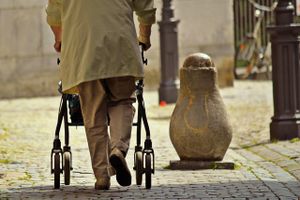
- Lead to a loss of personal freedom and to reduced quality of life.
- Precursors of falls and therefore of potentially severe injuries in elderly persons[1].

Causes of gait disorders include
- neurological, orthopedic, medical and psychiatric conditions and multifactorial etiology becomes more common with advancing age, making classification and management more complex.
- Any gait disorder should be thoroughly investigated in order to improve patient mobility and independence, to prevent falls and to detect the underlying causes as early as possible.
- Thorough clinical observation of gait, careful history taking focussed on gait and falls and physical, neurological and orthopedic examinations are basic steps in the categorization of gait disorders and serve as a guide for ancillary investigations and therapeutic interventions.
Gait Descriptions[edit | edit source]
This is not an exhaustive list.
- Antalgic gait a limp adopted so as to avoid pain on weight-bearing structures, characterized by a very short stance phase.
- Ataxic gait an unsteady, uncoordinated walk, with a wide base and the feet thrown out, coming down first on the heel and then on the toes with a double tap.This gait is associated with cerebellar disturbances and can be seen in patients with longstanding alcohol dependency. People with 'Sensory'Disturbances may present with a sensory ataxic gait. Presentation is a wide base of support, high steps, and slapping of feet on the floor in order to gain some sensory feedback. They may also need to rely on observation of foot placement and will often look at the floor during mobility due to a lack of proprioception
- Equine gait a walk accomplished mainly by flexing the hip joint; seen in crossed leg palsy.
- Parkinsonian Gait (seen in parkinson's disease and other neurologic conditions that affect the basal ganglia). Rigidity of joints results in reduced arm swing for balance. A stooped posture and flexed knees are a common presentation. Bradykinesia causes small steps that are shuffling in presentation. There may be occurrences of freezing or short rapid bursts of steps known as ‘festination’ and turning can be difficult.
- Trendelenburg gait, the gait characteristic of paralysis of the gluteus medius muscle, marked by a listing of the trunk toward the affected side at each step.
- Hemiplegic gait a gait involving flexion of the hip because of footdrop and circumduction of the leg.
- Steppage gait the gait in footdrop in which the advancing leg is lifted high in order that the toes may clear the ground. It is due to paralysis of the anterior tibial and fibular muscles, and is seen in lesions of the lower motor neuron, such as multiple neuritis, lesions of the anterior motor horn cells, and lesions of the cauda equina.
- Stuttering gait a walking disorder characterized by hesitancy that resembles stuttering; seen in some hysterical or schizophrenic patients as well as in patients with neurologic damage.
- Tabetic gait an ataxic gait in which the feet slap the ground; in daylight the patient can avoid some unsteadiness by watching his feet.
- Waddling gait exaggerated alternation of lateral trunk movements with an exaggerated elevation of the hip, suggesting the gait of a duck; characteristic of muscular dystrophy.
- Diplegic Gait (Spastic gait). Spasticity is normally associated with both lower limbs. Contractures of the adductor muscles can create a ‘scissor’ type gait with a narrowed base of support. Spasticity in the lower half of the legs results in plantarflexed ankles presenting in ‘tiptoe’ walking and often toe dragging. Excessive hip and knee flexion is required to overcome this
- Neuropathies Gaits. High stepping gait to gain floor clearance often due to foot drop[11][12][13][2]
Musculoskeletal Causes:
Pathological gait patterns resulting from musculoskeletal are often caused by soft tissue imbalance, joint alignment or bony abnormalities affect the gait pattern as a result[12].
Hip Pathology
- Arthritis is a common cause of pathological gait. An arthritic hip has reduced range of movement during swing phase which causes an exaggeration of movement in the opposite limb ‘hip hiking[12].
- Excessive Hip Flexion can significantly alter gait pattern most commonly due to; • Hip flexion contractures • IT band contractures, • Hip flexor spasticity, • Compensation for excessive knee flexion and ankle DF, • Hip pain • Compensation for excess ankle plantar flexion in mid swing. The deviation of stance phase will occur mainly on the affected side. The result is forward tilt of the trunk and increased demand on the hip extensors or increased lordosis of the spine with anterior pelvic tilt. A person with reduced spinal mobility will adopt a forward flexion position in order to alter their centre of gravity permanently during gait.
- Hip Abductor Weakness. The abductor muscles stabilise the pelvis to allow the opposite leg to lift during the swing phase. Weak abductor muscles will cause the hip to drop towards the side of the leg swinging forward. This is also known as Trendelenburg gait[13]
- Hip Adductor Contracture. During swing phase the leg crosses midline due to the weak adductor muscles, this is known as ‘scissor gait’[13]
- Weak Hip Extensors will cause a person to take a smaller step to lessen the hip flexion required for initial contact, resulting in a lesser force of contraction required from the extensors. Overall gait will be slower to allow time for limb stabilisation. Compensation is increased posterior trunk positioning to maintain alignment of the pelvis in relation to the trunk[13]
- Hip Flexor Weakness results in a smaller step length due to the weakness of the muscle to create the forward motion. Gait will likely be slower and may result in decreased floor clearance of the toes and create a drag
- Knee Pathologies
- Weak Quadriceps. The quadriceps role is to eccentrically control the knee during flexion through the stance phase. If these muscles are weak the hip extensors will compensate by bringing the limb back into a more extended position, reducing the amount of flexion at the knee during stance phase. Alternatively heel strike will occur earlier increasing the ankle of plantar flexion at the ankle, preventing the forward movement of the tibia, to help stabilise the knee joint[13].
- Severe Quadriceps Weakness or instability at the knee joint will present in hyperextension during the initial contact to stance phase. The knee joint will ‘snap’ back into hyperextension as the bodyweight moves forwards over the limb[13]
- Knee Flexion Contraction will cause a limping type gait pattern. The knee is restricted in extension, meaning heel strike is limited and step length reduced. To compensate the person is likely to ‘toe walk’ during stance phase. Knee flexion contractures of more than 30 degrees will be obvious during normal paced gait. Contractures less then this will be more evident with increased speeds[12][13].
Ankle Pathologies
- Ankle Dorsiflexion Weakness results in a lack of heel strike and decreased floor clearance. This leads to an increased step height and prolonged swing phase[13].
- Calf Tightening or Contractures due to a period of immobilisation or trauma will cause reduced heel strike due to restricted dorsiflexion. The compensated gait result will be ‘toe walking’ on stance phase, reduced step length and excessive knee and hip flexion during swing phase to ensure floor clearance[12].
Foot Pathologies
- Hallux Rigidus results in a lack of dorsiflexion of the great toe. The MPJ uses the windlass effect to raise the arch and stiffen the foot during dorsiflexion of the hallux. This stiffness increases the efficiency of the propulsion portion of the gait cycle. To be efficient in creating stiffness, the hallux should be able to dorsiflex at least 65 degrees.
Leg Length Discrepancy
- Leg length discrepancy can be as a result of an asymmetrical pelvic, tibia or femur length or for other reasons such as a scoliosis or contractures. The gait pattern will present as a pelvic dip to the shortened side during stance phase with possible ‘toe walking’ on that limb. The opposite leg is likely to increase its knee and hip flexion to reduce its length[12].
Antalgic Gait
- Antalgic gait due to knee pain presents with decreased weight bearing on the affected side. The knee remains in flexion and possible toe weight-bearing occurs during stance phase[12]
- Antalgic gait due to ankle pain may present with a reduced stride length and decreased weight bearing on the affected limb. If the problem is pain in the forefoot then toe-off will be avoided and heel weight-bearing used. If the pain is more in the heel, toe weight-bearing is more likely. General ankle pain may result in weight-bearing on the lateral border[12][13].
- Antalgic gait due to hip pain results in reduced stance phase on that side. The trunk is propelled quickly forwards with the opposite shoulder lifted in an attempt to even the weight distribution over the limb and reduce weight-bearing. Swing phase is also reduced[12].
Below are links to videos demonstrating normal gait and various gait abnormalities:
| [14] | [15] |
Gait Analysis[edit | edit source]
The analysis of the gait cycle is important in the biomechanical mobility examination to gain information about lower limb dysfunction in dynamic movement and loading.[16] When analysing the gait cycle, it is best to examine one joint at a time.[3] Objective and subjective methods can be used.[17][18]
Subjective[edit | edit source]
Our observation of gait is a subjective measure that we can use. We might ask the individual to walk normally, on insides and outsides of feet, in a straight line, running. All the time looking to compare sides and understanding of "normal". We also should observe the type of footwear the patient uses. A systematic review suggests shoes affect velocity, step time, and step length in younger children's gait[19].
Objective[edit | edit source]
An objective approach is quantitative and parameters like time, distance, and muscle activity will be measured. Other objective methods to assess the gait cycle that use equipment include:[20][18]
- Video Analysis and Treadmill
- Electronic and Computerized Apparatus
- Electronic Pedometers
- Satellite Positioning System[17]
Qualitative methods to assess and analyse gait include: [18]
- Rancho Los Amigos Hospital Rating List[21]
- Ten Meter Walking Test[22]
- 6 Minute Walk Test
- 2 Minute Walk Test
- Dynamic Gait Index
- Emory Functional Ambulation Profile[23]
- Timed Up and Go Test[24] This test is statistically associated with falling in men, but not in women.
- Functional Ambulation Categories[25]
- Tinetti-Test[26]
Clinical Bottom Line[edit | edit source]
Good knowledge of anatomy and biomechanics is important to understand the different phases of the gait cycle. When you know the normal pattern, you can see what’s going wrong!
References[edit | edit source]
- ↑ 1.0 1.1 Pirker W, Katzenschlager R. Gait disorders in adults and the elderly. Wiener Klinische Wochenschrift. 2017 Feb 1;129(3-4):81-95.Available from:https://www.ncbi.nlm.nih.gov/pmc/articles/PMC5318488/ (last accessed 27.6.2020)
- ↑ 2.0 2.1 Medical dictionary Gait speed Available from: https://medical-dictionary.thefreedictionary.com/gait+speed (last accessed 28.6.2020)
- ↑ 3.00 3.01 3.02 3.03 3.04 3.05 3.06 3.07 3.08 3.09 3.10 3.11 3.12 3.13 3.14 3.15 3.16 3.17 3.18 Shultz SJ et al. Examination of musculoskeletal injuries. 2nd ed, North Carolina: Human Kinetics, 2005. p55-60.
- ↑ 4.00 4.01 4.02 4.03 4.04 4.05 4.06 4.07 4.08 4.09 4.10 4.11 4.12 4.13 4.14 4.15 4.16 4.17 4.18 4.19 4.20 Loudon J, et al. The clinical orthopedic assessment guide. 2nd ed. Kansas: Human Kinetics, 2008. p.395-408.
- ↑ The biomechanics of running Tom F. Novacheck Motion Analysis Laboratory, Gillette Children’s Specialty Healthcare, Uni6ersity of Minnesota, 200 E. Uni6ersity A6e., St. Paul, MN 55101, USA Received 25 August 1997; accepted 22 September 1997 Available from:
- ↑ 6.0 6.1 Subotnick S. Sports medicine of the lower extremity. Harcourt (USA):Churchill Livingstone, 1999.
- ↑ Vaughan CL. Theories of bipedal walking: an odyssey. J Biomech 2001;36(2003):513-523.Available fromhttp://citeseerx.ist.psu.edu/viewdoc/download?doi=10.1.1.468.2414&rep=rep1&type=pdf
- ↑ Nicole Comninellis The Gait Cycle Animation Available from https://www.youtube.com/watch?time_continue=35&v=DP5-um6SvQI
- ↑ 9.0 9.1 9.2 Demos, Gait analysis, (http://www.ncbi.nlm.nih.gov/books/NBK27235/), 2004.
- ↑ Berger W, et al. Corrective reactions to stumbling in man: neuronal co-ordination of bilateral leg activity during gait. J Physiol 1984;357: 109-125.
- ↑ 11.0 11.1 Shi D, et al. Effect of anterior cruciate ligament reconstruction on biomechanical features of knee level in walking: a meta analysis. Chin Med J 2010;123(21):3137-3142.
- ↑ 12.0 12.1 12.2 12.3 12.4 12.5 12.6 12.7 12.8 12.9 Malanga G and Delisa J.A. Section One: Clinical Observation. Office of rehabilitation Research and Development No Date. http://www.rehab.research.va.gov/mono/gait/malanga.pdf (accessed 6 February 2010)
- ↑ 13.0 13.1 13.2 13.3 13.4 13.5 13.6 13.7 13.8 University of Washington. Pathologic Gait: Musculoskeletal http://courses.washington.edu/anatomy/KinesiologySyllabus/PathGait1Ortho.pdf (accessed 5 February 2015)
- ↑ onlinemedicalvideoAbnormal Gait Exam : Myopathic Gait Demonstration. Available fromhttps://www.youtube.com/watch?time_continue=5&v=b5rIEx9SsCo
- ↑ scfpta gait deviation published final 001.wmvAvailable fromhttps://www.youtube.com/watch?time_continue=5&v=b5rIEx9SsCo
- ↑ Langer PS, et al. A practical manual of clinical electrodynography. 2nd ed. Deer Park: The Langer Foundation for Biomechanics and Sports Medicine Research, 1989.
- ↑ 17.0 17.1 Terrier P, Schutz Y. How useful is satellite positioning system (GPS) to track gait parameters? A review. J Neuro Eng Rehab 2005;2:28.
- ↑ 18.0 18.1 18.2 Deckers JHM, et al. Ganganalyse en looptraining voor de paramedicus, Houten, Bohnfleu van Lonhum, 1996.
- ↑ Cranage S, Perraton L, Bowles KA, Williams C. The impact of shoe flexibility on gait, pressure and muscle activity of young children. A systematic review. Journal of Foot and Ankle Research. 2019 Dec 1;12(1):55.
- ↑ Frigo C, et al. Functionally oriented and clinically feasible quantitative gait analysis method. Med Biol Eng Comput 1998;36:179-185.
- ↑ Shumway-Cook A, Woollacott MH. Motor control: translating research into clinical practice. Lippincott Williams and Wilkins, 2007. p.408.
- ↑ Van Peppen RPS, KNGF-richtlijn Beroerte, 2004, Nederlands Tijdschrift voor Fysiotherapie.
- ↑ Baer RH, Wolf SL. Modified emory functional ambulation profile: an outcome measure for the rehabilitation of post stroke gait dysfunction. Stroke 2001;32(4):973-979.
- ↑ Potsiadlo D, Richardson S. The timed “Up and Go”: a test of functional mobility for frail elderly persons. J Am Geriatr Soc 1991;39(2):142-148.
- ↑ Shephard RJ, Taunton JE. Foot and ankle in sport and exercise, Toronto:Karger, 1987. p30-38.
- ↑ Bautmans I, et al. The feasibility of whole body vibration in institutionalised elderly persons and its influence on muscle performance, balance and mobility: a randomised controlled trial. BMC Geriatr 2005;5:17.

