Foot and Ankle Structure and Function
Original Editor - Vinit Kothekar.
Top Contributors - Vinit Kothekar, Wanda van Niekerk, Kim Jackson, Admin, Evan Thomas, Lucinda hampton, Chelsea Mclene, Candace Goh, Rachael Lowe, Simisola Ajeyalemi, Priyanka Chugh, WikiSysop, Rucha Gadgil, Jess Bell, Khloud Shreif, 127.0.0.1, Ewa Jaraczewska and Cath Young
Anatomy[edit | edit source]

Foot & ankle form a complex system which consists of 26 bones, 33 joints and more than 100 muscles, tendons and ligaments. It functions as a rigid structure for weight bearing and it can also function as a flexible structure to conform the uneven terrain. Foot and ankle provide various important functions which may include: support of body’s weight, providing balance, acts as a shock absorption, is the first contact to ground and transfer point for ground reaction force, compensates for proximal malalignment and may also substitute hand function in individuals with upper extremity amputations and paralysisCite error: Invalid <ref> tag; name cannot be a simple integer. Use a descriptive title. Foot is subdivided into rearfoot, midfoot and forefoot.
Talocrural (TC) joint[edit | edit source]
It is commonly known as ankle joint. Talocrural joint is formed between Tibia-fibula and Talus. Distal and inferior aspect of tibia – known as plafond – is connected to fibula via tibiofibular ligaments forming a strong mortise which articulates with the talar dome distally. Type of joint – hinge joint. Movements allowed – in sagittal plane, dorsiflexion and plantarflexion.
Subtalar (ST) joint[edit | edit source]

It is also known as talocalcaneal joint and is formed between talus and calcaneus. Three facets of talus - anterior, middle and posterior facets, articulates inferiorly with calcaneus.
Midtarsal (MT) joint[edit | edit source]
Also known as transverse tarsal joints or Chopart’s joint. It is S-shaped joint when viewed from above, and it consists of two joints – Talonavicular joint and Calcaneocuboid joint.
- Talonavicular (TN) JointIt is formed between anterior talar head and concavity on navicular bone. It does not have its own capsule and is the same one as of two anterior talocalcaneal articulations.
- Calcaneocuboid (CC) Joint – It is formed between anterior facet of calcaneus and posterior cuboid. Both articulating surface present convex and concave surface, with joint being convex vertically and concave transversely. Very little movement occur between joint segments.
Tarsometatarsal (TMT) Joint complex[edit | edit source]
Also known as Lisfranc’s joint. The distal tarsal rows including three cuneiform bones and cuboid articulate with base of metatarsals to form TMT complex. It is S-shaped joint and divided into 3 distinct columnsCite error: Invalid <ref> tag; name cannot be a simple integer. Use a descriptive title.
Medial – composed of 1st Metatarsal and medial Cuneiform.
Middle – composed of 2nd and 3rd Metatarsal and intermediate and lateral Cuneiform respectively.
Lateral – composed of 4th and 5th Metatarsal and Cuboid. It also divides the midfoot from forefoot.
Metatarsophalangeal (MTP) joints and Interphalangeal (IP) joints[edit | edit source]
MTP Joints are formed between metatarsal heads and corresponding base of proximal phalanx. Interphalangeal joints of the toes are formed between the phalanges of the toes. Each toe has proximal and distal IP joints except for the great toe which only has one IP joint.
| Joints | Type of Joints | Plane of movement | Motion |
|---|---|---|---|
| TC joint | Hinge joint | Sagittal plane | Dorsiflesxion & Plantarflexion |
| ST joint | Condyloid joint |
Mainly transverse plane Some sagittal plane |
Inversion & Eversion Dorsiflexion & Plantarflexion |
| MT joint |
TN joint - Ball & Socket joint CC joint - modified saddle joint |
Largely in transverse plane Some sagittal plane |
Inversion & Eversion Flexion & Extension |
| TMT joint | Plane joint | ||
| MTP joint | Condyloid joint |
Sagittal plane Some Transverse plane |
Flexion & Extension Abduction & Adduction |
| IP joint | Hinge joint | Sagittal plane | Flexion & Extension |
Kinematics[edit | edit source]
Talocrural joint[edit | edit source]
Tip of anterior malleoli is anterior and superior to lateral malleoli, which makes its axis oblique to both sagittal and frontal plane. The axis of rotation is approximately 13-18o laterally from frontal plane and at angle of 8-10o from transverse planeCite error: Invalid <ref> tag; name cannot be a simple integer. Use a descriptive title,Cite error: Invalid <ref> tag; name cannot be a simple integer. Use a descriptive title. Motion in other planes is required (like horizontal and frontal plane) to achieve a complete motion for plantarflexion and dorsiflexionCite error: Invalid <ref> tag; name cannot be a simple integer. Use a descriptive title. Normal available range for dorsiflexion is 0-30o and plantarflexion range is 0-55o.


Subtalar joint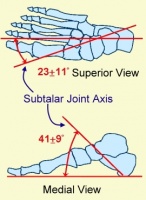 [edit | edit source]

The axis of subtalar joint lies about 42o upward of sagittal plane and about 16 to 23o medial to transverse planeCite error: Invalid <ref> tag; name cannot be a simple integer. Use a descriptive title,Cite error: Invalid <ref> tag; name cannot be a simple integer. Use a descriptive title. Literature presents vast range of subtalar motion ranging from 5 to 65o Cite error: Invalid <ref> tag; name cannot be a simple integer. Use a descriptive title. Average ROM for pronation is 5o and for supination is 20o. Inversion and Eversion ROM has been identified as 30o and 18o, respectivelyCite error: Invalid <ref> tag; name cannot be a simple integer. Use a descriptive title. Total inversion-eversion motion is about 2:1 or 3:2 ratio of inversion-to-eversion movementCite error: Invalid <ref> tag; name cannot be a simple integer. Use a descriptive title.
Midtarsal Joint[edit | edit source]
The midtarsal joint rotates at two axes due to its anatomy which makes its motion complex. Longitudinal axis lies about 15o superior to horizontal plane and about 10o medial to longitudinal plane. And oblique axis lies about 52o superior to horizontal plane and 57o from midline. Longitudinal axis is close to subtalar joint axis and oblique axis is similar to talocrural joint axis.

MT joint lockingCite error: Invalid <ref> tag; name cannot be a simple integer. Use a descriptive title, Cite error: Invalid <ref> tag; name cannot be a simple integer. Use a descriptive title – An important function of the foot is propulsion of weight during stance phase. This function is made possible by MT joint locking and unlocking. During heel strike foot needs to be flexible in order to adjust to the surface and MT joint unlocks to provide the flexibility. Whereas later on foot needs to act as rigid lever to propel the weight forward which is made possible by MT joint locking. During pronation/eversion of foot axis of TN and CC joint are parallel to each other which makes it easier for them to independently move and unlocking the MT joint. And the axes cross each other during supination/inversion which locks MT joint making it difficult to move. Blackwood et al concluded in the study that there is increased forefoot movement when calcaneus is everted. This is consisted with MT joint locking mechanism.
Lisfranc Joint Complex[edit | edit source]
The degree of sagittal motion for each TMT joint is presented belowCite error: Invalid <ref> tag; name cannot be a simple integer. Use a descriptive title.
| TMT Joint | Degree of Motion |
| 1st | 1.6o |
| 2nd | 0.6o |
| 3rd | 3.5o |
| 4th | 9.6o |
| 5th | 10.2o |
MTP and IP joints[edit | edit source]
The MTP joints are biaxial and move in sagittal and transverse plane. MTP joints present with a greater sagittal plane movement and very little transverse plane movement. At MTP joints, hyperextension is about 90o and flexion is about 30 to 50o. IP joints are hinge joints which limit motion in one direction.
Arthrokinematics[edit | edit source]
Influence on Kinetic Chain[edit | edit source]
Arches of Foot[edit | edit source]
Clinical Biomechanical Evaluation[edit | edit source]
References[edit | edit source]
- ↑ Midtarsal joint axis during pronation. Available from: https://vimeo.com/65147465

