Flexor Carpi Ulnaris Muscle
Original Editor Uchechukwu Chukwuemeka
Top Contributors - Uchechukwu Chukwuemeka, Ananya Bunglae Sudindar, Patti Cavaleri and Vidya Acharya
Original Editor -
Top Contributors - Uchechukwu Chukwuemeka, Ananya Bunglae Sudindar, Patti Cavaleri and Vidya Acharya
Description[edit | edit source]
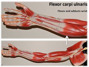
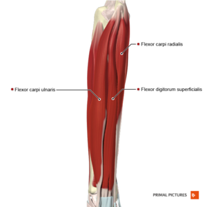
Flexor carpi ulnaris muscle (FCU) is the most medial flexor muscle in the superficial compartment of the forearm. It can adduct and flex the wrist at the same time; acts in tandem with flexor carpi radialis to flex the wrist and with the extensor carpi ulnaris to adduct the wrist. This muscle is the only muscle in the anterior compartment that is fully innervated by the ulnar nerve. [1]
Origin[edit | edit source]
It has a long linear origin from olecranon and posterior border of the ulna. In addition to that, FCU also has a humeral head origin from the medial epicondyle of the humerus.
Insertion[edit | edit source]
FCU inserts at the base of Pisiform bone, hook of hamate and base of 5th metacarpal. [1][2]
Innervation[edit | edit source]
FCU is innervated by the Ulnar nerve (C7, C8, T1)
Blood supply[edit | edit source]
The FCU is supplied by the ulnar collateral arteries along with the anterior and posterior ulnar recurrent arteries.
Function[edit | edit source]
FCU flexes and adducts the hand at the wrist joint.
Clinical Relevance[edit | edit source]
- The ulnar nerve gains access to the forearm by passing between the humeral and ulnar heads of the FCU. Within the cubital tunnel, compression and entrapment of the ulnar nerve may manifest, occurring between the aponeuroses of the two heads of the FCU. This anatomical scenario underscores the clinical significance of potential issues associated with ulnar nerve compression in the cubital tunnel.
- The FCU tendon insertion serves as a landmark in finding the ulnar nerve and ulnar artery, which are lateral to the tendon at the wrist joint.
- FCU shares a common tendon with the other wrist flexors and can be involved in various pathologies associated to them such as medial epicondylalgia.
- The FCU, situated in the forearm, is susceptible to involvement in Volkmann's contracture. In Volkmann's contracture, ischemic damage compromises the FCU, along with other muscles, leading to fibrosis and contracture. This constriction can result in deformities and impaired function in the affected limb.
Assessment[edit | edit source]
Manual Muscle Testing[edit | edit source]
The patient/client is seated with the posterior aspect of the forearm and hand flat on a table. The hand is then positioned to supination and extension.
The therapist is seated at the side of the upper limb being tested. The therapist stabilizes the patient's forearm and as well palpates the muscle and its tendon. With the other hand two to three fingers are placed on the radial side of the patient's hand at the 5th metacarpal and metacarpophalangeal joint of the patient.[5]
Instruction: The patient is instructed to abduct the little finger while flexing the wrist against the therapist's resistance. For further reading see...
References[edit | edit source]
- ↑ 1.0 1.1 Drake, RL, Vogl, W, Mitchell, AW, Gray, H. Gray's anatomy for Students 2nd ed. Philadelphia : Churchill Livingstone/Elsevier, 2010
- ↑ 2.0 2.1 Moore, KL, Dalley, AF, Agur, AM. Clinically oriented anatomy. 7th ed. Baltimore, MD: Lippincott Williams & Wilkins, 2014
- ↑ Reece CL, Susmarski A. Medial Epicondylitis. 2022 Apr 30. In: StatPearls [Internet]. Treasure Island (FL): StatPearls Publishing; 2022 Jan–.
- ↑ Mirza TM, Shrestha AB. Volkmann contracture.
- ↑ Hislop, HJ, Montgomery,J. Daniels and Worthingham's Muscle Testing: Techniques of Manual Examination. 8th ed. Missouri: Saunders Elsevier, 2007
- ↑ OTstudentVids. MMT of Flexor Carpi Radialis/Ulnaris and Extensor Carpi Uln. Available from: https://www.youtube.com/watch?v=31Wbe7xv8Jk [last accessed 29/3/2019]


