Dynamic Stabilisers of the Shoulder Complex: Difference between revisions
Amanda Ager (talk | contribs) No edit summary |
Amanda Ager (talk | contribs) mNo edit summary |
||
| Line 9: | Line 9: | ||
The stability of the shoulder joint, like any other joint in the body depends, on both static and dynamic stabilizers. Because of the vast range of motion of the shoulder complex (the most mobile joint of the human body), dynamic stabilizers are crucial for a strong sense of neuromuscular control throughout all movements and activities pertaining to the upper extremities. | The stability of the shoulder joint, like any other joint in the body depends, on both static and dynamic stabilizers. Because of the vast range of motion of the shoulder complex (the most mobile joint of the human body), dynamic stabilizers are crucial for a strong sense of neuromuscular control throughout all movements and activities pertaining to the upper extremities. | ||
Neuromuscular control in this context, can be understood as the “unconscious activation of dynamic restraints occurring in preparation for, and in response to, joint motion and loading for the purpose of maintaining functional joint stability.”<ref>Myers, J.B., C.A. Wassinger, and S.M. Lephart. Sensorimotor Contribution to Shoulder Joint Stability, in The Athlete’s Shoulder. 2009, Elsevier. p. 655-669.</ref>. Dynamic restraints result from neuromuscular control over the shoulder muscles, facilitated through motor control and [[Proprioception|proprioceptive input]]. | |||
''' | '''Static stabilizers''' include the [[Glenoid Labrum|joint labrum]] and capsuloligements components of the glenohumeral joint, as well as fascia tissues throughout the shoulder complex. There is also a theory that the neuromuscular bundle (nerves, veins, arteries) can also contribute to static stability. | ||
'''Dynamic stabilizers''' include the contractile tissues of the shoulder complex (tendons, muscles and tendon-muscular junctions). The most well known are the rotator cuff muscles ([[supraspinatus]], [[infraspinatus]], [[subscapularis]], [[Teres Minor|Teres minor]]), which collectively control the fine-tuning movement of the humeral head within the glenoid fossa (maintain centralization of the humeral head during static postures and dynamic movements). There are also the periscapsular muscles<ref>Curl LA, Warren RF. [https://www.ncbi.nlm.nih.gov/pubmed/?term=Glenohumeral+joint+stability%3A+selective+cutting+studies+on+the+static+capsular+restraints. Glenohumeral joint stability: selective cutting studies on the static capsular restraints.] Clinical Orthopaedics and Related Research®. 1996 Sep 1;330:54-65.</ref>, which are very important for homogeneous shoulder movements while avoiding biomechanical misalignments, such as a[[Supraspinatus tendinopathy|shoulder impingement]]. | |||
The dynamic stability of shoulder complex can be divided into: | |||
* Glenohumeral stability (Local) | |||
* Scapulothoracic stability (Global) | |||
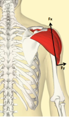
[[File:Deltoid action.png|thumb|241x241px|figure 1 | |||
line of action of three parts of deltoid follows line of pull of middle deltoid | line of action of three parts of deltoid follows line of pull of middle deltoid | ||
the deltoid force resolved into a very large translatory component (Fx) and a small rotatory component (Fy).<ref name=":0">Levangie PK, Norkin CC. Joint Structure and Function; A Comprehensive Analysis. 5th. Philadelphia: Fadavis Company. 2012.</ref>]] | the deltoid force resolved into a very large translatory component (Fx) and a small rotatory component (Fy).<ref name=":0">Levangie PK, Norkin CC. Joint Structure and Function; A Comprehensive Analysis. 5th. Philadelphia: Fadavis Company. 2012.</ref>]] | ||
== Glenohumeral joint stability == | == Glenohumeral joint stability == | ||
=== Deltoid | ==== '''Deltoid muscle''' ==== | ||
The deltoid muscle has a significant role as a stabilizer, and is generally accepted as a prime mover for glenohumeral joint during abduction, along with Supraspinatus. | |||
The deltoid is the primary muscle responsible for the abduction of the arm from 15 to 90 degrees. It also serves as a stabilizer of the humeral head, especially in instances of load carrying.<ref>Hallock GG. The hemideltoid muscle flap. Ann Plast Surg. 2000 Jan;44(1):18-22.</ref> | |||
From the biomechanical figure, the line of action (line of pull) of deltoid with the arm at the side of body, the parallel force component (fx) directed superiorly, is the largest of the three other components; resulting in a superior translation of the humeral head, and a small applied perpendicular force is directed towards rotating the humerus. Also, there is an inferior pull of force (fx), to offset the component of the middle deltoid which is active during arm elevation, as gravity cannot balance the force around the GH joint alone.<ref>Levangie PK, Norkin CC. Joint Structure and Function; A Comprehensive Analysis. 5th. Philadelphia: Fadavis Company. 2012.</ref> | |||
''<u>Blood supply of the deltoid:</u>'' The posterior circumflex humeral artery and the deltoid branch of the thoracoacromial artery are the vascular sources for the deltoid.<ref name=":1">Lam JH, Bordoni B. Anatomy, Shoulder and Upper Limb, Arm Abductor Muscles. [Updated 2020 Mar 31]. In: StatPearls [Internet]. Treasure Island (FL): StatPearls Publishing; 2020 Jan-. Available from: <nowiki>https://www.ncbi.nlm.nih.gov/books/NBK537148/</nowiki></ref> | |||
= | ''<u>Innervation of the deltoid</u>:'' The neural supply of the deltoid is via the axillary nerve (C5, C6) from the posterior cord of the brachial plexus.<ref name=":1" /> | ||
==== '''Rotator Cuff muscles''' ==== | |||
Rotator cuff not only abduct the shoulder it plays a role as a stabilizer muscles.<ref>Escamilla RF, Yamashiro K, Paulos L, Andrews JR. [https://www.ncbi.nlm.nih.gov/pubmed/19769415 Shoulder muscle activity and function in common shoulder rehabilitation exercises. Sports medicine.] 2009 Aug 1;39(8):663-85.</ref> | |||
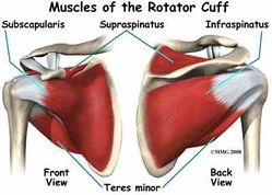
From figure 2 we can see all three muscles (teres minor, subscapularis, infraspinatus) in relation to their anatomical position and their muscle fiber direction from origin to insertion, tend to have a similar inferior line of pull<ref name=":0" /> and with the summation of three forces of rotator cuff, they nearly offset superior translation of humeral head created by deltoid. The wide range of motion of the shoulder is allowed by the variety of rotational moments of the cuff muscles<ref>Longo UG, Berton A, Papapietro N, Maffulli N, Denaro V. [https://www.ncbi.nlm.nih.gov/pubmed/?term=Biomechanics+of+the+rotator+cuff%3A+European+perspective.+InRotator+Cuff+Tea Biomechanics of the rotator cuff: European perspective. InRotator Cuff Tea]r 2012 (Vol. 57, pp. 10-17). Karger Publishers.</ref>. Teres minor, Infraspinatus as they are external rotators they contribute in abduction of arm by external rotation that participate clearing greater tubercle underneath the acromion. | |||
[[File:Muscles Rotator Cuff.jpg|thumb|figure 2|249x249px]] | |||
==== Supraspinatus muscle ==== | ==== Supraspinatus muscle ==== | ||
Regarding supraspinatus location more superior than the three other rotator cuff it has a line of pull superior that can't offset deltoid force. | Regarding supraspinatus location more superior than the three other rotator cuff it has a line of pull superior that can't offset deltoid force. | ||
| Line 37: | Line 48: | ||
Imbalance of one or more of these muscles consider a contribution cause to shoulder problems ([[Rotator Cuff Tendinopathy|impingement]], [[Shoulder Bursitis|bursitis]], instability ) | Imbalance of one or more of these muscles consider a contribution cause to shoulder problems ([[Rotator Cuff Tendinopathy|impingement]], [[Shoulder Bursitis|bursitis]], instability ) | ||
''<u>Blood supply of the supraspinatus:</u>'' The suprascapular artery delivers blood to the supraspinatus muscle.<ref name=":1" /> | |||
''<u>Innervation of the supraspinatus:</u>'' The neural supply of the supraspinatus is by the suprascapular nerve (C5, C6) from the upper trunk of the brachial plexus.<ref name=":1" /> | |||
== Scapulothoracic joint stability == | == Scapulothoracic joint stability == | ||
Revision as of 15:24, 18 April 2020
Original Editor - Khloud Shreif
Top Contributors - Khloud Shreif, Amanda Ager, Kim Jackson and Rishika Babburupage is still under construction
Introduction[edit | edit source]
The stability of the shoulder joint, like any other joint in the body depends, on both static and dynamic stabilizers. Because of the vast range of motion of the shoulder complex (the most mobile joint of the human body), dynamic stabilizers are crucial for a strong sense of neuromuscular control throughout all movements and activities pertaining to the upper extremities.
Neuromuscular control in this context, can be understood as the “unconscious activation of dynamic restraints occurring in preparation for, and in response to, joint motion and loading for the purpose of maintaining functional joint stability.”[1]. Dynamic restraints result from neuromuscular control over the shoulder muscles, facilitated through motor control and proprioceptive input.
Static stabilizers include the joint labrum and capsuloligements components of the glenohumeral joint, as well as fascia tissues throughout the shoulder complex. There is also a theory that the neuromuscular bundle (nerves, veins, arteries) can also contribute to static stability.
Dynamic stabilizers include the contractile tissues of the shoulder complex (tendons, muscles and tendon-muscular junctions). The most well known are the rotator cuff muscles (supraspinatus, infraspinatus, subscapularis, Teres minor), which collectively control the fine-tuning movement of the humeral head within the glenoid fossa (maintain centralization of the humeral head during static postures and dynamic movements). There are also the periscapsular muscles[2], which are very important for homogeneous shoulder movements while avoiding biomechanical misalignments, such as ashoulder impingement.
The dynamic stability of shoulder complex can be divided into:
- Glenohumeral stability (Local)
- Scapulothoracic stability (Global)

Glenohumeral joint stability[edit | edit source]
Deltoid muscle[edit | edit source]
The deltoid muscle has a significant role as a stabilizer, and is generally accepted as a prime mover for glenohumeral joint during abduction, along with Supraspinatus.
The deltoid is the primary muscle responsible for the abduction of the arm from 15 to 90 degrees. It also serves as a stabilizer of the humeral head, especially in instances of load carrying.[4]
From the biomechanical figure, the line of action (line of pull) of deltoid with the arm at the side of body, the parallel force component (fx) directed superiorly, is the largest of the three other components; resulting in a superior translation of the humeral head, and a small applied perpendicular force is directed towards rotating the humerus. Also, there is an inferior pull of force (fx), to offset the component of the middle deltoid which is active during arm elevation, as gravity cannot balance the force around the GH joint alone.[5]
Blood supply of the deltoid: The posterior circumflex humeral artery and the deltoid branch of the thoracoacromial artery are the vascular sources for the deltoid.[6]
Innervation of the deltoid: The neural supply of the deltoid is via the axillary nerve (C5, C6) from the posterior cord of the brachial plexus.[6]
Rotator Cuff muscles[edit | edit source]
Rotator cuff not only abduct the shoulder it plays a role as a stabilizer muscles.[7]
From figure 2 we can see all three muscles (teres minor, subscapularis, infraspinatus) in relation to their anatomical position and their muscle fiber direction from origin to insertion, tend to have a similar inferior line of pull[3] and with the summation of three forces of rotator cuff, they nearly offset superior translation of humeral head created by deltoid. The wide range of motion of the shoulder is allowed by the variety of rotational moments of the cuff muscles[8]. Teres minor, Infraspinatus as they are external rotators they contribute in abduction of arm by external rotation that participate clearing greater tubercle underneath the acromion.
Supraspinatus muscle[edit | edit source]
Regarding supraspinatus location more superior than the three other rotator cuff it has a line of pull superior that can't offset deltoid force.
Even though it still an effective stabilizer due to it's larger moment arm, it's capable to elevate glenohumeral joint near normal.[3]
From illustrated above we can consider deltoid and rotator cuff as one of a force couple of glenohumeral joint.
Imbalance of one or more of these muscles consider a contribution cause to shoulder problems (impingement, bursitis, instability )
Blood supply of the supraspinatus: The suprascapular artery delivers blood to the supraspinatus muscle.[6]
Innervation of the supraspinatus: The neural supply of the supraspinatus is by the suprascapular nerve (C5, C6) from the upper trunk of the brachial plexus.[6]
Scapulothoracic joint stability[edit | edit source]
For full rang shoulder elevation and reaching activities we need a force couples act on scapulothoracic joint for full rhythmic movement
Serratus anterior muscle and Trapezius[edit | edit source]
Serratus anterior[9][edit | edit source]
Serratus anterior muscle as a primary protractor muscle of scapula, it's capable of producing scapular protraction, upward rotation, external rotation and posterior tilting of the scapula with progress arm elevation.
The strong action of serratus as a protractor needs an apposite force to control this movement. Serratus anterior and trapezius muscles work as a primary force couple to rotate scapula upward.
Trapezius muscle[10][edit | edit source]
Trapezius has a retraction effect on scapula to counter the protraction and lateral pull of serratus anterior.
Upper trapezius[edit | edit source]
Take in consideration it's insertion to the clavicle, it produces upward rotation of scapula indirect and contributes to first degrees of clavicle elevation and retraction.
Middle trapezius[edit | edit source]
Contributes to offset the strong action of serratus anterior as a protractor and also produces an external rotation of scapula but it has a downward moment arm on scapula larger than it's upward moment arm.
Lower trapezius[edit | edit source]
With serratus anterior, lower trapezius is a primary upward rotator of scapula, it creates an upward rotation moment arm when the axis reaches acromioclavicular joint.
Serratus anterior and trapezius muscles they act agnostic for scapular movement and as a synergistic with glenohumeral forces[edit | edit source]
Rhomboid muscle and Teres major[edit | edit source]
- The rhomboid downward rotation action on scapula help to offset the undesired upward rotatory torque of teres major
- Rhomboid acts eccentrically to control change position of scapula during arm elevation so it contributes to counter the lateral translation force of serratus anterior.
- It has another stabilization role for scapula during adduction or extension.
Clinical presentation[edit | edit source]
For smooth synchronous movement of shoulder girdle we need the force couples of glenohumeral and scapulothoracic to work in proper timing and adequate forces to offset each other.
For example; the deltoid (middle fiber) acts to stabilize the humeral head against the glenoid cavity with small participation to abduct the arm, there are(subscapularis, teres minor, infraspinatus) their forces balance the deltoid action and along with supraspinatus that participates mainly in arm abduction.
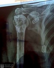
Deficit in these forces for example; insufficient activation of rotator cuff /deltoid over activation participate to narrowing coracoacormial space (figure 3) that compress tendons in this space ( rotator cuff tendinopathy /shoulder impingement)[12]
Scapular weaning, scapular dyskinesia that happen due to imbalance of scapulothoracic muscles, for example, serratus anterior, lower trapezius weakness, upper trapezius overactivation.
Physical therapy intervention[edit | edit source]
The role of physical therapy concentrates on to restore normal biomechanical alignment, the balance of force between muscles so the general treatment plane:
Strengthening for weak / inhibited muscles like serratus anterior, rotator cuff muscles, lower trapezius
Eccentric exercise for rotator cuff in case of rotator cuff tendinopathy to restore collagen alignment of tendon.
Muscles re-education to be activated in proper time is important to restore balance.
For more exercise this page below:
References[edit | edit source]
- ↑ Myers, J.B., C.A. Wassinger, and S.M. Lephart. Sensorimotor Contribution to Shoulder Joint Stability, in The Athlete’s Shoulder. 2009, Elsevier. p. 655-669.
- ↑ Curl LA, Warren RF. Glenohumeral joint stability: selective cutting studies on the static capsular restraints. Clinical Orthopaedics and Related Research®. 1996 Sep 1;330:54-65.
- ↑ 3.0 3.1 3.2 Levangie PK, Norkin CC. Joint Structure and Function; A Comprehensive Analysis. 5th. Philadelphia: Fadavis Company. 2012.
- ↑ Hallock GG. The hemideltoid muscle flap. Ann Plast Surg. 2000 Jan;44(1):18-22.
- ↑ Levangie PK, Norkin CC. Joint Structure and Function; A Comprehensive Analysis. 5th. Philadelphia: Fadavis Company. 2012.
- ↑ 6.0 6.1 6.2 6.3 Lam JH, Bordoni B. Anatomy, Shoulder and Upper Limb, Arm Abductor Muscles. [Updated 2020 Mar 31]. In: StatPearls [Internet]. Treasure Island (FL): StatPearls Publishing; 2020 Jan-. Available from: https://www.ncbi.nlm.nih.gov/books/NBK537148/
- ↑ Escamilla RF, Yamashiro K, Paulos L, Andrews JR. Shoulder muscle activity and function in common shoulder rehabilitation exercises. Sports medicine. 2009 Aug 1;39(8):663-85.
- ↑ Longo UG, Berton A, Papapietro N, Maffulli N, Denaro V. Biomechanics of the rotator cuff: European perspective. InRotator Cuff Tear 2012 (Vol. 57, pp. 10-17). Karger Publishers.
- ↑ Neumann DA, Camargo PR. Kinesiologic considerations for targeting activation of scapulothoracic muscles: part 1: serratus anterior. Brazilian journal of physical therapy. 2019 Feb 2.
- ↑ Camargo PR, Neumann DA. Kinesiologic considerations for targeting activation of scapulothoracic muscles–part 2: trapezius. Brazilian journal of physical therapy. 2019 Feb 3.
- ↑ Muscle and Motion. Force Couple , Scapular Force Couple. Available from: http://www.youtube.com/watch?v=YbbzQs7OBoY[last accessed 12/3/2020]
- ↑ <article> Joseph B. Myers, Ji-Hye Hwang, Maria R. Pasquale, J. Troy Blackburn and Scott M. Lephart. Rotator cuff coactivation ratios in participants with subacromial impingement syndrome. Journal of Science and Medicine in Sport, Volume 12, Issue 6, November 2009, Pages 603-608 </article>
- ↑ White Lion Athletics. Resistance Band Exercises: Best Exercises for Shoulder Rehab and Scapular Stabilzation. Available from: http://www.youtube.com/watch?v=Vez6-NTFkS8[last accessed 11/4/2020]


