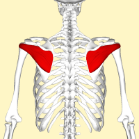
Infraspinatus
Original Editor - Wendy Walker
Lead Editors - Kate Sampson, Kim Jackson, Wendy Walker, Lilian Ashraf, 127.0.0.1, Oyemi Sillo, WikiSysop, Aya Alhindi, Naomi O'Reilly and George Prudden
Description[edit | edit source]
A thick, triangular muscle; one of the 4 muscles which comprise the Rotator Cuff of the shoulder.
Origin[edit | edit source]
The infraspinatus fossa of scapula, with some fibres arising from the infraspinatous fascia which covers the muscle and separates it from Teres Major and Teres Minor.[1]
Insertion[edit | edit source]
The posterior aspect of greater tuberosity of humerus, and the capsule of shoulder joint.[2]
Nerve Supply[edit | edit source]
Suprascapular Nerve (C5 & C6). It originates at the superior trunk of the brachial plexus. It runs laterally across the lateral cervical region to supply the infraspinatus and also the supraspinatus.[3]
Blood Supply[edit | edit source]
Suprascapular and circumflex scapular arteries.[4]
lymphatics[edit | edit source]
The lymphatic drainage is primarily of the subscapular (posterior) nodes. The subscapular nodes consist of 6 or 7 lymph nodes that lie along the posterior axillary fold. [3]
Action[edit | edit source]
Infraspinatus is:
- the main external rotator of the shoulder joint.
- It assists in producing shoulder extension.
- With the arm fixed, it abducts the inferior angle of the scapula.[2]
Function[edit | edit source]
It provides the primary muscle force for external rotation of the shoulder.
Along with the rest of the Rotator Cuff muscles it provides stability to the shoulder complex.[4]
Clinical Relevance[edit | edit source]
Atrophy in the supraspinatus and infraspinatus muscles, usually indicates compression on the suprascapular never near the scapular notch, such compression can be seen in overhead athletes and SLAP lesions. While isolated weakness or atrophy in the infraspinatus muscle can be seen in suprascapular nerve compression in the spinoglenoid notch, compression of the nerve in the spinoglenoid notch is usually due to a ganglion cyst.[5]
A study by Simons et al, described that infraspinatus muscle trigger points are associated with referred pain to the middle and anterior deltoid regions. Myofascial release techniques at the infraspinatus muscle may help shoulder pain.[6]
Assessment[edit | edit source]
To evaluate the infraspinatus muscle the arm is placed in neutral abduction or adduction position with the elbow flexed 90 degrees. Lateral rotation of the shoulder is done against resistance, the test is positive if there is pain or weakness. [7]
Resources[edit | edit source]
| [8] | [9] |
References[edit | edit source]
- ↑ Gray H. Anatomy of the Human Body. Philadelphia: Lea & Febiger, 1918.
- ↑ 2.0 2.1 Wheeles CR. Infraspinatus.
- ↑ 3.0 3.1 Williams JM, Sinkler MA, Obremskey W. Anatomy, shoulder and upper limb, infraspinatus muscle. StatPearls Publishing; 2023.
- ↑ 4.0 4.1 AnatomyExpert. 3D Infraspinatus- infraspinatus.
- ↑ Williams JM, Sinkler MA, Obremskey W. Anatomy, shoulder and upper limb, infraspinatus muscle. StatPearls [Internet]. 2020 May 24.
- ↑ Moccia D, Nackashi AA, Schilling R, Ward PJ. Fascial bundles of the infraspinatus fascia: anatomy, function, and clinical considerations. Journal of anatomy. 2016 Jan;228(1):176-83.
- ↑ Maruvada S, Madrazo-Ibarra A, Varacallo M. Anatomy, rotator cuff.
- ↑ Kenhub-Learn Human Anatomy. Infraspinatus Muscle - Origin, Insertion & Function - Human Anatomy | Kenhub [Internet]. Youtube; 2014
- ↑ AnatomyZone. Infraspinatus | Muscle Anatomy [Internet]. Youtube; 2018.


