Cervical Rib: Difference between revisions
No edit summary |
No edit summary |
||
| Line 22: | Line 22: | ||
* Compression of the sympathetic chain may cause [[Horner's Syndrome|Horner's syndrome]].<ref name=":1" /> | * Compression of the sympathetic chain may cause [[Horner's Syndrome|Horner's syndrome]].<ref name=":1" /> | ||
== Diagnosis | == Symptoms == | ||
On imaging, cervical ribs can be distinguished because their transverse processes are directed inferolaterally, whereas those of the adjacent thoracic spine are directed anterolaterally.<ref>Balan, Nisha Sharma, Anu (2008). ''Get through FRCR part 2B : rapid reporting of plain radiographs''. London: Royal Society of Medicine. ISBN .</ref><ref>Cervical Rib. Learning Radiology. Available from<nowiki/>https://learningradiology.com/notes/chestnotes/cervicalrib.htm [last accessed 05/11/2020] </ref> | '''Local Symptoms''' | ||
* Tender supraclavicular lump which is bony hard and is fixed when palpated. | |||
'''Sensory Symptoms''' | |||
* Tingling in hands or fingers; confined either to radial side or ulnar side or sometimes involve even whole hand. | |||
* Pain which may radiate down the arm. | |||
'''Vascular symptoms''' | |||
* Cold and clumsy extremities, particularly the fingers. | |||
* Skin colour changes to blue associated with trophic changes. | |||
* There is rare risk of gangrene. | |||
* Radial pulse becomes feeble or may even be absent. | |||
'''Motor Symptoms''' | |||
* Loss of hand grip. | |||
* Tendency of dropping things from the hand. | |||
* Wasting of palmar muscles<ref>Physiotherapy treatment.com. Cervical Rib Syndrome. Available from https://www.physiotherapy-treatment.com/cervical-rib.html [last accessed 05/11/2020]</ref><ref>Cervical Rib. Available from https://samarpanphysioclinic.com/2018/03/30/cervical-rib/ [last accessed 05/11/2020]</ref> | |||
== Diagnosis == | |||
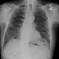
On '''imaging''', cervical ribs can be distinguished because their transverse processes are directed inferolaterally, whereas those of the adjacent thoracic spine are directed anterolaterally.<ref>Balan, Nisha Sharma, Anu (2008). ''Get through FRCR part 2B : rapid reporting of plain radiographs''. London: Royal Society of Medicine. ISBN .</ref><ref>Cervical Rib. Learning Radiology. Available from<nowiki/>https://learningradiology.com/notes/chestnotes/cervicalrib.htm [last accessed 05/11/2020] </ref> | |||
[[File:Cervical-ribs.jpg|center|thumb|200x200px]] | [[File:Cervical-ribs.jpg|center|thumb|200x200px]] | ||
== Management == | |||
'''1. medical t''' | |||
Anti-inflammatory drugs and | |||
analgesics | |||
this two given as a coservative treatment. | |||
'''2. surgical t''' | |||
surgery is essential in conditions of severe, progressive vascular and neurological signs and symptoms which are unbearable for the patients. It includes: | |||
* Removal of extra segment. | |||
* Dividing the scalene group of muscles. | |||
'''3. physiothrapy management:''' | |||
On the basis of symptoms of the patient, the regime of physiotherapy is planned. | |||
* For pain relief- short wave diathermy is used but it is contraindicated in case of sensory impairments. | |||
* To improve distal circulation- gripping exercise like ball sqizing, spring stretching. | |||
* To improve tone, power and endurance-Strengthening exercises of whole arm perticularly small muscles of the arm. | |||
* For posture Correction -In this, patient is guided to use mirror to see that his shoulders are in level, head is straight, looking forward | |||
* Specific exercises- To develop particular muscles groups for specific movements of shoulder girdle like elevation, retraction, and raising the arm overhead as these movements brings spontaneous relief. The important exercises are: | |||
1-Self resisted scapular elevation. | |||
2-Self resisted scapular adduction. | |||
3-Endurance training exercise for the shoulder girdle muscles. | |||
4-Progressive resistance exercises for shoulder girdle muscles with weight. | |||
* Deep Tissue Massage for TOS ( thoracic Outlet Syndrome). | |||
== References == | == References == | ||
Revision as of 08:46, 5 November 2020
Original Editor - Chelsea Mclene
Top Contributors - Chelsea Mclene, Aminat Abolade and Kim Jackson
Introduction[edit | edit source]
Cervical rib also known as "neck rib" or "supernumerary rib in cervical region" is an extra rib[1] that forms above first rib[2] which grows from the base of the neck just above the collarbone. It is a congenital overdevelopment of transverse process of cervical spine vertebra[3]. It can be on right, left or both sides and may be flaoting with no connection[4], fully formed bony rib or a thin strand of tissue fibre. They vary in size and shape.
In few cases, people having cervical rib may develop thoracic outlet syndrome[4] because of pressure on the nerves that may be caused by the presence of the rib. Partially formed extra rib may end in a swelling that shows as a lump in neck or it may tail off into a fibrous band of tissue that connects to the first proper rib[5]. Most cases are not clinically relevant and do not have symptoms. They are generally discovered incidentally during x-rays and CT scans.[6]
A cervical rib represents a persistent ossification of the C7 lateral costal element. During early development, this ossified costal element typically becomes re-absorbed. Failure of this process results in a variably elongated transverse process or complete rib that can be anteriorly fused with the T1 first rib below.[7]
Structure And Function[edit | edit source]
Associated Conditions[edit | edit source]
- Thoracic outlet syndrome due to compression of the lower trunk of the brachial plexus or subclavian artery.
- Compression of the brachial plexus may be identified by weakness of the muscles around the muscles in the hand.
- Compression of the subclavian artery is often diagnosed.
- Compression of the sympathetic chain may cause Horner's syndrome.[5]
Symptoms[edit | edit source]
Local Symptoms
- Tender supraclavicular lump which is bony hard and is fixed when palpated.
Sensory Symptoms
- Tingling in hands or fingers; confined either to radial side or ulnar side or sometimes involve even whole hand.
- Pain which may radiate down the arm.
Vascular symptoms
- Cold and clumsy extremities, particularly the fingers.
- Skin colour changes to blue associated with trophic changes.
- There is rare risk of gangrene.
- Radial pulse becomes feeble or may even be absent.
Motor Symptoms
Diagnosis[edit | edit source]
On imaging, cervical ribs can be distinguished because their transverse processes are directed inferolaterally, whereas those of the adjacent thoracic spine are directed anterolaterally.[10][11]
Management[edit | edit source]
1. medical t
Anti-inflammatory drugs and
analgesics
this two given as a coservative treatment.
2. surgical t
surgery is essential in conditions of severe, progressive vascular and neurological signs and symptoms which are unbearable for the patients. It includes:
- Removal of extra segment.
- Dividing the scalene group of muscles.
3. physiothrapy management:
On the basis of symptoms of the patient, the regime of physiotherapy is planned.
- For pain relief- short wave diathermy is used but it is contraindicated in case of sensory impairments.
- To improve distal circulation- gripping exercise like ball sqizing, spring stretching.
- To improve tone, power and endurance-Strengthening exercises of whole arm perticularly small muscles of the arm.
- For posture Correction -In this, patient is guided to use mirror to see that his shoulders are in level, head is straight, looking forward
- Specific exercises- To develop particular muscles groups for specific movements of shoulder girdle like elevation, retraction, and raising the arm overhead as these movements brings spontaneous relief. The important exercises are:
1-Self resisted scapular elevation.
2-Self resisted scapular adduction.
3-Endurance training exercise for the shoulder girdle muscles.
4-Progressive resistance exercises for shoulder girdle muscles with weight.
- Deep Tissue Massage for TOS ( thoracic Outlet Syndrome).
References[edit | edit source]
- ↑ Cervical Rib. Healthily. Available from https://www.livehealthily.com/neck-pain/cervical-rib [last accessed 05/11/2020]
- ↑ Cervical Rib. NHS. Available from https://www.nhs.uk/conditions/cervical-rib/ [ last accessed 05/11/2020]
- ↑ Fliegel BE, Menezes RG. Anatomy, Thorax, Cervical Rib.[updated 2020 Aug 22]. In:StatPearls[Internet].Treasure Island(FL): stat pearls publishing; 2020 jan.
- ↑ 4.0 4.1 Dr. Colin Tidy. Cervical Rib. Thoracic outlet syndrome. Patient. Available from https://patient.info/bones-joints-muscles/cervical-rib-thoracic-outlet-syndrome [last accessed 05/11/2020]]
- ↑ 5.0 5.1 Giles, Lynton G. F. (2009-01-01), Giles, Lynton G. F. (ed.), "Case 67 - Cervical ribs", 100 Challenging Spinal Pain Syndrome Cases (Second Edition), Edinburgh: Churchill Livingstone, pp. 311–314, doi:10.1016/b978-0-443-06716-7.00067-0, ISBN
- ↑ Guttentag, Adam; Salwen, Julia (1999). "Keep Your Eyes on the Ribs: The Spectrum of Normal Variants and Diseases That Involve the Ribs". RadioGraphics. 19 (5): 1125–1142. doi:10.1148/radiographics.19.5.g99se011125. PMID 10489169
- ↑ Tani, Edneia M.; Skoog, Lambert (2008-01-01), Bibbo, Marluce; Wilbur, David (eds.), "CHAPTER 22 - Salivary Glands and Rare Head and Neck Lesions", Comprehensive Cytopathology (Third Edition), Edinburgh: W.B. Saunders, pp. 607–632, ISBN.
- ↑ Physiotherapy treatment.com. Cervical Rib Syndrome. Available from https://www.physiotherapy-treatment.com/cervical-rib.html [last accessed 05/11/2020]
- ↑ Cervical Rib. Available from https://samarpanphysioclinic.com/2018/03/30/cervical-rib/ [last accessed 05/11/2020]
- ↑ Balan, Nisha Sharma, Anu (2008). Get through FRCR part 2B : rapid reporting of plain radiographs. London: Royal Society of Medicine. ISBN .
- ↑ Cervical Rib. Learning Radiology. Available fromhttps://learningradiology.com/notes/chestnotes/cervicalrib.htm [last accessed 05/11/2020]


