Brachial Plexus Injury: Difference between revisions
Aarti Sareen (talk | contribs) No edit summary |
Aarti Sareen (talk | contribs) No edit summary |
||
| Line 68: | Line 68: | ||
'''IV''': cord.<br> | '''IV''': cord.<br> | ||
'''3. Classification on anatomical location of injury''':<br> | '''3. Classification on anatomical location of injury'''<ref>Dodds SD et al.Perinatal Brachial Plexus Palsy. Curr Op Pediat 12: 40-47, 2000.</ref><ref>Kay SPJ. Obstetrical Brachial Palsy. Br J Plastic Surg 51: 43-50, 1998.</ref>:<br> | ||
*Upper plexus palsy (Erb’s palsy in the OBPI cases) involves C5-C6+/-C7roots. | *Upper plexus palsy (Erb’s palsy in the OBPI cases) involves C5-C6+/-C7roots. | ||
Revision as of 18:31, 22 December 2013
Introduction[edit | edit source]
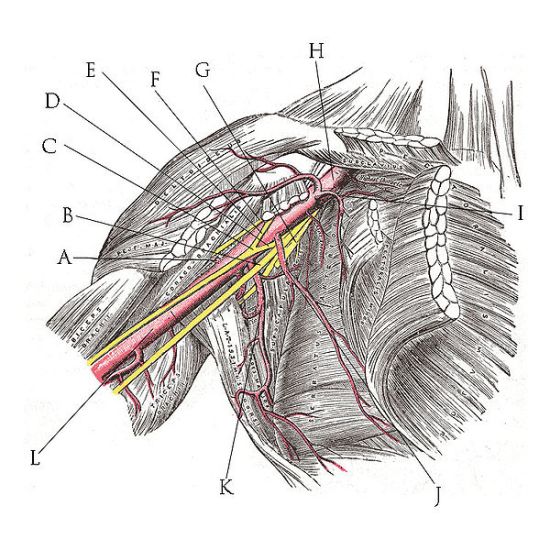
Brachial plexus is the network of nerves which runs through the cervical spine, neck,axilla and then into arm or it is a network of nerves passing through the cervico-axillary canal to reach axilla and innervates brachium (upper arm), antebrachium (forearm) and hand.It is a somatic nerve plexus formed by intercommunications among the ventral rami (roots) of the lower 4 cervical nerves (C5-C8) and the first thoracic nerve (T1).
Function
[edit | edit source]
The brachial plexus is responsible for cutaneous and muscular innervation of the entire upper limb, with two exceptions: the trapezius muscle innervated by the spinal accessory nerve (CN XI) and an area of skin near the axilla innervated by the intercostobrachial nerve.
Clinical Anatomy[edit | edit source]
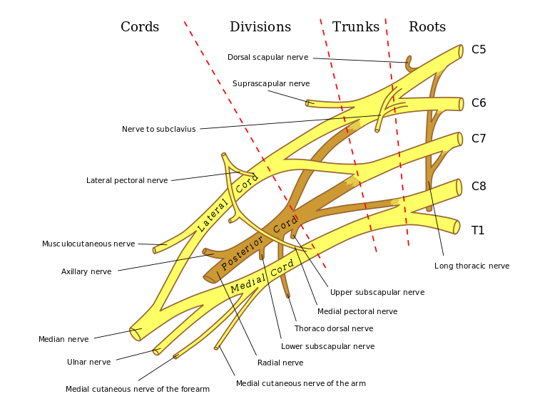
The plexus consists of roots, trunks, divisions,cords and branches.
1. ROOTS: These are consititued by the anterior primary rami of spinal nerves C5,6,7,8 and T1 with contributions from the anterior primary rami of C4 and T2. The origin of the plexus may shift one segment either upward or downward resulting in a PRE FIXED PLEUS or POST FIXED PLEXUS respectively. Ina prefixed plexus, the contribution by C4 is large and in that from T2 is often absent. In a post fixed plexus, the contribution by T1 is large, T2 is always present, C4 is absent, and C5 is reduced in size. The roots join to form trunks as follows:
2.TRUNKS: Upper trunk is formed by C5 & C6
Middle trunk is formed by C7
Lower trunk is formed by C8 & T1
3. DIVISIONS OF THE TRUNKS: Each trunk divides into ventral and dorsal divisions (which ultimately supply the anterior and posterior aspects of the limb). These divisions join to form cords.
4. CORDS: it forms 3 cords
- The Posterior Cord is formed from the three posterior divisions of the trunks (C5-C8,T1)
- The Lateral Cord is the anterior divisions from the upper and middle trunks (C5-C7)
- The Medial Cord is simply a continuation of the anterior division of the lower trunk (C8,T1)
5. BRANCHES: The specific branches of each cord can be seen at this page
Mechanism of injury[edit | edit source]
Injury to brachial plexus can occur in many ways. These include the contact sports, road traffic accident, motorvehicle accident or during birth. The network of nerves is fragile and can be damaged by pressure,stretching, or cutting. Stretching can occur when the head and neck are forced away from the shoulder, such as might happen in a fall off a motorcycle. If severe enough, the nerves can actually avulse, or tear out of, their roots in the neck. Pressure could occur from crushing of the brachial plexus between the collarbone and first rib, or swelling in this area from injured muscles or other structures[1].Although these are but a common few events, there is one of two mechanisms of injury that remain constant during the point of injury.[2]. The two mechanisms that can occur are traction and heavy impact[3]. These two methods disturb the nerves of the brachial plexus and cause the injury[4].
TRACTION: Traction, also known as stretch injury, is one of the mechanisms that cause brachial plexus injury. The nerves of the brachial plexus are damaged due to the forced pull by the widening of the shoulder and neck.Traction occurs from severe movement and causes a pull or tension among the nerves. There are two types of traction: downward traction and upward traction. In downward traction there is tension of the arm which forces the angle of the neck and shoulder to become broader. This tension is forced and can cause lesions of the upper roots and trunk of the nerves of the brachial plexus. Upward traction also results in the broadening of the neck and shoulder angle but this time the nerves of T1 and C8 are torn away.[5]
IMPACT: Heavy impact to the shoulder is the second common mechanism to causing injury to the brachial plexus. Depending on the severity of the impact, lesions can occur at all nerves in the brachial plexus. The location of impact also affects the severity of the injury and depending on the location the nerves of the brachial plexus may be ruptured or avulsed. Some forms of impact that affect the injury to the brachial plexus are shoulder dislocation, clavicle fractures, hyperextension of the arm and sometimes delivery at birth.[6] During the delivery of a baby, the shoulder of the baby may graze against the pelvic bone of the mother. During this process, the brachial plexus can receive damage resulting in injury.This is very low compared to the other identified brachial plexus injuries.[7]
Classification of injuries[edit | edit source]
The various classifications of brachial plexus injury are as follows:
- Leffert classification of brachial plexus injury
- Millesi classification of brachial plexus injury
- Classification on anatomical location of injury
1. Leffert classification of brachial plexus injury[8]: It is based on etiology and level of injury and is as follows
- I Open (usually from stabbing)
- II Closed (usually from motorcycle accident)
- IIa Supraclavicular
- preganglionic:
- avulsion of nerve roots, usually from high speed injuries
with other injuries and LOC;
- no proximal stump, no neuroma formation (neg Tinel's)
- pseudomeningocele, denervation of neck muscles are common
- horner's sign (ptosis, miosis, anhydrosis)
- postgangionic:
- roots remain intact;
- usually from traction injuries;
- there are proximal stump and neuroma formation (pos Tinel's)
- deep dorsal neck muscles are intact, and pseudomeningoceles
will not develop;
- Infraclavicular Lesion:
- usually involves branches from the trunks (supraclavicular);
- function is affected based on trunk involved;
- Trunk Injured Functional Loss
Upper biceps , shoulder muscles
Middle Wrist and Finger Extension;
Lower Wrist and Finger Flexion;
- III Radiation induced
- IV Obstetric
- IVa Erb's (upper root) - waiter's tip hand;
- IVb Klumpke (lower root)
2. Millesi classification of brachial plexus injury[9]: It is mainly divided into 4
I: supraganglionic.
II: infraganglionic.
III: trunk.
IV: cord.
3. Classification on anatomical location of injury[10][11]:
- Upper plexus palsy (Erb’s palsy in the OBPI cases) involves C5-C6+/-C7roots.
- Lower plexus palsy (Klumpke’s palsy) involves C8-T1 roots (and sometimes also C7)
- Total plexus lesions involve all nerve roots C5-T1
- Some authors have included a fourth type,an intermediate type that primarily involves the C7 root.[12][13]
Recent Resources[edit | edit source]
References[edit | edit source]
- ↑ American Society of Surgery of Hand. Available from www.assh.org/Public/HandConditions/Documents/Web_Version_PDF/BrachPlex.pdf
- ↑ Midha, Rajiv, MD. "Neurosurgery." Epidemiology of Brachial Plexus Injuries in a Multitrauma Po... :. Congress of Neurological Surgeons, June 1997. Web. 29 Jan. 2013.
- ↑ Narakas, A.O. "The Treatment of Brachial Plexus Injuries." Link.springer.com. International Orthopaedics, June 1985. Web. 28 Jan. 2013.
- ↑ Hems, TE.; Mahmood, F. (Jun 2012). "Injuries of the terminal branches of the infraclavicular brachial plexus: patterns of injury, management and outcome.". J Bone Joint Surg Br 94 (6): 799–804.
- ↑ Coene, L.N.J.E.M. "Mechanisms of Brachial Plexus Lesions." ScienceDirect.com. Clinical Neurology and Neurosurgery, 25 Mar. 2003. Web. 29 Jan. 2013.
- ↑ Jeyaseelan, L.; Singh, VK.; Ghosh, S.; Sinisi, M.; Fox, M. (Jan 2013). "Iatropathic brachial plexus injury: A complication of delayed fixation of clavicle fractures.". Bone Joint J 95–B (1): 106–10.
- ↑ Joyner, Benny, Mary Ann Soto, and Henry M. Adam. "Brachial Plexus Injury." Brachial Plexus Injury. Pediatrics in Review, 1 June 2006. Web. 29 Jan. 2013.
- ↑ Clifford R. Wheeless.Wheeless' Textbook of Orthopaedics.Duke's Orthopaedics.
- ↑ Andrew Hodges.A-Z of Plastic Surgery.Oxford University Press.
- ↑ Dodds SD et al.Perinatal Brachial Plexus Palsy. Curr Op Pediat 12: 40-47, 2000.
- ↑ Kay SPJ. Obstetrical Brachial Palsy. Br J Plastic Surg 51: 43-50, 1998.
- ↑ Al-qattan MM. Self-mutilation in Children with Obstetric Brachial Pexus Palsy. J Hand Surg Br 24B; 5: 547-549, 1999
- ↑ Shenaq SM et al. Brachial Plexus Birth Injuries and Current Management. Clin Plast Surg 25; 4: 527-536, 1998


