Bladder Anatomy
Original Editor - User Name
Top Contributors - Khloud Shreif and David Olukayode
Description[edit | edit source]
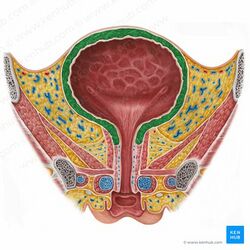
IT is a hollow muscular, elastic, subperitoneal organ, , located at the lesser pelvic cavity, it collect the urine that is filtered through the kidney through ureters to be evacuated/ voided later on.
It can accommodate about 1000 ml of urine with average about 400-600 ml that vary from one to another. The outer layer of bladder wall formed of smooth involuntary muscle fibers (detrusor muscles), it contract at urination to empty the bladder.
Anatomical parts of the bladder[edit | edit source]
When it is filled it is like an oval, formed of four parts. The neck is the constricted part, located inferiorly and leads to urethra. The apex is anterosuperior, points to the abdominal wall. The base/ fundus located posteriorly, it is a triangular-shaped its tip points backward. The body of the bladder is the main part of the bladder and located between the apex and fundus[1].
The trigon is a triangular region at the base of the bladder, lined with smooth muscle, collagen, and elastin to a lesser degree. Located between the internal-urethral meatus and the two ureteral orifices[2].
Urethral sphincters[edit | edit source]
Internal urethral sphincter
It is formed of smooth involuntary muscle fibbers under the control of the autonomic nervous system, it contains alpha-adrenergic receptors its stimulation helps to fill the bladder. In female it is formed by bladder neck and the proximal urethra (urethra in female is about 4 -5 cm while in males it is bout 20cm in length)[3].
External urethral sphincter
It is consisted of skeletal muscle fibers, it is under voluntary control, and contains nicotinic receptors[3].
Innervation[edit | edit source]
Nerve[edit | edit source]
Parasympathetic (s2-s4): responds to stretch receptors of the bladder wall and it is responsible for detrusor contraction to empty the bladder. The pudendal nerve is a somatic parasympathetic innervates the external urethral sphincter and helps to voluntary control emptying the bladder.
Sympathetic (t12-l2): store the urine as it cause relaxation of the detrusor muscle of the bladder.
Artery[edit | edit source]
Branches from internal iliac artery; superior and inferior vesicle arteries.
Small branches from obturator and inferior gluteal artery.
Lymphatic drainage[edit | edit source]
The neck and fundus of bladder through internal, sacral and common iliac lymph nodes.
The superolateral aspect of the bladder through the external iliac lymph nodes.
Clinical relevance[edit | edit source]
Detrusor sphincter dyssynergia
Atonic bladder
Neurogenic bladder
Treatment[edit | edit source]
Resources[edit | edit source]
- ↑ Shermadou ES, Rahman S, Leslie SW. Anatomy, abdomen and pelvis, bladder.2018.
- ↑ Hickling DR, Sun TT, Wu XR. Anatomy and physiology of the urinary tract: relation to host defense and microbial infection. Urinary tract infections: Molecular pathogenesis and clinical management. 2017 Feb 15:1-25.
- ↑ 3.0 3.1 Sam P, Jiang J, LaGrange CA. Anatomy, abdomen and pelvis, sphincter urethrae. InStatPearls [Internet] 2021 Nov 5. StatPearls Publishing.


