Assessment and Treatment of the Thoracic Spine: Difference between revisions
No edit summary |
No edit summary |
||
| Line 145: | Line 145: | ||
File:Exercises to enhance thoracic rotation.jpg|thumb|Figure 7. Thoracic rotation exercises. </gallery> | File:Exercises to enhance thoracic rotation.jpg|thumb|Figure 7. Thoracic rotation exercises. </gallery> | ||
Exercises to stabilise the thoracic spine are shown in figures 8, 9 | Exercises to stabilise the thoracic spine are shown in figures 8, 9 and 10. Initially it is beneficial to use closed kinetic chain exercises (Figures 8 and 9) before aiming to increase strength through range (Figure 10). Figure 11 shows moderately advanced kinetic chain exercises. | ||
<gallery widths="250px" heights="350px"> | <gallery widths="250px" heights="350px"> | ||
| Line 152: | Line 152: | ||
File:Thoracic motor control strengthening through range exercises.jpg|thumb|Figure 10. Thoracic motor control exercises - strengthening through range. </gallery> | File:Thoracic motor control strengthening through range exercises.jpg|thumb|Figure 10. Thoracic motor control exercises - strengthening through range. </gallery> | ||
== A Lot is Unkonwn == | |||
* Most ‘research’ is anecdotal / youtube and social media | |||
* Information from other spinal areas has been ‘tansferred’ to the thoracic spine, not recognising the unique differences of the thorax | |||
* These are many deep thoracic muscles whose contribution to thoracic motor control is unknown, such as semispinalis thoracic and rotatores | |||
* It is also not known what role the thoracic spine has in proprioception | |||
== Summary == | |||
Assessment:<ref name=":0" /> | |||
* Use anatomical knowledge and biomechanical insights to look at poor motion habits | |||
* Don’t get bogged down by looking at tiny faults | |||
Management:<ref name=":0" /> | |||
* It can be beneficial to use specific techniques to mobilise restricted areas, but this will not result in macro changes | |||
* Postural adjustments can, however, be used to change systems and make macro changes | |||
* Thoracic rotation is essential for movement, sports and performance - it is, therefore, important to utilise exercises that promote rotation mobility (optimal range of motion), stability (motor control) and strength | |||
* Exercise therapy should be used in conjunction with manual therapy to achieve an optimal outcome | |||
* Management should not just focus on thoracic stiffness - it is perhaps more important to address thoracic motor control and segmental stability | |||
== References == | == References == | ||
Revision as of 10:46, 4 September 2021
Top Contributors - Jess Bell, Kim Jackson, Kirenga Bamurange Liliane, Tarina van der Stockt, Lucinda hampton, Olajumoke Ogunleye, Aminat Abolade and Merinda Rodseth
Subjective Assessment[edit | edit source]
Detailed information on the assessment of the thoracic spine is available here, but specific questions to consider in the subjective assessment include:[1]
- How did the problem begin? How long has it been a problem?
- How has the pain progressed over time?
- Is there a history of overload or trauma?
- Does the patient have pain with breathing? And during which part of the breath does this occur?
- What effect does coughing and / or sneezing have?
- Can the patient lie on the affected side at night?
- How is the pain behaving and what is the level of irritability?
- Consider in particular more stiffness
- If there is stiffness for a prolonged period in the morning, and a history of enthesopathies, the patient may have a spondyloarthropathy[1][2]
- What are the specific functional impairments? (e.g. during sport, activities of daily living)
- What is the patient’s medical history?
- Essential to know due to spinal masqueraders in the thoracic spine
- Are there any psychosocial factors contributing to the pain?
- The sympathetic nervous system is prominent in the thoracic region[1]
- Are there any red flags?[3]
Pain can be caused by inflammation, or it may originate in the cartilage, ligaments, bone (fracture) or the nerve root:[1]
- Nerve root or facet pain may be described as lancinating / nauseating and it may radiate and follow angulation of rib
- Costochondritis tends to be described as a deep, boring, aching pain in the chest wall that may radiate (often posteriorly or to the neck)
- Rib fracture or intercostal strain is typically described as a sudden, sharp / piercing pain, which is then aggravated by laughing, sneezing, coughing, deep breaths or any type of straining manoeuvre
- When patients have immobility, they tend to report stiffness, restricted movement, or a sense of feeling ‘stuck’
Objective Assessment[edit | edit source]
The objective examination is guided by findings in the subjective interview. Remember:[1]
- It is essential to understand which structures are loaded during each test
- Keep tests to a minimum
- “Less is more” to avoid flaring up the patient
- Consider combinations of tests
- The best ‘special test’ is the one the patient demonstrates to you
- Consider the diaphragm
During the assessment, the therapist should develop a sound hypothesis. “If you can’t find it, you can’t assess it and you can’t treat it”.[1]
Objective Testing[edit | edit source]

The following tests should be included in an objective examination of the thoracic spine:[1]
- Static and dynamic postural assessment:
- Watch how the patient moves / drifts / hinges
- Consider different types of postural dysfunctions[4](see Figure 1)
- Is the postural change a primary problem or related to something else in the chain (e.g. lumbopelvic dysfunction)
- Breathing mechanics
- ROM tests from neutral
- Look for intersegmental restrictions
- Assess from behind
- Inter-ring and articular palpation during motion
- Motor control and strength tests
- Sitting
- Puppy lie
- 4 point
- Neurodynamic tests
- Neurological
- Thoracic dermatome testing (see Figure 2)
- Palpation, including the clavicle and first rib

Active Thoracic Movement Tests[edit | edit source]
During the active movement tests:[1]
- Assess the patient in sitting in order to isolate the thoracic spine (by blocking the lumbar spine)
- Assess for asymmetry of movement and any segmental cause of restriction
- Remember that motor control is also important in the thoracic region, not just stiffness
Thoracic Flexion and Extension[edit | edit source]
- Flexion: feel for anterior tilt of the ribs
- Extension: feel for posterior rib tilt
- NB extension is the most limited movement in the thoracic spine[9]
Thoracic Rotation[edit | edit source]
Rotation is one of the most useful tests for the thoracic spine. During rotation movements, it is possible to identify:[1]
- Stiffness
- Sequencing issues
Thoracic Side Bend / Lateral Shift[edit | edit source]
Assess the patient from behind - it is only necessary to look from the front if you wish to specifically assess the ribs from this position. During thoracic side bend there is:[1]
- Contralateral rib translation
- Ribs approximate on the ipsilateral side
- Ribs separate on the contralateral side
Treatment[edit | edit source]
Sleep[edit | edit source]
Sleep is the most powerful antioxidant.[1] It is recognised that there is a bi-directional relationship between pain and sleep.[10]
Thoracic Manipulation[edit | edit source]
It is still not known if / why thoracic manipulation works, but it has been found that thoracic manipulation can decrease pain, improve mobility and enhance a patient’s feeling of health.[11]
- There is no evidence that one manipulation is better than another
- There is no evidence that thoracic manipulation has a long-term effect, so if it is used, it should be in combination with specific rehabilitation
- Joint position, direction, velocity and force are all variables that should be considered[1]
Postural Correction and Motor Control[edit | edit source]
Correcting a patient’s posture can also have a positive impact on a patient’s pain.[1]
- Consider the centre of gravity
- Look for areas of muscle spasm or hyperactivity
Iliocostalis Release[edit | edit source]
Iliocostalis is the most lateral of the erector spinae.[12] Patients with significant thoracic kyphosis and lumbar lordosis may have increased activity of iliocostalis. This muscle can be released under the 10th and 11th ribs.[1]
Posterior-Anterior Glides[edit | edit source]
Patients with inverted thoracic spines, rotated spine and increased kyphosis will likely find posterior-anterior (PA) glides of the spinous processes irritable as they are highly nociceptive.[1]
Instead, it can be beneficial to perform a PA glide on the rib angle, thus mobilising 13 articulations per thoracic ring. For individuals with inverted spines, this will create an anterior-posterior (AP) movement on the spinous process.[1]
For patients with increased kyphosis, a PA glide on the rib angle while performing an AP glide on the anterior shoulder / coracoid can be effective. A rotatory technique for patients with kyphosis can be effective.[1]
Exercise Therapy[edit | edit source]
A survey of thoracic spine management trends in the UK found that exercise is used widely as a treatment modality despite limited supporting evidence.[13]
In terms of exercise prescription, speed, starting positions, dosage and load progression have not been investigated. However, exercises which aim to stretch, mobilise and stabilise can be beneficial in clinical practice.[1]
Specific Exercises[edit | edit source]
Stretches for the thoracic spine are shown in Figure 3. Figure 4 shows stretches that can specifically address an inverted thoracic spine.
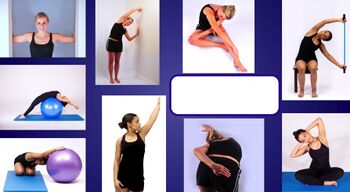
Figure 3. Exercises to stretch the thoracic spine.

Figure 4. Stretch to address an inverted thoracic spine.


Figures 5 and 6 show exercises to mobilise the thoracic spine, while Figure 7 focuses specifically on rotation of the thoracic spine.
- Exercises to mobilise the thoracic spine.jpg
Figure 5. Exercises to mobilise the thoracic spine.
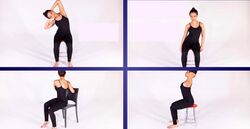
Figure 6. Right side flexion, lateral translation, rotation and extension.
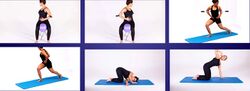
Figure 7. Thoracic rotation exercises.


Exercises to stabilise the thoracic spine are shown in figures 8, 9 and 10. Initially it is beneficial to use closed kinetic chain exercises (Figures 8 and 9) before aiming to increase strength through range (Figure 10). Figure 11 shows moderately advanced kinetic chain exercises.
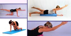
Figure 8. Thoracic motor control exercises.
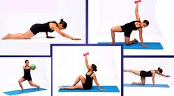
Figure 9. Thoracic motor control exercise progressions.
- Thoracic motor control strengthening through range exercises.jpg
Figure 10. Thoracic motor control exercises - strengthening through range.


A Lot is Unkonwn[edit | edit source]
- Most ‘research’ is anecdotal / youtube and social media
- Information from other spinal areas has been ‘tansferred’ to the thoracic spine, not recognising the unique differences of the thorax
- These are many deep thoracic muscles whose contribution to thoracic motor control is unknown, such as semispinalis thoracic and rotatores
- It is also not known what role the thoracic spine has in proprioception
Summary[edit | edit source]
Assessment:[1]
- Use anatomical knowledge and biomechanical insights to look at poor motion habits
- Don’t get bogged down by looking at tiny faults
Management:[1]
- It can be beneficial to use specific techniques to mobilise restricted areas, but this will not result in macro changes
- Postural adjustments can, however, be used to change systems and make macro changes
- Thoracic rotation is essential for movement, sports and performance - it is, therefore, important to utilise exercises that promote rotation mobility (optimal range of motion), stability (motor control) and strength
- Exercise therapy should be used in conjunction with manual therapy to achieve an optimal outcome
- Management should not just focus on thoracic stiffness - it is perhaps more important to address thoracic motor control and segmental stability
References[edit | edit source]
- ↑ 1.00 1.01 1.02 1.03 1.04 1.05 1.06 1.07 1.08 1.09 1.10 1.11 1.12 1.13 1.14 1.15 1.16 1.17 1.18 1.19 1.20 Bell-Jenje T. Assessment and Treatment of the Thoracic Spine Course. Physioplus, 2021.
- ↑ Martey C. Co-morbidities within Spondyloarthritis Course. Physioplus, 2020.
- ↑ Finucane LM, Downie A, Mercer C, Greenhalgh SM, Boissonnault WG, Pool-Goudzwaard AL et al. International framework for red flags for potential serious spinal pathologies. J Orthop Sports Phys Ther. 2020;50(7):350-72.
- ↑ Czaprowski D, Stoliński Ł, Tyrakowski M, Kozinoga M, Kotwicki T. Non-structural misalignments of body posture in the sagittal plane. Scoliosis Spinal Disord. 2018;13:6.
- ↑ Jones MR, Prabhakar A, Viswanath O, Urits I, Green JB, Kendrick JB et al. Thoracic outlet syndrome: a comprehensive review of pathophysiology, diagnosis, and treatment. Pain Ther. 2019;8(1):5-18.
- ↑ Li N, Dierks G, Vervaeke HE, Jumonville A, Kaye AD, Myrcik D et al. Thoracic outlet syndrome: a narrative review. J Clin Med. 2021;10(5):962.
- ↑ John Gibbons. Upper Limb Tension Test (ULTT) for the Median Nerve (C5-T1 Brachial plexus). Available from: https://www.youtube.com/watch?v=fhsrNKWVh0s [last accessed 4/9/2021]
- ↑ ohn Gibbons. Upper Limb Tension Test - Radial Nerve (C5-T1 Brachial Plexus). Available from: https://www.youtube.com/watch?v=VngRTMhAlGE [last accessed 4/9/2021]
- ↑ Wilke HJ, Herkommer A, Werner K, Liebsch C. In vitro analysis of the segmental flexibility of the thoracic spine. PLoS One. 2017;12(5):e0177823.
- ↑ Haack M, Simpson N, Sethna N, Kaur S, Mullington J. Sleep deficiency and chronic pain: potential underlying mechanisms and clinical implications. Neuropsychopharmacology. 2020;45(1):205-16.
- ↑ Takatalo J, Leinonen T, Rytkönen M, Häkkinen A, Ylinen J. The effect of thoracic spine manipulation on thoracic spine pain and mobility – Preliminary results of RCT. Manual Therapy. 2016;25:e161.
- ↑ Henson B, Kadiyala B, Edens MA. Anatomy, Back, Muscles. [Updated 2021 Aug 10]. In: StatPearls [Internet]. Treasure Island (FL): StatPearls Publishing; 2021 Jan-. Available from: https://www.ncbi.nlm.nih.gov/books/NBK537074/
- ↑ Heneghan NR, Gormley S, Hallam C, Rushton A. Management of thoracic spine pain and dysfunction: A survey of clinical practice in the UK. Musculoskelet Sci Pract. 2019;39:58-66.

