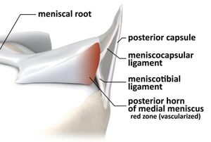
Coronary Ligaments of the Knee
This article is currently under review and may not be up to date. Please come back soon to see the finished work! (29 May 2024)
Original Editor -Simisola Ajeyalemi andRewan Elsayed Elkanafany
Top Contributors - Chloe Waller, Simisola Ajeyalemi, Rewan Elsayed Elkanafany, Kim Jackson and Tien Yunn How
Description[edit | edit source]
The coronary ligaments, also known as the meniscotibial ligaments, are part of the fibrous capsule of the knee joint. They are made up of the medial coronary ligament and the lateral coronary ligament. They connect the inferior edges of the menisci to the of the tibial plateau[1].
Function[edit | edit source]
The coronary ligaments support rotational stability of the knee and prevent anterior tibial translation[2].
Clinical relevance[edit | edit source]
Meniscotibial ligament strain is a common cause of knee pain in middle-aged sportspeople[3]. Coronary ligament injuries may occur either as a rupture in its mid-substance or as an avulsion. In a study by Peltier et al[4] they concluded that lesions of the meniscotibial ligament may increase rotatory instability of the knee. Injury to the meniscotibial ligament attachment of the posterior horn of the medial meniscus is suggested by recent literature to be associated with ramp lesions.[5] Ramp lesions are reported to increase forces on the anterior cruciate ligament,. [4]
Assessment[edit | edit source]
Depending on the severity of the injury, patients presenting with a coronary ligament injury will typically experience pain that is frequently sharp with sudden movements and may or may not be accompanied by swelling. Most of the time, the patient is still able to walk, and both flexion and extension ranges of motion are complete, but with discomfort at end range. A severe injury may result in an effusion that limits full end ranges.
A thorough history of the injury and physical examination are using adequate in diagnosing an injury but if the their is still doubt an MRI can confirm the diagnosis.
A useful test to assess a coronary ligament injury is the McMurray test
Treatment[edit | edit source]
Treatment will vary according to the injury, its degree and whether any other structures have been affected as well .
References[edit | edit source]
- ↑ Guy S, Ferreira A, Carrozzo A, Delaloye JR, Cavaignac E, Vieira TD, Sonnery-Cottet B. Isolated Meniscotibial Ligament Rupture: The Medial Meniscus "Belt Lesion". Arthrosc Tech. 2022 Jan 13;11(2):e133-e138.
- ↑ Feger J, Knipe H, Knipe H, et al. Meniscotibial ligaments. Available from: https://radiopaedia.org/articles/meniscotibial-ligaments(Accessed on 21 Nov 2022)
- ↑ Millar AP. Meniscotibial ligament strains: a prospective survey. British journal of sports medicine. 1991 Jun 1;25(2):94-5.
- ↑ 4.0 4.1 A. Peltier, T. Lording, L. Maubisson, R. Ballis, P. Neyret, S. Lustig. The role of the meniscotibial ligament in posteromedial rotational knee stability Knee Surg Sports Traumatol Arthrosc 2015 23:2967–2973
- ↑ Sonnery-Cottet B, Conteduca J, Thaunat M, Gunepin FX, Seil R. Hidden lesions of the posterior horn of the medial meniscus: a systematic arthroscopic exploration of the concealed portion of the knee. Am J Sports Med. 2014;42:921-926.
- ↑ Advanced Massage Techniques School.Knee Coronary Ligaments TestAvailable from https://www.youtube.com/watch?v=zMQl73Mw-rM


