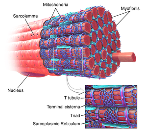
Myofibril
Original Editor - Lucinda hampton
Top Contributors - Lucinda hampton and Vidya Acharya
Introduction[edit | edit source]
Myofibrils are long contractile fibres, groups of which extend in parallel columns along the length of striated myocytes (single multinucleated cells that combine to form the muscle).
The myofibrils are made up of thick and thin myofilaments, which give the muscle its striped appearance. The thick filaments are composed strands of the protein myosin, and the thin filaments are strands of the protein actin, along with two other muscle regulatory proteins, tropomyosin and troponin. Under the influence of adenosine triphosphate (ATP), actin and myosin form a contractile compound, actomyosin, which is required for muscle contraction.
Myofibrils are made up of repeating subunits called sarcomeres. The sarcomeres are responsible for muscle contractions.[1][2].
Sub Heading 2[edit | edit source]
Individual myofibrils (approximately 1 μm in diameter) comprise approximately 80% of the volume of a whole muscle. The number of myofibrils ranges from 50 per myocyte in the muscles of a fetus to approximately 2000 per myocyte in the muscles of an untrained adult. The hypertrophy and atrophy of adult skeletal muscle are associated with certain types of training and disuse and result from the regulation of the number of myofibrils per fiber. Training and disuse have negligible effects on the number of fibers in mammals.
Sub Heading 3[edit | edit source]
Resources[edit | edit source]
- bulleted list
- x
or
- numbered list
- x
References[edit | edit source]
- ↑ Britannica Myofibril Available;https://www.britannica.com/science/myofibril (accessed 9.7.2022)
- ↑ Biology Dictionary Myofibril Available:https://biologydictionary.net/myofibril/ (accessed 9.7.2022)


