Functional Anatomy of the Hip-Muscles and Fascia
Original Editor - Ewa Jaraczewska
Top Contributors - Ewa Jaraczewska, Jess Bell, Kim Jackson and Lucinda hampton
Description[edit | edit source]
One theory assumes that the human body has two muscular systems: local and global. The local muscular system acts close to the joint axis, provides joint compression, and is responsible for this joint stability. The global system contains superficial muscles generating greater torque and greater moment arm.[1]However it is the muscle architecture and the line of action that determines the muscles' primary role. Large forces produced by the muscles and small changes in their length create joint compression thus producing active stabilisation of the joint.[1]
Daily activities require the hip joint to withstand high forces which is possible due to the contribution of the individual muscles surrounding the joint. [2]Active stability provided by the hip muscles can increase passive stability in the normal hip as well as the hip with structural abnormality. [1]
Muscles[edit | edit source]
External Rotators[edit | edit source]
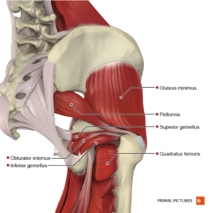
Quadratus femoris: from ischial tuberosity to the intertrochanteric crest of the femur
Obturator internus and externus : from obturator membrane and ischiopubic rams to greater trochanter (internus) or intertrochanteric fossa of the femur
Gemelli (superior and inferior): from ischial spine(superior) or ischial tuberosity (inferior) to greater trochanter and obturator internus tendon
Piriformis: from anterior surface of the sacrum and sacrotuberous ligament to greater trochanter
The roles of these muscles are as follow:
- Active stabilisers of the hip joint. The primary role includes femoral head stabilisation in the acetabulum
- When deep external rotators are resected during hip arthroplasty with posterior surgical approach there is an increased rate of prosthetic dislocation and functional deficits. With capsular repairs, the dislocation rate is lower.
- Resisted external rotation of the hip with extension activates piriformis, but his line of force is not conducive to enhance joint compression.
Internal Rotators[edit | edit source]
Gluteus medius and Gluteus minimus are primary abductors and assist with internal rotation
Tensor Fascia Latae: from anterior superior iliac spine to iliotibial track.
- Located between the deep and superficial layers of the ITB
- Works in different movement planes: assist with hip abduction in the frontal plane, performs hip flexion in saggital plane, completes internal rotation in transverse plane together with anterior gluteus medium and gluteus minimus [3]
Abductors[edit | edit source]
Gluteus minimus: from the outer surface of the ilium, between the anterior and posterior gluteal lines to the greater trochanter.
Its function include:
- Stabilisation of the hip and pelvis through modulation of the joint capsule
- Stabilisation of the femoral head in the acetabulum
- Rotation and flexion of the hip
- Prevention of anterior dislocation and migration of the femoral head in superior and medial direction
- Proprioceptive role
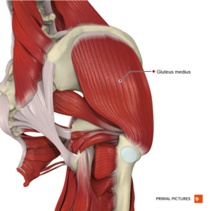
Gluteus medius: from the outer surface of the ilium, between the iliac crest, and the anterior and posterior gluteal lines to the greater trochanter. The muscle has three segments; anterior, posterior, middle or superficial, each with a specific orientation of the muscle fibres.
- Primary abductor of the hip
- Stabiliser of the pelvis and hip,
- Preventing the pelvis from adduction in single leg stance.
- Important stabiliser of the pelvis on the hip by contracting prior to and after foot contact regardless of the walking speed.[1]
Piriformis: externally (laterally) rotates the femur during the hip extension and abducts the femur during hip flexion.[4] The piriformis muscle acts as an auxiliary muscle and shows coactivation during pelvic floor muscles contracture.[5]
Adductors[edit | edit source]
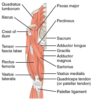
Adductor longus: Pubic bone between the crest and symphysis to linea aspera of the femur
Adductor brevis: Body and inferior ramus of the pubis to linea aspera of the femur
Adductor magnus: Ischial tuberosity and inferior ramus of the pubis to linea aspera and the adductor tubercle
Gracilis: Inferior pubic ramus to medial side of the tibial tuberosity
Pectineus: Pectineal line of the pubis and pubic tubercle to pectineal line of the femur
- Hip adductors contribute to static balance performance[6]
- Adductor longus provides some medial rotation
- Adductor magnus extends the hip through his attachment on the ischial tuberosity
- In open chain activation, the primary function is hip adduction
- In closed chain activation, hip adductors help to stabilise the pelvis and lower extremity during the stance phase of gait.
- Secondary roles of hip adductors include hip flexion and rotation.[7]
- Gracilis and semitendinosus create the conjoined tendons known as the pes anserinus.[8]
Flexors[edit | edit source]
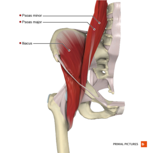
Iliopsoas has three portions:
Iliacus: lateral edge of the sacrum and iliac fossa to lesser trochanter of the femur
Psoas major: transverse processes of vertebrae T12–L5 to lesser trochanter of the femur
Psoas minor: vertebral bodies of T12–L1 to iliopubic ramus
- Psoas major and Iliacus are separately innervated. They are active throughout hip flexion
- Iliacus and both psoas muscles have a role similar to that of the rotator cuff muscles at the shoulder. They affect hip joint stability by creating a tension in musculotendinous units as they pass over the anterior aspect of the hip joint[1]
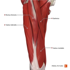
Rectus femoris: Anterior-inferior iliac spine, superior rim of the femoral acetabulum to base of the patella
Sartorius: Anterior superior iliac spine to upper medial side of the tibia.
- Serves as both a hip and knee flexor
- Flexes the hip and externally rotate it
Extensors[edit | edit source]
Gluteus maximus: ilium, sacrum, coccyx, and the sacrotuberous ligament to gluteal tuberosity of the femur and iliotibial band.
- One of the primary hip extensors
- The strongest and biggest muscle in the body
- Prone to weakness and inhibition[9]
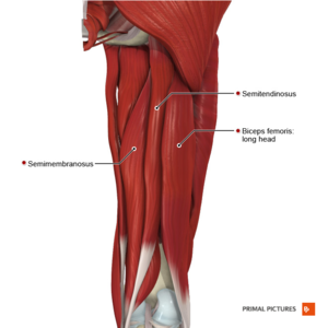
Biceps Femoris: long head: ischial tuberosity; short head: upper supra-condylar line and linea aspera to lateral tibial condyle and head of the fibula.
- The biceps femoris long head is the most affected muscle in the hamstring strain injury[10]
Semimembranosus: Ischial tuberosity to superomedial surface of the tibia
Semitendinosus: Ischial tuberosity to medial condyle of the tibia
Fascia[edit | edit source]
Clinical relevance[edit | edit source]
Resources[edit | edit source]
- ↑ 1.0 1.1 1.2 1.3 1.4 Retchford TH, Crossley KM, Grimaldi A, Kemp JL, Cowan SM. Can local muscles augment stability in the hip? A narrative literature review. J Musculoskelet Neuronal Interact. 2013 Mar 1;13(1):1-2.
- ↑ Correa TA, Crossley KM, Kim HJ, Pandy MG. Contributions of individual muscles to hip joint contact force in normal walking. Journal of biomechanics. 2010 May 28;43(8):1618-22.
- ↑ Besomi Molina M. Towards the investigation of the tensor fascia lata muscle and iliotibial band function in runners: the relevance of the why and the how. The University of Queensland, Australia. A thesis submitted for the degree of Doctor of Philosophy at The University of Queensland in 2020.
- ↑ Chang C, Jeno SH, Varacallo M. Anatomy, bony pelvis and lower limb, piriformis muscle. StatPearls [Internet]. 2020 Nov 12.
- ↑ Wang Z, Zhu Y, Han D, Huang Q, Maruyama H, Onoda K. Effect of hip external rotator muscle contraction on pelvic floor muscle function and the piriformis. International Urogynecology Journal. 2021 Nov 29:1-7.
- ↑ Porto JM, Freire Junior RC, Bocarde L, Fernandes JA, Marques NR, Rodrigues NC, de Abreu DC. Contribution of hip abductor–adductor muscles on static and dynamic balance of community-dwelling older adults. Aging clinical and experimental research. 2019 May;31(5):621-7.
- ↑ Kiel J, Kaiser K. Adductor strain. Available at https://europepmc.org/article/nbk/nbk493166 (last access 23.02.2022)
- ↑ Walters BB, Varacallo M. Anatomy, Bony Pelvis and Lower Limb, Thigh Sartorius Muscle. In: StatPearls. StatPearls Publishing, Treasure Island (FL); 2021
- ↑ Buckthorpe M, Stride M, Della Villa F. Assessing and treating gluteus maximus weakness–a clinical commentary. International journal of sports physical therapy. 2019 Jul;14(4):655.
- ↑ Llurda-Almuzara L, Labata-Lezaun N, López-de-Celis C, Aiguadé-Aiguadé R, Romaní-Sánchez S, Rodríguez-Sanz J, Fernández-de-Las-Peñas C, Pérez-Bellmunt A. Biceps femoris activation during hamstring strength exercises: a systematic review. International Journal of Environmental Research and Public Health. 2021 Jan;18(16):8733.
